Loading
PDBj
MenuPDBj@FacebookPDBj@TwitterPDBj@YouTubewwPDB FoundationwwPDB
RCSB PDBPDBeBMRBAdv. SearchSearch help
PDB: 11 results

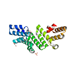
4P29
DownloadVisualize
BU of 4p29 by Molmil
Crystal structure of the LpoA N-terminal domain from Haemophilus influenzae
Descriptor: CHLORIDE ION, GLYCEROL, LpoA, ...
Authors:Vijayalakshmi, J, Saper, M.A.
Deposit date:2014-03-02
Release date:2015-03-04
Last modified:2023-12-27
Method:X-RAY DIFFRACTION (1.95006478 Å)
Cite:Structural analyses of theHaemophilus influenzaepeptidoglycan synthase activator LpoA suggest multiple conformations in solution.
J.Biol.Chem., 292, 2017
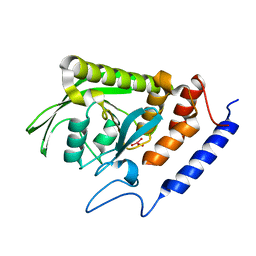
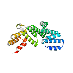
6DCJ
DownloadVisualize
BU of 6dcj by Molmil
LpoA N-terminal domain from Haemophilus influenzae; monoclinic form at 1.35 A resolution
Descriptor: CHLORIDE ION, Penicillin-binding protein activator LpoA
Authors:Vijayalakshmi, J, Saper, M.A.
Deposit date:2018-05-07
Release date:2019-05-01
Last modified:2023-10-11
Method:X-RAY DIFFRACTION (1.35 Å)
Cite:Crystal structures of the amino-terminal domain of LpoA from Escherichia coli and Haemophilus influenzae.
Acta Crystallogr.,Sect.F, 75, 2019
2I42
DownloadVisualize
BU of 2i42 by Molmil
Crystal structure of Yersinia protein tyrosine phosphatase complexed with vanadate, a transition state analogue
Descriptor: Tyrosine-protein phosphatase, VANADATE ION
Authors:Vijayalakshmi, J, Saper, M.A.
Deposit date:2006-08-21
Release date:2006-09-19
Last modified:2023-08-30
Method:X-RAY DIFFRACTION (2.2 Å)
Cite:Crystal structure of Yersinia protein tyrosine phosphatase complexed with vanadate, a transition state analogue
To be Published
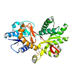
3CKM
DownloadVisualize
BU of 3ckm by Molmil
LpoA (YraM) C-domain from Haemophilus influenzae, a regulator of PBP1A
Descriptor: BETA-MERCAPTOETHANOL, SULFATE ION, YRAM (HI1655)
Authors:Vijayalakshmi, J, Saper, M.A.
Deposit date:2008-03-16
Release date:2008-05-13
Last modified:2017-11-08
Method:X-RAY DIFFRACTION (1.35 Å)
Cite:Structure of YraM, a protein essential for growth of Haemophilus influenzae.
Proteins, 73, 2008
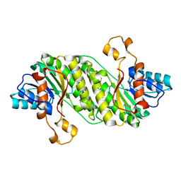
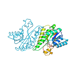
3F1L
DownloadVisualize
BU of 3f1l by Molmil
The 0.95 A structure of an oxidoreductase, yciK from E.coli
Descriptor: Uncharacterized oxidoreductase yciK
Authors:Vijayalakshmi, J, Woodard, R.W.
Deposit date:2008-10-28
Release date:2008-11-18
Last modified:2023-09-06
Method:X-RAY DIFFRACTION (0.95 Å)
Cite:Structure of an oxidoreductase, yciK from E. coli, in two crystal forms - NADP+ unbound structure at 0.95 A resolution
to be published
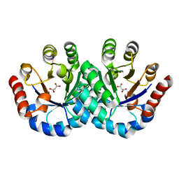
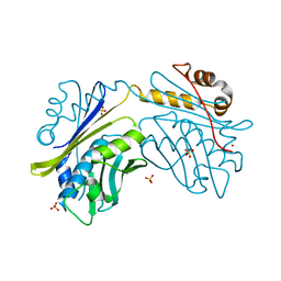
3F4W
DownloadVisualize
BU of 3f4w by Molmil
The 1.65A Crystal Structure of 3-hexulose-6-phosphate synthase from Salmonella typhimurium
Descriptor: MALONATE ION, Putative hexulose 6 phosphate synthase
Authors:Vijayalakshmi, J, Woodard, R.W.
Deposit date:2008-11-03
Release date:2008-11-25
Last modified:2023-09-06
Method:X-RAY DIFFRACTION (1.65 Å)
Cite:The 1.65A Crystal Structure of 3-hexulose-6-phosphate synthase from Salmonella typhimurium
To be Published
3F1K
DownloadVisualize
BU of 3f1k by Molmil
Crystal Structure of yciK from E. coli, an oxidoreductase, complexed with NADP+ at 2.6A resolution
Descriptor: NADP NICOTINAMIDE-ADENINE-DINUCLEOTIDE PHOSPHATE, Uncharacterized oxidoreductase yciK
Authors:Vijayalakshmi, J, Woodard, R.W.
Deposit date:2008-10-28
Release date:2008-11-18
Last modified:2023-12-27
Method:X-RAY DIFFRACTION (2.6 Å)
Cite:Structure of an oxidoreductase, yciK from E. coli, in two crystal forms - NADP+ unbound structure at 0.95 A resolution
to be published
1HW7
DownloadVisualize
BU of 1hw7 by Molmil
HSP33, HEAT SHOCK PROTEIN WITH REDOX-REGULATED CHAPERONE ACTIVITY
Descriptor: 2-(N-MORPHOLINO)-ETHANESULFONIC ACID, HEAT SHOCK PROTEIN HSP33, SULFATE ION, ...
Authors:Vijayalakshmi, J, Mukhergee, M.K, Graumann, J, Jakob, U, Saper, M.A.
Deposit date:2001-01-09
Release date:2001-05-16
Last modified:2024-04-03
Method:X-RAY DIFFRACTION (2.2 Å)
Cite:The 2.2 A crystal structure of Hsp33: a heat shock protein with redox-regulated chaperone activity.
Structure, 9, 2001
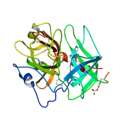
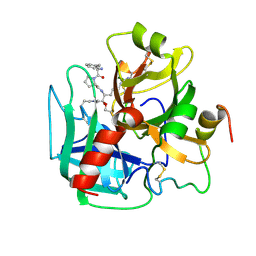
1HAH
DownloadVisualize
BU of 1hah by Molmil
THE ISOMORPHOUS STRUCTURES OF PRETHROMBIN2, HIRUGEN-AND PPACK-THROMBIN: CHANGES ACCOMPANYING ACTIVATION AND EXOSITE BINDING TO THROMBIN
Descriptor: 2-acetamido-2-deoxy-beta-D-glucopyranose, ALPHA-THROMBIN (LARGE SUBUNIT), ALPHA-THROMBIN (SMALL SUBUNIT), ...
Authors:Tulinsky, A, Vijayalakshmi, J.
Deposit date:1994-06-27
Release date:1994-12-20
Last modified:2020-07-29
Method:X-RAY DIFFRACTION (2.3 Å)
Cite:The isomorphous structures of prethrombin2, hirugen-, and PPACK-thrombin: changes accompanying activation and exosite binding to thrombin.
Protein Sci., 3, 1994
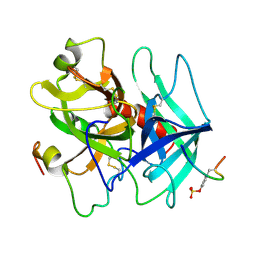
1HAG
DownloadVisualize
BU of 1hag by Molmil
THE ISOMORPHOUS STRUCTURES OF PRETHROMBIN2, HIRUGEN-AND PPACK-THROMBIN: CHANGES ACCOMPANYING ACTIVATION AND EXOSITE BINDING TO THROMBIN
Descriptor: 2-acetamido-2-deoxy-beta-D-glucopyranose, HIRUGEN, PRETHROMBIN 2
Authors:Tulinsky, A, Vijayalakshmi, J.
Deposit date:1994-06-27
Release date:1994-12-20
Last modified:2020-07-29
Method:X-RAY DIFFRACTION (2 Å)
Cite:The isomorphous structures of prethrombin2, hirugen-, and PPACK-thrombin: changes accompanying activation and exosite binding to thrombin.
Protein Sci., 3, 1994
1HAI
DownloadVisualize
BU of 1hai by Molmil
THE ISOMORPHOUS STRUCTURES OF PRETHROMBIN2, HIRUGEN-AND PPACK-THROMBIN: CHANGES ACCOMPANYING ACTIVATION AND EXOSITE BINDING TO THROMBIN
Descriptor: 2-acetamido-2-deoxy-beta-D-glucopyranose, ALPHA-THROMBIN (LARGE SUBUNIT), ALPHA-THROMBIN (SMALL SUBUNIT), ...
Authors:Tulinsky, A, Vijayalakshmi, J.
Deposit date:1994-06-27
Release date:1994-12-20
Last modified:2020-07-29
Method:X-RAY DIFFRACTION (2.4 Å)
Cite:The isomorphous structures of prethrombin2, hirugen-, and PPACK-thrombin: changes accompanying activation and exosite binding to thrombin.
Protein Sci., 3, 1994

218853

PDB entries from 2024-04-24

PDB statisticsPDBj update infoContact PDBjnumon