Loading
PDBj
MenuPDBj@FacebookPDBj@TwitterPDBj@YouTubewwPDB FoundationwwPDB
RCSB PDBPDBeBMRBAdv. SearchSearch help
PDB: 143 results

7KV6
DownloadVisualize
BU of 7kv6 by Molmil
Surface glycan-binding protein B from Bacteroides fluxus in complex with mixed-linkage glucotriose
Descriptor: GUANIDINE, PKD domain protein, beta-D-glucopyranose-(1-4)-beta-D-glucopyranose-(1-3)-beta-D-glucopyranose
Authors:Tamura, K, Brumer, H, Van Petegem, F.
Deposit date:2020-11-26
Release date:2021-02-17
Last modified:2024-03-06
Method:X-RAY DIFFRACTION (1.6 Å)
Cite:Distinct protein architectures mediate species-specific beta-glucan binding and metabolism in the human gut microbiota.
J.Biol.Chem., 296, 2021
7KWB
DownloadVisualize
BU of 7kwb by Molmil
Surface glycan-binding protein B from Bacteroides thetaiotaomicron
Descriptor: BtSGBP-B
Authors:Tamura, K, Brumer, H, Van Petegem, F.
Deposit date:2020-11-30
Release date:2021-02-17
Last modified:2024-03-06
Method:X-RAY DIFFRACTION (2.6 Å)
Cite:Distinct protein architectures mediate species-specific beta-glucan binding and metabolism in the human gut microbiota.
J.Biol.Chem., 296, 2021
7KV3
DownloadVisualize
BU of 7kv3 by Molmil
Surface glycan-binding protein A from Bacteroides thetaiotaomicron in complex with laminarihexaose
Descriptor: RagB/SusD family nutrient uptake outer membrane protein, SULFATE ION, beta-D-glucopyranose-(1-3)-beta-D-glucopyranose-(1-3)-beta-D-glucopyranose-(1-3)-beta-D-glucopyranose-(1-3)-beta-D-glucopyranose-(1-3)-beta-D-glucopyranose
Authors:Tamura, K, Brumer, H, Van Petegem, F.
Deposit date:2020-11-26
Release date:2021-02-17
Last modified:2024-03-06
Method:X-RAY DIFFRACTION (2.05 Å)
Cite:Distinct protein architectures mediate species-specific beta-glucan binding and metabolism in the human gut microbiota.
J.Biol.Chem., 296, 2021
7KV4
DownloadVisualize
BU of 7kv4 by Molmil
Surface glycan-binding protein A from Bacteroides fluxus
Descriptor: SusD family protein
Authors:Tamura, K, Brumer, H, Van Petegem, F.
Deposit date:2020-11-26
Release date:2021-02-17
Last modified:2024-03-06
Method:X-RAY DIFFRACTION (1.84 Å)
Cite:Distinct protein architectures mediate species-specific beta-glucan binding and metabolism in the human gut microbiota.
J.Biol.Chem., 296, 2021
7KWC
DownloadVisualize
BU of 7kwc by Molmil
Surface glycan-binding protein B (truncated) from Bacteroides thetaiotaomicron
Descriptor: BtSGBP-B_31-285
Authors:Tamura, K, Brumer, H, Van Petegem, F.
Deposit date:2020-11-30
Release date:2021-02-17
Last modified:2024-03-06
Method:X-RAY DIFFRACTION (2.611 Å)
Cite:Distinct protein architectures mediate species-specific beta-glucan binding and metabolism in the human gut microbiota.
J.Biol.Chem., 296, 2021
3FYQ
DownloadVisualize
BU of 3fyq by Molmil
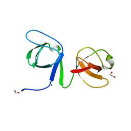
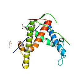
Structure of Drosophila melanogaster talin IBS2 domain (residues 1981-2168)
Descriptor: CG6831-PA (Talin)
Authors:Cheung, T.Y.S, Fairchild, M.J, Zarivach, R, Tanentzapf, G, Van Petegem, F.
Deposit date:2009-01-22
Release date:2009-02-03
Last modified:2018-01-24
Method:X-RAY DIFFRACTION (1.95 Å)
Cite:Crystal structure of the talin integrin binding domain 2.
J.Mol.Biol., 387, 2009
6ULB
DownloadVisualize
BU of 6ulb by Molmil
Sex Hormone-binding globulin mutant E176K in complex with Danazol
Descriptor: CALCIUM ION, Danazol, Sex hormone-binding globulin
Authors:Round, P.W, Das, S, Van Petegem, F.
Deposit date:2019-10-07
Release date:2020-10-21
Last modified:2023-10-11
Method:X-RAY DIFFRACTION (1.75 Å)
Cite:Structural and biochemical analyses of danazol interactions with sex hormone-binding globulin and effects on androgen action
To Be Published
6VHO
DownloadVisualize
BU of 6vho by Molmil
Glycoside hydrolase family 16 endo-glucanase from Bacteroides ovatus in complex with G4G4G3G-NHCOCH2Br
Descriptor: Glycosyl hydrolase family 16, beta-D-glucopyranose-(1-4)-beta-D-glucopyranose-(1-3)-N-acetyl-beta-D-glucopyranosylamine
Authors:Tamura, K, Brumer, H, van Petegem, F.
Deposit date:2020-01-10
Release date:2021-01-13
Last modified:2023-10-11
Method:X-RAY DIFFRACTION (2.148 Å)
Cite:Orthogonal Active-Site Labels for Mixed-Linkage endo-beta-Glucanases.
Acs Chem.Biol., 16, 2021
6UY7
DownloadVisualize
BU of 6uy7 by Molmil
Crystal structure of the STAC3 tandem SH3 domains - P269R
Descriptor: 1,2-ETHANEDIOL, SH3 and cysteine-rich domain-containing protein 3
Authors:Rufenach, B, Van Petegem, F.
Deposit date:2019-11-12
Release date:2020-06-17
Last modified:2023-10-11
Method:X-RAY DIFFRACTION (2.105 Å)
Cite:Multiple Sequence Variants in STAC3 Affect Interactions with CaV1.1 and Excitation-Contraction Coupling.
Structure, 28, 2020
6UY9
DownloadVisualize
BU of 6uy9 by Molmil
Crystal structure of the STAC3 tandem SH3 domains - P269R, W284S
Descriptor: 1,2-ETHANEDIOL, SH3 and cysteine-rich domain-containing protein 3, SODIUM ION
Authors:Rufenach, B, Van Petegem, F.
Deposit date:2019-11-12
Release date:2020-06-17
Last modified:2023-10-11
Method:X-RAY DIFFRACTION (1.6 Å)
Cite:Multiple Sequence Variants in STAC3 Affect Interactions with CaV1.1 and Excitation-Contraction Coupling.
Structure, 28, 2020
4KEJ
DownloadVisualize
BU of 4kej by Molmil
Crystal structure of mouse Ryanodine Receptor 2 (1-217) disease mutant R169Q
Descriptor: Ryanodine receptor 2
Authors:Kimlicka, L, Van Petegem, F.
Deposit date:2013-04-25
Release date:2013-09-04
Last modified:2023-09-20
Method:X-RAY DIFFRACTION (2.553 Å)
Cite:Type 2 Ryanodine Receptor Domain A Contains a Unique and Dynamic alpha-Helix That Transitions to a beta-Strand in a Mutant Linked with a Heritable Cardiomyopathy.
J.Mol.Biol., 425, 2013
4KEI
DownloadVisualize
BU of 4kei by Molmil
Crystal structure of mouse Ryanodine Receptor 2 (1-217) disease mutant P164S
Descriptor: Ryanodine receptor 2
Authors:Kimlicka, L, Van Petegem, F.
Deposit date:2013-04-25
Release date:2013-09-04
Last modified:2023-09-20
Method:X-RAY DIFFRACTION (2.406 Å)
Cite:Type 2 Ryanodine Receptor Domain A Contains a Unique and Dynamic alpha-Helix That Transitions to a beta-Strand in a Mutant Linked with a Heritable Cardiomyopathy.
J.Mol.Biol., 425, 2013
4KEK
DownloadVisualize
BU of 4kek by Molmil
Crystal structure of mouse Ryanodine Receptor 2 (1-217) disease mutant R176Q
Descriptor: Ryanodine receptor 2, SULFATE ION
Authors:Kimlicka, L, Van Petegem, F.
Deposit date:2013-04-25
Release date:2013-09-04
Last modified:2023-09-20
Method:X-RAY DIFFRACTION (2.146 Å)
Cite:Type 2 Ryanodine Receptor Domain A Contains a Unique and Dynamic alpha-Helix That Transitions to a beta-Strand in a Mutant Linked with a Heritable Cardiomyopathy.
J.Mol.Biol., 425, 2013
4DJC
DownloadVisualize
BU of 4djc by Molmil
1.35 A crystal structure of the NaV1.5 DIII-IV-Ca/CaM complex
Descriptor: CALCIUM ION, Calmodulin, ISOPROPYL ALCOHOL, ...
Authors:Sarhan, M.F, Tung, C.-C, Van Petegem, F, Ahern, C.A.
Deposit date:2012-02-01
Release date:2012-02-22
Last modified:2024-02-28
Method:X-RAY DIFFRACTION (1.35 Å)
Cite:Crystallographic basis for calcium regulation of sodium channels.
Proc.Natl.Acad.Sci.USA, 109, 2012
4ETT
DownloadVisualize
BU of 4ett by Molmil
Crystal structure of rabbit ryanodine receptor 1 mutant E2764K
Descriptor: GLYCEROL, Ryanodine receptor 1
Authors:Yuchi, Z, Lau, K, Van Petegem, F.
Deposit date:2012-04-24
Release date:2012-06-13
Last modified:2023-09-13
Method:X-RAY DIFFRACTION (2.195 Å)
Cite:Disease mutations in the ryanodine receptor central region: crystal structures of a phosphorylation hot spot domain.
Structure, 20, 2012
6B29
DownloadVisualize
BU of 6b29 by Molmil
Crystal structure of the second SH3 domain of STAC3 (309-364)
Descriptor: SH3 and cysteine-rich domain-containing protein 3
Authors:Wong King Yuen, S.M, Van Petegem, F.
Deposit date:2017-09-19
Release date:2017-10-25
Last modified:2024-03-13
Method:X-RAY DIFFRACTION (1.3 Å)
Cite:Structural insights into binding of STAC proteins to voltage-gated calcium channels.
Proc. Natl. Acad. Sci. U.S.A., 114, 2017
4ERV
DownloadVisualize
BU of 4erv by Molmil
Crystal structure of human ryanodine receptor 3 (2597-2800)
Descriptor: GLYCEROL, Ryanodine receptor 3, SULFATE ION
Authors:Yuchi, Z, Lau, K, Van Petegem, F.
Deposit date:2012-04-20
Release date:2012-06-13
Last modified:2024-02-28
Method:X-RAY DIFFRACTION (1.75 Å)
Cite:Disease mutations in the ryanodine receptor central region: crystal structures of a phosphorylation hot spot domain.
Structure, 20, 2012
4ESU
DownloadVisualize
BU of 4esu by Molmil
Crystal structure of rabbit ryanodine receptor 1 mutant S2776M
Descriptor: GLYCEROL, Ryanodine receptor 1
Authors:Yuchi, Z, Lau, K, Van Petegem, F.
Deposit date:2012-04-23
Release date:2012-06-13
Last modified:2023-09-13
Method:X-RAY DIFFRACTION (1.59 Å)
Cite:Disease mutations in the ryanodine receptor central region: crystal structures of a phosphorylation hot spot domain.
Structure, 20, 2012
6B27
DownloadVisualize
BU of 6b27 by Molmil
Crystal structure of human STAC2 Tandem SH3 Domains (296-411) in complex with a CaV1.1 II-III loop peptide
Descriptor: CHLORIDE ION, SH3 and cysteine-rich domain-containing protein 2, SULFATE ION, ...
Authors:Wong King Yuen, S.M, Van Petegem, F.
Deposit date:2017-09-19
Release date:2017-10-25
Last modified:2024-03-13
Method:X-RAY DIFFRACTION (1.73 Å)
Cite:Structural insights into binding of STAC proteins to voltage-gated calcium channels.
Proc. Natl. Acad. Sci. U.S.A., 114, 2017
6DLG
DownloadVisualize
BU of 6dlg by Molmil
Crystal structure of a SHIP1 surface entropy reduction mutant
Descriptor: ISOPROPYL ALCOHOL, Phosphatidylinositol 3,4,5-trisphosphate 5-phosphatase 1
Authors:Gardill, B.R, Cheung, S.T, Mui, A.L, Van Petegem, F.
Deposit date:2018-06-01
Release date:2019-06-05
Last modified:2024-03-13
Method:X-RAY DIFFRACTION (1.499 Å)
Cite:Interleukin-10 and Small Molecule SHIP1 Allosteric Regulators Trigger Anti-Inflammatory Effects Through SHIP1/STAT3 Complexes
Biorxiv, 2020
4I2S
DownloadVisualize
BU of 4i2s by Molmil
Crystal structure of rabbit ryanodine receptor 1 (residues 1-536) disease mutant I404M
Descriptor: GLYCEROL, Ryanodine receptor 1
Authors:Kimlicka, L, Van Petegem, F.
Deposit date:2012-11-22
Release date:2013-02-20
Last modified:2023-09-20
Method:X-RAY DIFFRACTION (2.5 Å)
Cite:Disease mutations in the ryanodine receptor N-terminal region couple to a mobile intersubunit interface.
Nat Commun, 4, 2013
4I8M
DownloadVisualize
BU of 4i8m by Molmil
Crystal structure of rabbit Ryanodine receptor 1 (residues 1-536) disease mutant V219I
Descriptor: GLYCEROL, Ryanodine receptor 1
Authors:Kimlicka, L, Van Petegem, F.
Deposit date:2012-12-03
Release date:2013-02-20
Last modified:2023-09-20
Method:X-RAY DIFFRACTION (2.8 Å)
Cite:Disease mutations in the ryanodine receptor N-terminal region couple to a mobile intersubunit interface.
Nat Commun, 4, 2013
4I96
DownloadVisualize
BU of 4i96 by Molmil
Crystal structure of the N-terminal two domains of the skeletal muscle ryanodine receptor (rabbit RyR1) residues 217-536
Descriptor: Ryanodine receptor 1
Authors:Kimlicka, L, Tung, C.C, Van Petegem, F.
Deposit date:2012-12-04
Release date:2013-02-20
Last modified:2023-09-20
Method:X-RAY DIFFRACTION (2.73 Å)
Cite:Disease mutations in the ryanodine receptor N-terminal region couple to a mobile intersubunit interface.
Nat Commun, 4, 2013
4I7I
DownloadVisualize
BU of 4i7i by Molmil
Crystal structure of rabbit ryanodine receptor 1 (residues 1-536) disease mutant L14R
Descriptor: GLYCEROL, Ryanodine receptor 1
Authors:Kimlicka, L, Van Petegem, F.
Deposit date:2012-11-30
Release date:2013-02-20
Last modified:2023-09-20
Method:X-RAY DIFFRACTION (2.9 Å)
Cite:Disease mutations in the ryanodine receptor N-terminal region couple to a mobile intersubunit interface.
Nat Commun, 4, 2013
4I1E
DownloadVisualize
BU of 4i1e by Molmil
Crystal structure of Rabbit Ryanodine Receptor 1 (residues 1-536) disease mutant G249R
Descriptor: Ryanodine receptor 1
Authors:Kimlicka, L, Van Petegem, F.
Deposit date:2012-11-20
Release date:2013-02-20
Last modified:2024-02-28
Method:X-RAY DIFFRACTION (2.4 Å)
Cite:Disease mutations in the ryanodine receptor N-terminal region couple to a mobile intersubunit interface.
Nat Commun, 4, 2013

220472

건을2024-05-29부터공개중

PDB statisticsPDBj update infoContact PDBjnumon