9IPB
 
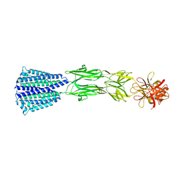
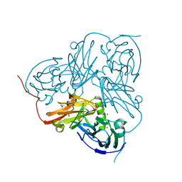
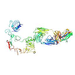
 | | Local refinement structure of sEGFR and 528 Fv (from LH-type bispecific diabody Ex3) complex | | Descriptor: | 2-acetamido-2-deoxy-beta-D-glucopyranose, 2-acetamido-2-deoxy-beta-D-glucopyranose-(1-4)-2-acetamido-2-deoxy-beta-D-glucopyranose, 528 Fv from LH-type bispecific diabody Ex3, ... | | Authors: | Sato, K, Uehara, S, Tsugita, A, Matsui, T, Asano, R, Makabe, K, Yokoyama, T, Tanaka, Y. | | Deposit date: | 2024-07-10 | | Release date: | 2025-05-28 | | Last modified: | 2025-08-06 | | Method: | ELECTRON MICROSCOPY (2.93 Å) | | Cite: | Bispecific antibody-antigen complex structures reveal activity enhancement by domain rearrangement.
Cell Rep, 44, 2025
|
|
9IPA
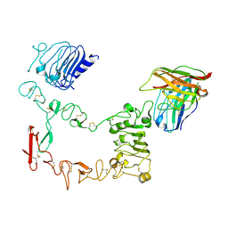
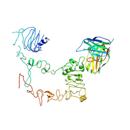
 | | Poly-alanine model for HL-type bispecific diabody Ex3 composed of 528 and OKT3 Fvs in ternary complex with sEGFR and CD3gamma-epsilon (open conformation) | | Descriptor: | Epidermal growth factor receptor, HL-type bispecific diabody Ex3, T-cell surface glycoprotein CD3 gamma chain,T-cell surface glycoprotein CD3 epsilon chain | | Authors: | Sato, K, Uehara, S, Tsugita, A, Matsui, T, Asano, R, Makabe, K, Yokoyama, T, Tanaka, Y. | | Deposit date: | 2024-07-10 | | Release date: | 2025-05-28 | | Last modified: | 2025-08-06 | | Method: | ELECTRON MICROSCOPY (3.85 Å) | | Cite: | Bispecific antibody-antigen complex structures reveal activity enhancement by domain rearrangement.
Cell Rep, 44, 2025
|
|
9IPE
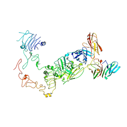
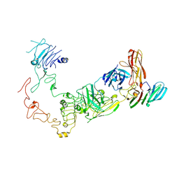
 | | Poly-alanine model for LH-type bispecific diabody Ex3 composed of 528 and OKT3 Fvs in ternary complex with sEGFR and CD3gamma-epsilon (open conformation) | | Descriptor: | Epidermal growth factor receptor, LH-type bispecific diabody Ex3, T-cell surface glycoprotein CD3 gamma chain,T-cell surface glycoprotein CD3 epsilon chain | | Authors: | Sato, K, Uehara, S, Tsugita, A, Matsui, T, Asano, R, Makabe, K, Yokoyama, T, Tanaka, Y. | | Deposit date: | 2024-07-10 | | Release date: | 2025-05-28 | | Last modified: | 2025-08-06 | | Method: | ELECTRON MICROSCOPY (3.31 Å) | | Cite: | Bispecific antibody-antigen complex structures reveal activity enhancement by domain rearrangement.
Cell Rep, 44, 2025
|
|
9IP8
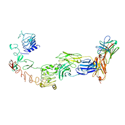
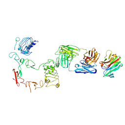
 | | Poly-alanine model for HL-type bispecific diabody Ex3 composed of 528 and OKT3 Fvs in ternary complex with sEGFR and CD3gamma-epsilon (closed conformation) | | Descriptor: | Epidermal growth factor receptor, HL-type bispecific diabody Ex3, T-cell surface glycoprotein CD3 gamma chain,T-cell surface glycoprotein CD3 epsilon chain | | Authors: | Sato, K, Uehara, S, Tsugita, A, Matsui, T, Asano, R, Makabe, K, Yokoyama, T, Tanaka, Y. | | Deposit date: | 2024-07-10 | | Release date: | 2025-05-28 | | Last modified: | 2025-08-06 | | Method: | ELECTRON MICROSCOPY (3.91 Å) | | Cite: | Bispecific antibody-antigen complex structures reveal activity enhancement by domain rearrangement.
Cell Rep, 44, 2025
|
|
7CMG
 | | The Structure of the periplasmic domain of PorM | | Descriptor: | Por secretion system protein porM/gldM | | Authors: | Sato, K, Okada, K, Nakayam, K, Imada, K. | | Deposit date: | 2020-07-27 | | Release date: | 2020-09-02 | | Last modified: | 2023-11-29 | | Method: | X-RAY DIFFRACTION (3.7 Å) | | Cite: | PorM, a core component of bacterial type IX secretion system, forms a dimer with a unique kinked-rod shape.
Biochem.Biophys.Res.Commun., 532, 2020
|
|
9JL6
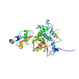
 | | Cryo-EM structure of chalcone synthase (CHS) from Physcomitrella patens in the presence of CHIL | | Descriptor: | Chalcone synthase | | Authors: | Sato, K, Yokoyama, T, Tanaka, Y, Imaizumi, R, Yasuda, A, Yanai, T, Yamashita, S, Waki, T, Tsunashima, M, Nakayama, T. | | Deposit date: | 2024-09-18 | | Release date: | 2025-10-01 | | Method: | ELECTRON MICROSCOPY (2.18 Å) | | Cite: | Cryo-EM structure of chalcone synthase (CHS) from Physcomitrella patens in the presence of CHIL
To Be Published
|
|
1K37
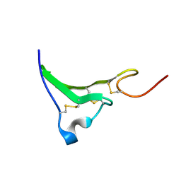 | | NMR Structure of human Epiregulin | | Descriptor: | Epiregulin | | Authors: | Sato, K, Miura, K, Tada, M, Aizawa, T, Miyamoto, K, Kawano, K. | | Deposit date: | 2001-10-02 | | Release date: | 2003-09-30 | | Last modified: | 2024-10-23 | | Method: | SOLUTION NMR | | Cite: | Solution structure of epiregulin and the effect of its C-terminal domain for receptor binding affinity
Febs Lett., 553, 2003
|
|
1K36
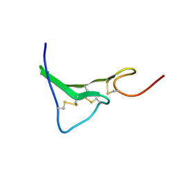 | | NMR Structure of human Epiregulin | | Descriptor: | Epiregulin | | Authors: | Sato, K, Miura, K, Tada, M, Aizawa, T, Miyamoto, K, Kawano, K. | | Deposit date: | 2001-10-02 | | Release date: | 2003-09-30 | | Last modified: | 2024-11-20 | | Method: | SOLUTION NMR | | Cite: | Solution structure of epiregulin and the effect of its C-terminal domain for receptor binding affinity
Febs Lett., 553, 2003
|
|
2VN3
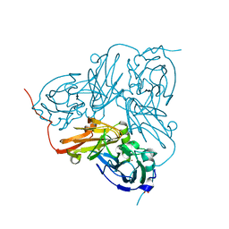 | | Nitrite Reductase from Alcaligenes xylosoxidans | | Descriptor: | COPPER (II) ION, DISSIMILATORY COPPER-CONTAINING NITRITE REDUCTASE, SULFATE ION, ... | | Authors: | Sato, K, Firbank, S.J, Li, C, Banfield, M.J, Dennison, C. | | Deposit date: | 2008-01-30 | | Release date: | 2008-12-23 | | Last modified: | 2023-12-13 | | Method: | X-RAY DIFFRACTION (2.35 Å) | | Cite: | The Importance of the Long Type 1 Copper-Binding Loop of Nitrite Reductase for Structure and Function.
Chemistry, 14, 2008
|
|
2VMJ
 | | Type 1 Copper-Binding Loop of Nitrite Reductase mutant: 130- CAPEGMVPWHVVSGM-144 to 130-CTPHPFM-136 | | Descriptor: | 4-(2-HYDROXYETHYL)-1-PIPERAZINE ETHANESULFONIC ACID, DISSIMILATORY COPPER-CONTAINING NITRITE REDUCTASE, ZINC ION | | Authors: | Sato, K, Firbank, S.J, Li, C, Banfield, M.J, Dennison, C. | | Deposit date: | 2008-01-28 | | Release date: | 2008-12-23 | | Last modified: | 2023-12-13 | | Method: | X-RAY DIFFRACTION (2.5 Å) | | Cite: | The Importance of the Long Type 1 Copper-Binding Loop of Nitrite Reductase for Structure and Function.
Chemistry, 14, 2008
|
|
9IP7
 | | Local refinement structure of sEGFR and 528 Fv (from HL-type bispecific diabody Ex3) complex | | Descriptor: | 2-acetamido-2-deoxy-beta-D-glucopyranose, 2-acetamido-2-deoxy-beta-D-glucopyranose-(1-4)-2-acetamido-2-deoxy-beta-D-glucopyranose, 528 Fv from HL-type bispecific diabody Ex3, ... | | Authors: | Sato, K, Uehara, S, Tsugita, A, Matsui, T, Asano, R, Makabe, K, Yokoyama, T, Tanaka, Y. | | Deposit date: | 2024-07-10 | | Release date: | 2025-05-28 | | Last modified: | 2025-08-06 | | Method: | ELECTRON MICROSCOPY (3.21 Å) | | Cite: | Bispecific antibody-antigen complex structures reveal activity enhancement by domain rearrangement.
Cell Rep, 44, 2025
|
|
9IP9
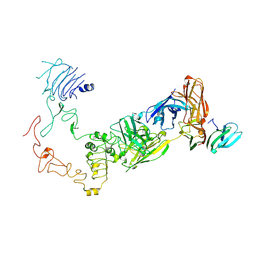
 | | Poly-alanine model for HL-type bispecific diabody Ex3 composed of 528 and OKT3 Fvs in ternary complex with sEGFR and CD3gamma-epsilon (middle conformation) | | Descriptor: | Epidermal growth factor receptor, HL-type bispecific diabody Ex3, T-cell surface glycoprotein CD3 gamma chain,T-cell surface glycoprotein CD3 epsilon chain | | Authors: | Sato, K, Uehara, S, Tsugita, A, Matsui, T, Asano, R, Makabe, K, Yokoyama, T, Tanaka, Y. | | Deposit date: | 2024-07-10 | | Release date: | 2025-05-28 | | Last modified: | 2025-08-06 | | Method: | ELECTRON MICROSCOPY (3.64 Å) | | Cite: | Bispecific antibody-antigen complex structures reveal activity enhancement by domain rearrangement.
Cell Rep, 44, 2025
|
|
9IPC
 | | Poly-alanine model for LH-type bispecific diabody Ex3 composed of 528 and OKT3 Fvs in ternary complex with sEGFR and CD3gamma-epsilon (closed conformation) | | Descriptor: | Epidermal growth factor receptor, LH-type bispecific diabody Ex3, T-cell surface glycoprotein CD3 gamma chain,T-cell surface glycoprotein CD3 epsilon chain | | Authors: | Sato, K, Uehara, S, Tsugita, A, Matsui, T, Asano, R, Makabe, K, Yokoyama, T, Tanaka, Y. | | Deposit date: | 2024-07-10 | | Release date: | 2025-05-28 | | Last modified: | 2025-08-06 | | Method: | ELECTRON MICROSCOPY (3.4 Å) | | Cite: | Bispecific antibody-antigen complex structures reveal activity enhancement by domain rearrangement.
Cell Rep, 44, 2025
|
|
9IPD
 | | Poly-alanine model for LH-type bispecific diabody Ex3 composed of 528 and OKT3 Fvs in ternary complex with sEGFR and CD3gamma-epsilon (middle conformation) | | Descriptor: | Epidermal growth factor receptor, LH-type bispecific diabody Ex3, T-cell surface glycoprotein CD3 gamma chain,T-cell surface glycoprotein CD3 epsilon chain | | Authors: | Sato, K, Uehara, S, Tsugita, A, Matsui, T, Asano, R, Makabe, K, Yokoyama, T, Tanaka, Y. | | Deposit date: | 2024-07-10 | | Release date: | 2025-05-28 | | Last modified: | 2025-08-06 | | Method: | ELECTRON MICROSCOPY (3.29 Å) | | Cite: | Bispecific antibody-antigen complex structures reveal activity enhancement by domain rearrangement.
Cell Rep, 44, 2025
|
|
2Z0D
 | |
2Z0E
 | |
2ZZP
 | |
1QCM
 | | AMYLOID BETA PEPTIDE (25-35), NMR, 20 STRUCTURES | | Descriptor: | AMYLOID BETA PEPTIDE | | Authors: | Kohno, T, Kobayashi, K, Maeda, T, Sato, K, Takashima, A. | | Deposit date: | 1996-07-19 | | Release date: | 1997-07-07 | | Last modified: | 2024-05-22 | | Method: | SOLUTION NMR | | Cite: | Three-dimensional structures of the amyloid beta peptide (25-35) in membrane-mimicking environment.
Biochemistry, 35, 1996
|
|
2P5J
 | | sPLA2 inhibitor pip 17 | | Descriptor: | pip17 | | Authors: | Thwin, M.M, Satyanarayanajois, D.S, Nagarajarao, L.M, Sato, K, Gopalakrishnakone, P.P, Arjunan, P. | | Deposit date: | 2007-03-15 | | Release date: | 2007-11-13 | | Last modified: | 2024-05-22 | | Method: | SOLUTION NMR | | Cite: | Novel Peptide Inhibitors of Human Secretory Phospholipase A2 with Antiinflammatory Activity: Solution Structure and Molecular Modeling.
J.Med.Chem., 50, 2007
|
|
2P5H
 | | sPLA2 inhibitor 9 | | Descriptor: | pip9 | | Authors: | Thwin, M.M, Satyanarayanajois, D.S, Nagarajarao, L.M, Sato, K, Gopalakrishnakone, P.P, Arjunan, P. | | Deposit date: | 2007-03-15 | | Release date: | 2007-11-13 | | Last modified: | 2024-05-22 | | Method: | SOLUTION NMR | | Cite: | Novel Peptide Inhibitors of Human Secretory Phospholipase A2 with Antiinflammatory Activity: Solution Structure and Molecular Modeling.
J.Med.Chem., 50, 2007
|
|
4Z5T
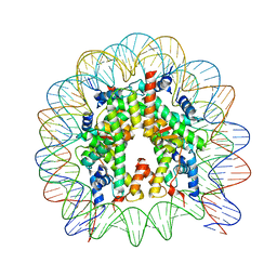 | | The nucleosome containing human H3.5 | | Descriptor: | DNA (146-MER), Histone H2A type 1-B/E, Histone H2B type 1-J, ... | | Authors: | Urahama, T, Harada, A, Maehara, K, Horikoshi, N, Sato, K, Sato, Y, Shiraishi, K, Sugino, N, Osakabe, A, Tachiwana, H, Kagawa, W, Kimura, H, Ohkawa, Y, Kurumizaka, H. | | Deposit date: | 2015-04-03 | | Release date: | 2016-02-10 | | Last modified: | 2023-11-08 | | Method: | X-RAY DIFFRACTION (2.8 Å) | | Cite: | Histone H3.5 forms an unstable nucleosome and accumulates around transcription start sites in human testis.
Epigenetics Chromatin, 9, 2016
|
|
8JUD
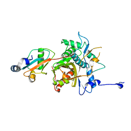
 | | Crystal structure of human MMP-7 in complex with inhibitor | | Descriptor: | CALCIUM ION, Matrilysin, Peptide Inhibitor, ... | | Authors: | Kamitani, M, Abe-Sato, K, Oka, Y. | | Deposit date: | 2023-06-26 | | Release date: | 2023-11-01 | | Last modified: | 2023-11-22 | | Method: | X-RAY DIFFRACTION (1.5 Å) | | Cite: | Structure-Based Optimization and Biological Evaluation of Potent and Selective MMP-7 Inhibitors for Kidney Fibrosis.
J.Med.Chem., 66, 2023
|
|
8JUG
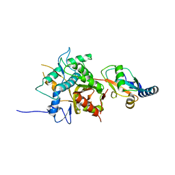
 | | Crystal structure of human MMP-7 in complex with inhibitor | | Descriptor: | CALCIUM ION, Matrilysin, Peptide Inhibitor, ... | | Authors: | Kamitani, M, Abe-Sato, K, Oka, Y. | | Deposit date: | 2023-06-26 | | Release date: | 2023-11-01 | | Last modified: | 2023-11-22 | | Method: | X-RAY DIFFRACTION (1.3 Å) | | Cite: | Structure-Based Optimization and Biological Evaluation of Potent and Selective MMP-7 Inhibitors for Kidney Fibrosis.
J.Med.Chem., 66, 2023
|
|
8JUF
 | | Crystal structure of human MMP-7 in complex with inhibitor | | Descriptor: | CALCIUM ION, Matrilysin, Peptide Inhibitor, ... | | Authors: | Kamitani, M, Abe-Sato, K, Oka, Y. | | Deposit date: | 2023-06-26 | | Release date: | 2023-11-01 | | Last modified: | 2023-11-22 | | Method: | X-RAY DIFFRACTION (1.39 Å) | | Cite: | Structure-Based Optimization and Biological Evaluation of Potent and Selective MMP-7 Inhibitors for Kidney Fibrosis.
J.Med.Chem., 66, 2023
|
|
5Y1A
 | | HBP35 of Porphyromonas gingivalis | | Descriptor: | 35 kDa hemin binding protein | | Authors: | Kakuda, S, Suzuki, M, Sato, K. | | Deposit date: | 2017-07-20 | | Release date: | 2018-07-25 | | Last modified: | 2024-03-27 | | Method: | X-RAY DIFFRACTION (1.8 Å) | | Cite: | Immunoglobulin-like domains of the cargo proteins are essential for protein stability during secretion by the type IX secretion system.
Mol. Microbiol., 110, 2018
|
|


