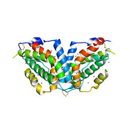
3H5V
 
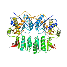 | | Crystal structure of the GluR2-ATD | | 分子名称: | 2-acetamido-2-deoxy-beta-D-glucopyranose-(1-4)-2-acetamido-2-deoxy-beta-D-glucopyranose, Glutamate receptor 2 | | 著者 | Jin, R, Singh, S.K, Gu, S, Furukawa, H, Sobolevsky, A, Zhou, J, Jin, Y, Gouaux, E. | | 登録日 | 2009-04-22 | | 公開日 | 2009-06-09 | | 最終更新日 | 2020-07-29 | | 実験手法 | X-RAY DIFFRACTION (2.33 Å) | | 主引用文献 | Crystal structure and association behaviour of the GluR2 amino-terminal domain.
Embo J., 28, 2009
|
|
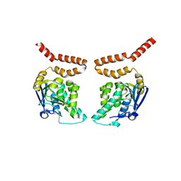
3HCW
 | | CRYSTAL STRUCTURE OF PROBABLE maltose operon transcriptional repressor malR FROM STAPHYLOCOCCUS AREUS | | 分子名称: | GLYCEROL, Maltose operon transcriptional repressor | | 著者 | Patskovsky, Y, Toro, R, Morano, C, Freeman, J, Chang, S, Sauder, J.M, Burley, S.K, Almo, S.C, New York SGX Research Center for Structural Genomics (NYSGXRC) | | 登録日 | 2009-05-06 | | 公開日 | 2009-05-19 | | 最終更新日 | 2024-02-21 | | 実験手法 | X-RAY DIFFRACTION (2.2 Å) | | 主引用文献 | Crystal Structure of Maltose Operon Transcriptional Repressor from Staphylococcus Aureus
To be Published
|
|
3HEQ
 | | Human prion protein variant D178N with M129 | | 分子名称: | CADMIUM ION, Major prion protein | | 著者 | Lee, S, Antony, L, Hartmann, R, Knaus, K.J, Surewicz, K, Surewicz, W.K, Yee, V.C. | | 登録日 | 2009-05-10 | | 公開日 | 2010-01-12 | | 最終更新日 | 2021-10-13 | | 実験手法 | X-RAY DIFFRACTION (1.8 Å) | | 主引用文献 | Conformational diversity in prion protein variants influences intermolecular beta-sheet formation.
Embo J., 29, 2010
|
|
3HGP
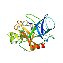 | | Structure of porcine pancreatic elastase complexed with a potent peptidyl inhibitor FR130180 determined by high resolution crystallography | | 分子名称: | 4-[[(2S)-3-methyl-1-oxo-1-[(2S)-2-[[(3S)-1,1,1-trifluoro-4-methyl-2-oxo-pentan-3-yl]carbamoyl]pyrrolidin-1-yl]butan-2-yl]carbamoyl]benzoic acid, CALCIUM ION, Elastase-1, ... | | 著者 | Tamada, T, Kinoshita, T, Kuroki, R, Tada, T. | | 登録日 | 2009-05-14 | | 公開日 | 2009-07-28 | | 最終更新日 | 2023-11-01 | | 実験手法 | X-RAY DIFFRACTION (0.94 Å) | | 主引用文献 | Combined High-Resolution Neutron and X-ray Analysis of Inhibited Elastase Confirms the Active-Site Oxyanion Hole but Rules against a Low-Barrier Hydrogen Bond
J.Am.Chem.Soc., 131, 2009
|
|
3HID
 | | Crystal structure of adenylosuccinate synthetase from Yersinia pestis CO92 | | 分子名称: | 1-(2-METHOXY-ETHOXY)-2-{2-[2-(2-METHOXY-ETHOXY]-ETHOXY}-ETHANE, Adenylosuccinate synthetase | | 著者 | Zhang, R, Zhou, M, Peterson, S, Anderson, W, Joachimiak, A, Center for Structural Genomics of Infectious Diseases (CSGID) | | 登録日 | 2009-05-19 | | 公開日 | 2009-06-02 | | 最終更新日 | 2024-02-21 | | 実験手法 | X-RAY DIFFRACTION (1.6 Å) | | 主引用文献 | The crystal structure of the adenylosuccinate synthetase from Yersinia pestis CO92
To be Published
|
|
3HI1
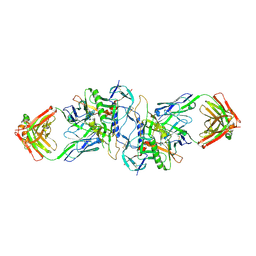 | | Structure of HIV-1 gp120 (core with V3) in Complex with CD4-Binding-Site Antibody F105 | | 分子名称: | 2-acetamido-2-deoxy-beta-D-glucopyranose, F105 Heavy Chain, F105 Light Chain, ... | | 著者 | Kwon, Y.D, Chen, L, Zhou, T, Wu, X, O'Dell, S, Cavacini, L, Hessell, A.J, Pancera, M, Tang, M, Xu, L, Yang, Z, Zhang, M.-Y, Arthos, J, Burton, D.R, Dimitrov, D, Nabel, G.J, Posner, M, Sodroski, J, Wyatt, R, Mascola, J.R, Kwong, P.D. | | 登録日 | 2009-05-18 | | 公開日 | 2009-11-17 | | 最終更新日 | 2023-09-06 | | 実験手法 | X-RAY DIFFRACTION (2.9 Å) | | 主引用文献 | Structural basis of immune evasion at the site of CD4 attachment on HIV-1 gp120.
Science, 326, 2009
|
|
7R8V
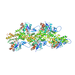 | | Cryo-EM structure of the ADP state actin filament | | 分子名称: | ADENOSINE-5'-DIPHOSPHATE, Actin, alpha skeletal muscle, ... | | 著者 | Gong, R, Espinosa de los Reyes, S, Reynolds, M.J, Gurel, P, Alushin, G.M. | | 登録日 | 2021-06-27 | | 公開日 | 2021-07-28 | | 最終更新日 | 2022-08-03 | | 実験手法 | ELECTRON MICROSCOPY (2.82 Å) | | 主引用文献 | Structural basis for tunable control of actin dynamics by myosin-15 in mechanosensory stereocilia.
Sci Adv, 8, 2022
|
|
7R91
 | | cryo-EM structure of the rigor state wild type myosin-15-F-actin complex | | 分子名称: | ADENOSINE-5'-DIPHOSPHATE, Actin, alpha skeletal muscle, ... | | 著者 | Gong, R, Reynolds, M.J, Gurel, P, Alushin, G.M. | | 登録日 | 2021-06-28 | | 公開日 | 2021-07-28 | | 最終更新日 | 2022-08-03 | | 実験手法 | ELECTRON MICROSCOPY (2.83 Å) | | 主引用文献 | Structural basis for tunable control of actin dynamics by myosin-15 in mechanosensory stereocilia.
Sci Adv, 8, 2022
|
|
7RB9
 | | Cryo-EM structure of the rigor state Jordan myosin-15-F-actin complex | | 分子名称: | ADENOSINE-5'-DIPHOSPHATE, Actin, alpha skeletal muscle, ... | | 著者 | Gong, R, Reynolds, M.J, Gurel, P, Alushin, G.M. | | 登録日 | 2021-07-05 | | 公開日 | 2021-07-28 | | 最終更新日 | 2022-08-03 | | 実験手法 | ELECTRON MICROSCOPY (3.76 Å) | | 主引用文献 | Structural basis for tunable control of actin dynamics by myosin-15 in mechanosensory stereocilia.
Sci Adv, 8, 2022
|
|
3HES
 | | Human prion protein variant F198S with M129 | | 分子名称: | CADMIUM ION, Major prion protein | | 著者 | Lee, S, Antony, L, Hartmann, R, Knaus, K.J, Surewicz, K, Surewicz, W.K, Yee, V.C. | | 登録日 | 2009-05-10 | | 公開日 | 2010-01-12 | | 最終更新日 | 2021-10-13 | | 実験手法 | X-RAY DIFFRACTION (2 Å) | | 主引用文献 | Conformational diversity in prion protein variants influences intermolecular beta-sheet formation.
Embo J., 29, 2010
|
|
3HG7
 | | CRYSTAL STRUCTURE OF D-isomer specific 2-hydroxyacid dehydrogenase family protein from Aeromonas salmonicida subsp. salmonicida A449 | | 分子名称: | D-isomer specific 2-hydroxyacid dehydrogenase family protein, GLYCEROL | | 著者 | Malashkevich, V.N, Toro, R, Morano, C, Sauder, J.M, Burley, S.K, Almo, S.C, New York SGX Research Center for Structural Genomics (NYSGXRC) | | 登録日 | 2009-05-13 | | 公開日 | 2009-05-26 | | 最終更新日 | 2024-02-21 | | 実験手法 | X-RAY DIFFRACTION (1.8 Å) | | 主引用文献 | CRYSTAL STRUCTURE OF D-isomer specific 2-hydroxyacid dehydrogenase family protein from Aeromonas salmonicida subsp. salmonicida A449
To be Published
|
|
7RB8
 | | cryo-EM structure of the ADP state wild type myosin-15-F-actin complex | | 分子名称: | ADENOSINE-5'-DIPHOSPHATE, Actin, alpha skeletal muscle, ... | | 著者 | Gong, R, Reynolds, M.J, Gurel, P, Alushin, G.M. | | 登録日 | 2021-07-05 | | 公開日 | 2021-07-28 | | 最終更新日 | 2022-08-03 | | 実験手法 | ELECTRON MICROSCOPY (3.63 Å) | | 主引用文献 | Structural basis for tunable control of actin dynamics by myosin-15 in mechanosensory stereocilia.
Sci Adv, 8, 2022
|
|
3HIA
 | | Crystal structure of the choline binding domain of Spr1274 in Streptococcus pneumoniae | | 分子名称: | Choline binding protein, PHOSPHOCHOLINE, SULFATE ION | | 著者 | Zhang, Z.-Y, Li, W.-Z, Jiang, Y.-L, Bao, R, Zhou, C.-Z, Chen, Y.-X. | | 登録日 | 2009-05-19 | | 公開日 | 2009-08-04 | | 最終更新日 | 2023-11-01 | | 実験手法 | X-RAY DIFFRACTION (2.38 Å) | | 主引用文献 | Crystal structure of the choline binding domain of Spr1274 in Streptococcus pneumoniae
To be Published
|
|
3HIX
 | | Crystal Structure of the Rhodanese_3 like domain from Anabaena sp Alr3790 protein. Northeast Structural Genomics Consortium Target NsR437i | | 分子名称: | Alr3790 protein, MANGANESE (II) ION | | 著者 | Vorobiev, S, Chen, Y, Forouhar, F, Maglaqui, M, Ciccosanti, C, Mao, L, Xiao, R, Acton, T.B, Montelione, G.T, Tong, L, Hunt, J.F, Northeast Structural Genomics Consortium (NESG) | | 登録日 | 2009-05-20 | | 公開日 | 2009-05-26 | | 最終更新日 | 2011-07-13 | | 実験手法 | X-RAY DIFFRACTION (1.92 Å) | | 主引用文献 | Crystal Structure of the Rhodanese_3 like domain from Anabaena sp Alr3790 protein.
To be Published
|
|
3HJ7
 | | Crystal structure of TILS C-terminal domain | | 分子名称: | CHLORIDE ION, tRNA(Ile)-lysidine synthase | | 著者 | Nakanishi, K, Bonnefond, L, Kimura, S, Suzuki, T, Ishitani, R, Nureki, O. | | 登録日 | 2009-05-21 | | 公開日 | 2009-10-20 | | 最終更新日 | 2023-11-01 | | 実験手法 | X-RAY DIFFRACTION (2.2 Å) | | 主引用文献 | Structural basis for translational fidelity ensured by transfer RNA lysidine synthetase.
Nature, 461, 2009
|
|
3HJG
 | | Crystal structure of putative alpha-ribazole-5'-phosphate phosphatase CobC FROM Vibrio parahaemolyticus | | 分子名称: | Putative alpha-ribazole-5'-phosphate phosphatase CobC, SULFATE ION | | 著者 | Patskovsky, Y, Toro, R, Morano, C, Rutter, M, Iizuka, M, Sauder, J.M, Burley, S.K, Almo, S.C, New York SGX Research Center for Structural Genomics (NYSGXRC) | | 登録日 | 2009-05-21 | | 公開日 | 2009-06-02 | | 最終更新日 | 2024-02-21 | | 実験手法 | X-RAY DIFFRACTION (2.8 Å) | | 主引用文献 | Crystal structure of putative alpha-ribazole-5' -phosphate phosphatase FROM Vibrio parahaemolyticus
To be Published
|
|
3HHD
 | | Structure of the Human Fatty Acid Synthase KS-MAT Didomain as a Framework for Inhibitor Design. | | 分子名称: | CHLORIDE ION, Fatty acid synthase | | 著者 | Pappenberger, G.M, Benz, J, Thoma, R, Rudolph, M.G. | | 登録日 | 2009-05-15 | | 公開日 | 2010-02-09 | | 最終更新日 | 2023-09-06 | | 実験手法 | X-RAY DIFFRACTION (2.15 Å) | | 主引用文献 | Structure of the human fatty acid synthase KS-MAT didomain as a framework for inhibitor design.
J.Mol.Biol., 397, 2010
|
|
3HDT
 | | Crystal structure of putative kinase from Clostridium symbiosum ATCC 14940 | | 分子名称: | CHLORIDE ION, PHOSPHATE ION, putative kinase | | 著者 | Chang, C, Freeman, L, Jedrzejczak, R, Joachimiak, A, Midwest Center for Structural Genomics (MCSG) | | 登録日 | 2009-05-07 | | 公開日 | 2009-05-26 | | 最終更新日 | 2017-11-01 | | 実験手法 | X-RAY DIFFRACTION (2.79 Å) | | 主引用文献 | Crystal structure of putative kinase from Clostridium symbiosum ATCC 14940
To be Published
|
|
3HGT
 | |
3HHC
 | |
3HD7
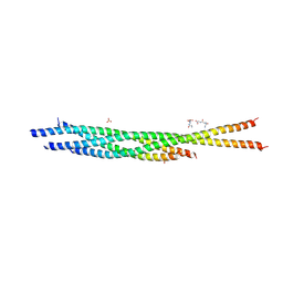 | | HELICAL EXTENSION OF THE NEURONAL SNARE COMPLEX INTO THE MEMBRANE, spacegroup C 1 2 1 | | 分子名称: | SULFATE ION, Synaptosomal-associated protein 25, Syntaxin-1A, ... | | 著者 | Stein, A, Weber, G, Wahl, M.C, Jahn, R. | | 登録日 | 2009-05-07 | | 公開日 | 2009-07-14 | | 最終更新日 | 2024-03-20 | | 実験手法 | X-RAY DIFFRACTION (3.4 Å) | | 主引用文献 | Helical extension of the neuronal SNARE complex into the membrane
Nature, 460, 2009
|
|
3HI8
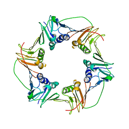 | | Crystal structure of proliferating cell nuclear antigen (PCNA) from Haloferax volcanii | | 分子名称: | Proliferating cell nuclear antigen PcnA | | 著者 | Morgunova, E, Gray, F.C, MacNeill, S.A, Ladenstein, R. | | 登録日 | 2009-05-19 | | 公開日 | 2009-09-29 | | 最終更新日 | 2023-09-06 | | 実験手法 | X-RAY DIFFRACTION (3.202 Å) | | 主引用文献 | Structural insights into the adaptation of proliferating cell nuclear antigen (PCNA) from Haloferax volcanii to a high-salt environment.
Acta Crystallogr.,Sect.D, 65, 2009
|
|
3HIN
 | | CRYSTAL STRUCTURE OF putative enoyl-CoA hydratase from Rhodopseudomonas palustris CGA009 | | 分子名称: | Putative 3-hydroxybutyryl-CoA dehydratase | | 著者 | Malashkevich, V.N, Toro, R, Morano, C, Sauder, J.M, Burley, S.K, Almo, S.C, New York SGX Research Center for Structural Genomics (NYSGXRC) | | 登録日 | 2009-05-20 | | 公開日 | 2009-06-02 | | 最終更新日 | 2024-02-21 | | 実験手法 | X-RAY DIFFRACTION (2 Å) | | 主引用文献 | CRYSTAL STRUCTURE OF putative enoyl-CoA hydratase from Rhodopseudomonas palustris CGA009
To be Published
|
|
3HIE
 | |
3HG9
 | | CRYSTAL STRUCTURE OF putative pilM protein from Pseudomonas aeruginosa 2192 | | 分子名称: | NICKEL (II) ION, PilM | | 著者 | Malashkevich, V.N, Toro, R, Sauder, J.M, Burley, S.K, Almo, S.C, New York SGX Research Center for Structural Genomics (NYSGXRC) | | 登録日 | 2009-05-13 | | 公開日 | 2009-05-26 | | 最終更新日 | 2024-02-21 | | 実験手法 | X-RAY DIFFRACTION (2.4 Å) | | 主引用文献 | CRYSTAL STRUCTURE OF putative pilM protein from Pseudomonas aeruginosa 2192
To be Published
|
|


