4DWM
 
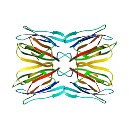
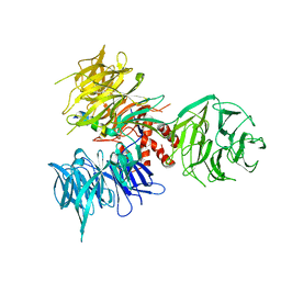
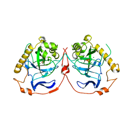
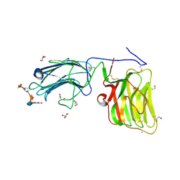
 | | Crystal structure of the complex of type I Ribosome inactivating protein with N-acetylglucosamine at 1.62 A resolution | | 分子名称: | 2-acetamido-2-deoxy-beta-D-glucopyranose, GLYCEROL, rRNA N-glycosidase | | 著者 | Yamini, S, Pandey, S, Sinha, M, Kaur, P, Sharma, S, Singh, T.P. | | 登録日 | 2012-02-25 | | 公開日 | 2012-03-07 | | 最終更新日 | 2023-11-08 | | 実験手法 | X-RAY DIFFRACTION (1.62 Å) | | 主引用文献 | Crystal structure of the complex of type I Ribosome inactivating protein with N-acetylglucosamine at 1.62 A resolution
To be Published
|
|
4QBT
 | |
4MLK
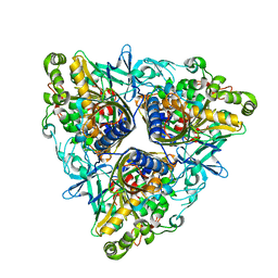
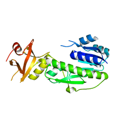
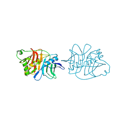
 | | 3.05A resolution structure of CT584 from Chlamydia trachomatis | | 分子名称: | CT584 protein | | 著者 | Hickey, J, Lovell, S, Kemege, K, Barta, M.L, Battaile, K.P, Hefty, P.S. | | 登録日 | 2013-09-06 | | 公開日 | 2013-11-13 | | 最終更新日 | 2024-02-28 | | 実験手法 | X-RAY DIFFRACTION (3.051 Å) | | 主引用文献 | Structure of CT584 from Chlamydia trachomatis refined to 3.05 angstrom resolution.
Acta Crystallogr.,Sect.F, 69, 2013
|
|
1UH1
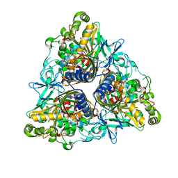
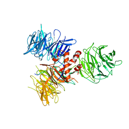
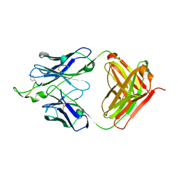
 | | Crystal structure of jacalin- GalNAc-beta(1-3)-Gal-alpha-O-Me complex | | 分子名称: | 2-acetamido-2-deoxy-beta-D-galactopyranose-(1-3)-methyl alpha-D-galactopyranoside, Agglutinin alpha chain, Agglutinin beta-3 chain, ... | | 著者 | Jeyaprakash, A.A, Katiyar, S, Swaminathan, C.P, Sekar, K, Surolia, A, Vijayan, M. | | 登録日 | 2003-06-23 | | 公開日 | 2003-09-23 | | 最終更新日 | 2023-10-25 | | 実験手法 | X-RAY DIFFRACTION (2.8 Å) | | 主引用文献 | Structural Basis of the Carbohydrate Specificities of Jacalin: An X-ray and Modeling Study
J.MOL.BIOL., 332, 2003
|
|
7ARS
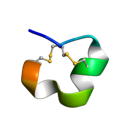
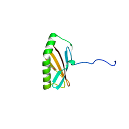
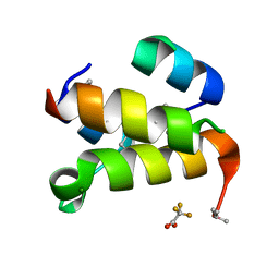 | | The de novo designed hybrid alpha/beta-miniprotein (with Se-Methionine) | | 分子名称: | MAGNESIUM ION, alpha/beta-peptide, trifluoroacetic acid | | 著者 | Bejger, M, Fortuna, P, Drewniak-Switalska, M, Rypniewski, W, Berlicki, L. | | 登録日 | 2020-10-26 | | 公開日 | 2021-07-14 | | 最終更新日 | 2024-05-01 | | 実験手法 | X-RAY DIFFRACTION (1.15 Å) | | 主引用文献 | A computationally designed beta-amino acid-containing miniprotein.
Chem.Commun.(Camb.), 57, 2021
|
|
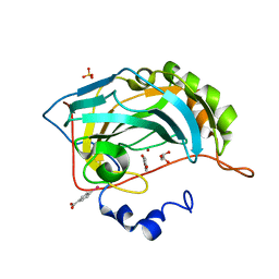
1UGW
 | | Crystal structure of jacalin- Gal complex | | 分子名称: | Agglutinin alpha chain, Agglutinin alpha-chain, Agglutinin beta-3 chain, ... | | 著者 | Jeyaprakash, A.A, Katiyar, S, Swaminathan, C.P, Sekar, K, Surolia, A, Vijayan, M. | | 登録日 | 2003-06-22 | | 公開日 | 2003-09-23 | | 最終更新日 | 2023-10-25 | | 実験手法 | X-RAY DIFFRACTION (1.7 Å) | | 主引用文献 | Structural Basis of the Carbohydrate Specificities of Jacalin: An X-ray and Modeling Study
J.MOL.BIOL., 332, 2003
|
|
2XR8
 | |
2XRX
 | |
2MD6
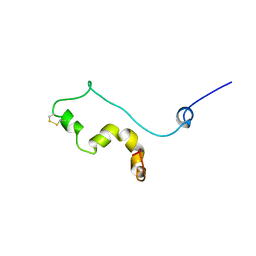
 | | NMR SOLUTION STRUCTURE OF ALPHA CONOTOXIN LO1A FROM Conus longurionis | | 分子名称: | ALPHA CONOTOXIN LO1A | | 著者 | Maiti, M, Lescrinier, E, Herdewijn, P, Lebbe, E.K.M, Peigneur, S, D'Souza, L, Tytgat, J. | | 登録日 | 2013-09-01 | | 公開日 | 2014-03-05 | | 最終更新日 | 2023-06-14 | | 実験手法 | SOLUTION NMR | | 主引用文献 | Structure-Function Elucidation of a New alpha-Conotoxin, Lo1a, from Conus longurionis.
J.Biol.Chem., 289, 2014
|
|
7WA4
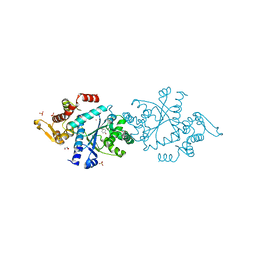
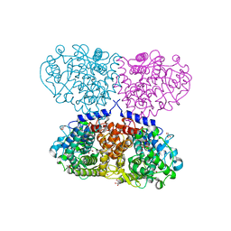
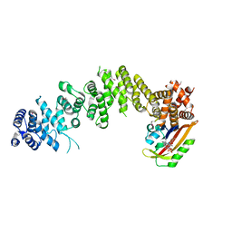 | | Crystal structure of GIGANTEA in complex with LKP2 | | 分子名称: | Adagio protein 2, FLAVIN MONONUCLEOTIDE, Protein GIGANTEA | | 著者 | Pathak, D, Dahal, P, Kwon, E, Kim, D.Y. | | 登録日 | 2021-12-12 | | 公開日 | 2022-04-27 | | 最終更新日 | 2022-05-04 | | 実験手法 | X-RAY DIFFRACTION (3.5 Å) | | 主引用文献 | Structural analysis of the regulation of blue-light receptors by GIGANTEA.
Cell Rep, 39, 2022
|
|
1H4Z
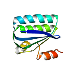 | | Structure of the Anti-Sigma Factor Antagonist SpoIIAA in its Unphosphorylated Form | | 分子名称: | ANTI-SIGMA F FACTOR ANTAGONIST | | 著者 | Seavers, P.R, Lewis, R.J, Brannigan, J.A, Verschueren, K.H.G, Murshudov, G.N, Wilkinson, A.J. | | 登録日 | 2001-05-16 | | 公開日 | 2001-07-06 | | 最終更新日 | 2023-12-13 | | 実験手法 | X-RAY DIFFRACTION (2.74 Å) | | 主引用文献 | Structure of the Bacillus Cell Fate Determinant Spoiiaa in Phosphorylated and Unphosphorylated Forms
Structure, 9, 2001
|
|
4Q93
 | |
1TVS
 | |
3I7K
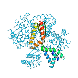
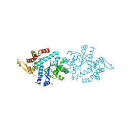
 | | Crystal Structure of DDB1 in Complex with the H-Box Motif of WHX | | 分子名称: | DNA damage-binding protein 1, X protein | | 著者 | Li, T, Robert, E.I, Breugel, P.C.V, Strubin, M, Zheng, N. | | 登録日 | 2009-07-08 | | 公開日 | 2009-12-08 | | 最終更新日 | 2023-09-06 | | 実験手法 | X-RAY DIFFRACTION (2.8 Å) | | 主引用文献 | A promiscuous alpha-helical motif anchors viral hijackers and substrate receptors to the CUL4-DDB1 ubiquitin ligase machinery.
Nat.Struct.Mol.Biol., 17, 2010
|
|
1YRW
 | |
3I8E
 | | Crystal Structure of DDB1 in Complex with the H-Box Motif of WDR42A | | 分子名称: | DNA damage-binding protein 1, WD repeat-containing protein 42A | | 著者 | Li, T, Robert, E.I, Breugel, P.C.V, Strubin, M, Zheng, N. | | 登録日 | 2009-07-09 | | 公開日 | 2009-12-08 | | 最終更新日 | 2023-09-06 | | 実験手法 | X-RAY DIFFRACTION (3.4 Å) | | 主引用文献 | A promiscuous alpha-helical motif anchors viral hijackers and substrate receptors to the CUL4-DDB1 ubiquitin ligase machinery.
Nat.Struct.Mol.Biol., 17, 2010
|
|
2INW
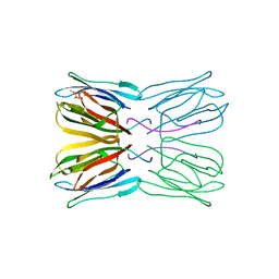
 | | Crystal structure of Q83JN9 from Shigella flexneri at high resolution. Northeast Structural Genomics Consortium target SfR137. | | 分子名称: | PHOSPHATE ION, Putative structural protein | | 著者 | Kuzin, A.P, Su, M, Jayaraman, S, Vorobiev, S.M, Wang, D, Jiang, M, Cunningham, K, Ma, L.-C, Xiao, R, Liu, J, Baran, M, Swapna, G.V.T, Acton, T.B, Rost, B, Montelione, G.T, Tong, L, Hunt, J.F, Northeast Structural Genomics Consortium (NESG) | | 登録日 | 2006-10-09 | | 公開日 | 2006-10-24 | | 最終更新日 | 2011-07-13 | | 実験手法 | X-RAY DIFFRACTION (1.5 Å) | | 主引用文献 | Crystal Structures of Phd-Doc, HigA, and YeeU Establish Multiple Evolutionary Links between Microbial Growth-Regulating Toxin-Antitoxin Systems.
Structure, 18, 2010
|
|
2IK2
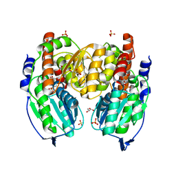
 | | Yeast inorganic pyrophosphatase variant D115E with magnesium and phosphate | | 分子名称: | Inorganic pyrophosphatase, MAGNESIUM ION, PHOSPHATE ION | | 著者 | Oksanen, E, Ahonen, A.K, Tuominen, H, Tuominen, V, Lahti, R, Goldman, A, Heikinheimo, P. | | 登録日 | 2006-10-02 | | 公開日 | 2007-02-13 | | 最終更新日 | 2023-08-30 | | 実験手法 | X-RAY DIFFRACTION (1.8 Å) | | 主引用文献 | A Complete Structural Description of the Catalytic Cycle of Yeast Pyrophosphatase.
Biochemistry, 46, 2007
|
|
5OO2
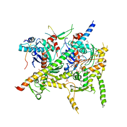
 | | Crystal structure of Mycolicibacterium hassiacum glucosylglycerate hydrolase (MhGgH) E419A variant in complex with glucosylglycolate | | 分子名称: | (alpha-D-glucopyranosyloxy)acetic acid, GLYCEROL, SERINE, ... | | 著者 | Cereija, T.B, Macedo-Ribeiro, S, Pereira, P.J.B. | | 登録日 | 2017-08-05 | | 公開日 | 2018-08-29 | | 最終更新日 | 2024-01-17 | | 実験手法 | X-RAY DIFFRACTION (2.06 Å) | | 主引用文献 | The structural characterization of a glucosylglycerate hydrolase provides insights into the molecular mechanism of mycobacterial recovery from nitrogen starvation.
Iucrj, 6, 2019
|
|
1TZC
 | | Crystal structure of phosphoglucose/phosphomannose isomerase from Pyrobaculum aerophilum in complex with 5-phosphoarabinonate | | 分子名称: | 5-PHOSPHOARABINONIC ACID, GLYCEROL, SULFATE ION, ... | | 著者 | Swan, M.K, Hansen, T, Schoenheit, P, Davies, C. | | 登録日 | 2004-07-09 | | 公開日 | 2004-07-20 | | 最終更新日 | 2024-04-03 | | 実験手法 | X-RAY DIFFRACTION (1.45 Å) | | 主引用文献 | A novel phosphoglucose/phosphomannose isomease from the crenarchaeon Pyrobaculum aerophilum is a member of the PGI superfamily: structural evidence at 1.16 A resolution
J.Biol.Chem., 279, 2004
|
|
4I6M
 | | Structure of Arp7-Arp9-Snf2(HSA)-RTT102 subcomplex of SWI/SNF chromatin remodeler. | | 分子名称: | Actin-like protein ARP9, Actin-related protein 7, PHOSPHATE ION, ... | | 著者 | Schubert, H.L, Cairns, B.R, Hill, C.P. | | 登録日 | 2012-11-29 | | 公開日 | 2013-02-13 | | 最終更新日 | 2017-08-16 | | 実験手法 | X-RAY DIFFRACTION (2.801 Å) | | 主引用文献 | Structure of an actin-related subcomplex of the SWI/SNF chromatin remodeler.
Proc.Natl.Acad.Sci.USA, 110, 2013
|
|
5IK5
 | | Laminin A2LG45 C-form, G6/7 bound. | | 分子名称: | 2-acetamido-2-deoxy-beta-D-glucopyranose-(1-4)-2-acetamido-2-deoxy-beta-D-glucopyranose, 7-hydroxy-4-methyl-2H-chromen-2-one, CALCIUM ION, ... | | 著者 | Briggs, D.C, Hohenester, E, Campbell, K.P. | | 登録日 | 2016-03-03 | | 公開日 | 2016-08-10 | | 最終更新日 | 2024-01-10 | | 実験手法 | X-RAY DIFFRACTION (1.39 Å) | | 主引用文献 | Structural basis of laminin binding to the LARGE glycans on dystroglycan.
Nat.Chem.Biol., 12, 2016
|
|
5IL5
 | |
3IDN
 | |
4E3G
 | |


