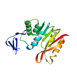
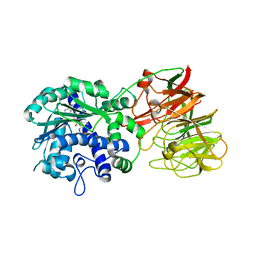
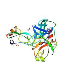
3BHU
 
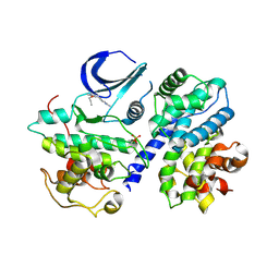 | | Structure of phosphorylated Thr160 CDK2/cyclin A in complex with the inhibitor meriolin 5 | | 分子名称: | 4-(4-propoxy-1H-pyrrolo[2,3-b]pyridin-3-yl)pyrimidin-2-amine, Cell division protein kinase 2, Cyclin-A2, ... | | 著者 | Echalier, A, Bettayeb, K, Ferandin, Y, Lozach, O, Clement, M, Valette, A, Liger, F, Marquet, B, Morris, J.C, Endicott, J.A, Joseph, B, Meijer, L. | | 登録日 | 2007-11-29 | | 公開日 | 2008-02-12 | | 最終更新日 | 2017-10-25 | | 実験手法 | X-RAY DIFFRACTION (2.3 Å) | | 主引用文献 | Meriolins, a new class of cell death inducing kinase inhibitors with enhanced selectivity for cyclin-dependent kinases
Cancer Res., 67, 2007
|
|
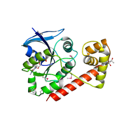
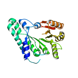
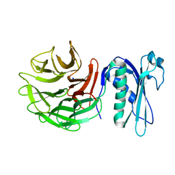
3B0W
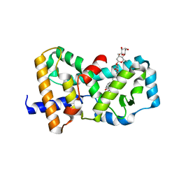 | | Crystal structure of the orphan nuclear receptor ROR(gamma)t ligand-binding domain in complex with digoxin | | 分子名称: | DIGOXIN, Nuclear receptor ROR-gamma | | 著者 | Fujita-Sato, S, Ito, S, Isobe, T, Ohyama, T, Wakabayashi, K, Morishita, K, Ando, O, Isono, F. | | 登録日 | 2011-06-17 | | 公開日 | 2011-07-06 | | 最終更新日 | 2023-11-01 | | 実験手法 | X-RAY DIFFRACTION (2.2 Å) | | 主引用文献 | Structural Basis of Digoxin That Antagonizes ROR{gamma}t Receptor Activity and Suppresses Th17 Cell Differentiation and Interleukin (IL)-17 Production
J.Biol.Chem., 286, 2011
|
|
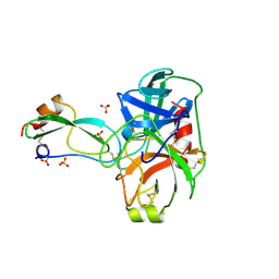
2ZNR
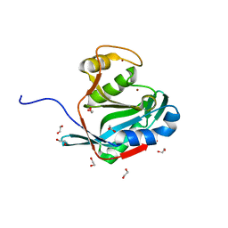 | | Crystal structure of the DUB domain of human AMSH-LP | | 分子名称: | 1,2-ETHANEDIOL, AMSH-like protease, PRASEODYMIUM ION, ... | | 著者 | Sato, Y, Azusa, Y, Yamagata, A, Mimura, H, Wang, X, Yamashita, M, Ookata, K, Nureki, O, Iwai, K, Komada, M, Fukai, S. | | 登録日 | 2008-05-01 | | 公開日 | 2008-09-02 | | 最終更新日 | 2024-03-13 | | 実験手法 | X-RAY DIFFRACTION (1.2 Å) | | 主引用文献 | Structural basis for specific cleavage of Lys 63-linked polyubiquitin chains
Nature, 455, 2008
|
|
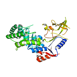
3A1U
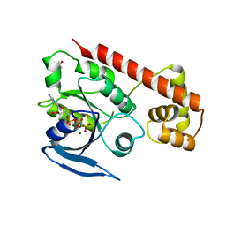 | | Crystal structue of the cytosolic domain of T. maritima FeoB iron iransporter in GMPPNP form | | 分子名称: | (4R)-2-METHYLPENTANE-2,4-DIOL, Iron(II) transport protein B, MAGNESIUM ION, ... | | 著者 | Hattori, M, Ishitani, R, Nureki, O. | | 登録日 | 2009-04-22 | | 公開日 | 2009-09-22 | | 最終更新日 | 2023-11-01 | | 実験手法 | X-RAY DIFFRACTION (1.8 Å) | | 主引用文献 | Structural basis of novel interactions between the small-GTPase and GDI-like domains in prokaryotic FeoB iron transporter
Structure, 17, 2009
|
|
3A27
 | | Crystal structure of M. jannaschii TYW2 in complex with AdoMet | | 分子名称: | S-ADENOSYLMETHIONINE, Uncharacterized protein MJ1557 | | 著者 | Umitsu, M, Nishimasu, H, Ishitani, R, Nureki, O. | | 登録日 | 2009-04-28 | | 公開日 | 2009-09-15 | | 最終更新日 | 2023-11-01 | | 実験手法 | X-RAY DIFFRACTION (2.005 Å) | | 主引用文献 | Structural basis of AdoMet-dependent aminocarboxypropyl transfer reaction catalyzed by tRNA-wybutosine synthesizing enzyme, TYW2
Proc.Natl.Acad.Sci.USA, 106, 2009
|
|
3A1S
 | | Crystal structue of the cytosolic domain of T. maritima FeoB iron iransporter in GDP form I | | 分子名称: | (4R)-2-METHYLPENTANE-2,4-DIOL, (4S)-2-METHYL-2,4-PENTANEDIOL, GUANOSINE-5'-DIPHOSPHATE, ... | | 著者 | Hattori, M, Ishitani, R, Nureki, O. | | 登録日 | 2009-04-22 | | 公開日 | 2009-09-22 | | 最終更新日 | 2011-07-13 | | 実験手法 | X-RAY DIFFRACTION (1.5 Å) | | 主引用文献 | Structural basis of novel interactions between the small-GTPase and GDI-like domains in prokaryotic FeoB iron transporter
Structure, 17, 2009
|
|
3A2K
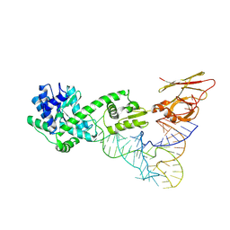 | | Crystal structure of TilS complexed with tRNA | | 分子名称: | bacterial tRNA, tRNA(Ile)-lysidine synthase | | 著者 | Nakanishi, K, Bonnefond, L, Ishitani, R, Nureki, O. | | 登録日 | 2009-05-23 | | 公開日 | 2009-10-20 | | 最終更新日 | 2018-01-24 | | 実験手法 | X-RAY DIFFRACTION (3.65 Å) | | 主引用文献 | Structural basis for translational fidelity ensured by transfer RNA lysidine synthetase.
Nature, 461, 2009
|
|
3B76
 | | Crystal structure of the third PDZ domain of human ligand-of-numb protein-X (LNX1) in complex with the C-terminal peptide from the coxsackievirus and adenovirus receptor | | 分子名称: | 1,2-ETHANEDIOL, E3 ubiquitin-protein ligase LNX, SODIUM ION | | 著者 | Ugochukwu, E, Burgess-Brown, N, Berridge, G, Elkins, J, Bunkoczi, G, Pike, A.C.W, Sundstrom, M, Arrowsmith, C.H, Weigelt, J, Edwards, A.M, Gileadi, O, von Delft, F, Doyle, D, Structural Genomics Consortium (SGC) | | 登録日 | 2007-10-30 | | 公開日 | 2007-11-13 | | 最終更新日 | 2023-08-30 | | 実験手法 | X-RAY DIFFRACTION (1.75 Å) | | 主引用文献 | Crystal structure of the third PDZ domain of human ligand-of-numb protein-X (LNX1) in complex with the C-terminal peptide from the coxsackievirus and adenovirus receptor.
To be Published
|
|
3B6I
 | | WrbA from Escherichia coli, native structure | | 分子名称: | FLAVIN MONONUCLEOTIDE, Flavoprotein wrbA, POLYETHYLENE GLYCOL (N=34) | | 著者 | Andrade, S.L.A, Patridge, E.V, Ferry, J.G, Einsle, O. | | 登録日 | 2007-10-29 | | 公開日 | 2007-12-11 | | 最終更新日 | 2023-08-30 | | 実験手法 | X-RAY DIFFRACTION (1.66 Å) | | 主引用文献 | Crystal structure of the NADH:quinone oxidoreductase WrbA from Escherichia coli.
J.Bacteriol., 189, 2007
|
|
2ZW9
 | | Crystal structure of tRNA wybutosine synthesizing enzyme TYW4 | | 分子名称: | Leucine carboxyl methyltransferase 2, S-ADENOSYLMETHIONINE | | 著者 | Suzuki, Y, Noma, A, Suzuki, T, Ishitani, R, Nureki, O. | | 登録日 | 2008-12-01 | | 公開日 | 2009-06-02 | | 最終更新日 | 2024-03-13 | | 実験手法 | X-RAY DIFFRACTION (2.5 Å) | | 主引用文献 | Structural basis of tRNA modification with CO2 fixation and methylation by wybutosine synthesizing enzyme TYW4.
Nucleic Acids Res., 37, 2009
|
|
2Z2U
 | |
3BFU
 | | Structure of the ligand-binding core of GluR2 in complex with the agonist (R)-TDPA at 1.95 A resolution | | 分子名称: | (2R)-2-amino-3-(4-hydroxy-1,2,5-thiadiazol-3-yl)propanoic acid, Glutamate receptor 2 | | 著者 | Beich-Frandsen, M, Mirza, O, Vestergaard, B, Gajhede, M, Kastrup, J.S. | | 登録日 | 2007-11-23 | | 公開日 | 2008-10-14 | | 最終更新日 | 2023-11-01 | | 実験手法 | X-RAY DIFFRACTION (1.95 Å) | | 主引用文献 | Structures of the ligand-binding core of iGluR2 in complex with the agonists (R)- and (S)-2-amino-3-(4-hydroxy-1,2,5-thiadiazol-3-yl)propionic acid explain their unusual equipotency.
J.Med.Chem., 51, 2008
|
|
3C3J
 | | Crystal structure of tagatose-6-phosphate ketose/aldose isomerase from Escherichia coli | | 分子名称: | Putative tagatose-6-phosphate ketose/aldose isomerase | | 著者 | Zhang, R, Skarina, T, Egorova, O, Savchenko, A, Edwards, A.M, Joachimiak, A, Midwest Center for Structural Genomics (MCSG) | | 登録日 | 2008-01-28 | | 公開日 | 2008-02-19 | | 最終更新日 | 2011-07-13 | | 実験手法 | X-RAY DIFFRACTION (1.8 Å) | | 主引用文献 | The crystal structure of the tagatose-6-phosphate ketose/aldose isomerase from Escherichia coli.
To be Published
|
|
3BTD
 | | The Crystal Structures of the Complexes Between the Bovine Beta-Trypsin and Ten P1 Variants of BPTI. | | 分子名称: | CALCIUM ION, PROTEIN (BOVINE PANCREATIC TRYPSIN INHIBITOR), PROTEIN (TRYPSIN), ... | | 著者 | Helland, R, Otlewski, J, Sundheim, O, Dadlez, M, Smalas, A.O. | | 登録日 | 1999-03-11 | | 公開日 | 2000-03-13 | | 最終更新日 | 2023-08-30 | | 実験手法 | X-RAY DIFFRACTION (1.9 Å) | | 主引用文献 | The crystal structures of the complexes between bovine beta-trypsin and ten P1 variants of BPTI.
J.Mol.Biol., 287, 1999
|
|
3BTN
 | | Crystal structure of antizyme inhibitor, an ornithine decarboxylase homologous protein | | 分子名称: | Antizyme inhibitor 1 | | 著者 | Dym, O, Unger, T, Albeck, S, Kahana, C, Israel Structural Proteomics Center (ISPC) | | 登録日 | 2007-12-30 | | 公開日 | 2008-04-15 | | 最終更新日 | 2024-02-21 | | 実験手法 | X-RAY DIFFRACTION (2.05 Å) | | 主引用文献 | Crystallographic and biochemical studies revealing the structural basis for antizyme inhibitor function.
Protein Sci., 17, 2008
|
|
3BN0
 | |
3BNG
 | | W. succinogenes NrfA Y218F | | 分子名称: | ACETATE ION, CALCIUM ION, Cytochrome c-552, ... | | 著者 | Lukat, P, Einsle, O. | | 登録日 | 2007-12-14 | | 公開日 | 2008-02-26 | | 最終更新日 | 2023-11-01 | | 実験手法 | X-RAY DIFFRACTION (1.5 Å) | | 主引用文献 | Binding and Reduction of Sulfite by Cytochrome c Nitrite Reductase
Biochemistry, 47, 2008
|
|
3BW6
 | | Crystal structure of the longin domain of yeast Ykt6 | | 分子名称: | SULFATE ION, Synaptobrevin homolog YKT6 | | 著者 | Pylypenko, O, Schonichen, A, Ludwig, D, Ungermann, C, Goody, R.S, Rak, A, Geyer, M. | | 登録日 | 2008-01-08 | | 公開日 | 2008-04-01 | | 最終更新日 | 2024-02-21 | | 実験手法 | X-RAY DIFFRACTION (2.5 Å) | | 主引用文献 | Farnesylation of the SNARE protein Ykt6 increases its stability and helical folding.
J.Mol.Biol., 377, 2008
|
|
3BTK
 | | THE CRYSTAL STRUCTURES OF THE COMPLEXES BETWEEN BOVINE BETA-TRYPSIN AND TEN P1 VARIANTS OF BPTI | | 分子名称: | CALCIUM ION, PROTEIN (PANCREATIC TRYPSIN INHIBITOR), PROTEIN (TRYPSIN), ... | | 著者 | Helland, R, Otlewski, J, Sundheim, O, Dadlez, M, Smalas, A.O. | | 登録日 | 1999-03-10 | | 公開日 | 2000-03-13 | | 最終更新日 | 2023-08-30 | | 実験手法 | X-RAY DIFFRACTION (1.85 Å) | | 主引用文献 | The crystal structures of the complexes between bovine beta-trypsin and ten P1 variants of BPTI.
J.Mol.Biol., 287, 1999
|
|
3BTT
 | | THE CRYSTAL STRUCTURES OF THE COMPLEXES BETWEEN BOVINE BETA-TRYPSIN AND TEN P1 VARIANTS OF BPTI | | 分子名称: | CALCIUM ION, PROTEIN (PANCREATIC TRYPSIN INHIBITOR), PROTEIN (TRYPSIN), ... | | 著者 | Helland, R, Otlewski, J, Sundheim, O, Dadlez, M, Smalas, A.O. | | 登録日 | 1999-03-10 | | 公開日 | 2000-03-13 | | 最終更新日 | 2023-08-30 | | 実験手法 | X-RAY DIFFRACTION (1.9 Å) | | 主引用文献 | The crystal structures of the complexes between bovine beta-trypsin and ten P1 variants of BPTI.
J.Mol.Biol., 287, 1999
|
|
3BQY
 | | Crystal structure of a possible TetR family transcriptional regulator from Streptomyces coelicolor A3(2). | | 分子名称: | ACETIC ACID, PHOSPHATE ION, Putative TetR family transcriptional regulator | | 著者 | Cuff, M.E, Skarina, T, Kagan, O, Edwards, A.M, Savchenko, A, Joachimiak, A, Midwest Center for Structural Genomics (MCSG) | | 登録日 | 2007-12-20 | | 公開日 | 2008-01-15 | | 最終更新日 | 2017-10-25 | | 実験手法 | X-RAY DIFFRACTION (1.95 Å) | | 主引用文献 | Structure of a possible TetR family transcriptional regulator from Streptomyces coelicolor A3(2).
TO BE PUBLISHED
|
|
6PNV
 | | 1.42 Angstrom Resolution Crystal Structure of Translocation Protein TolB from Salmonella enterica | | 分子名称: | POTASSIUM ION, SODIUM ION, Tol-Pal system protein TolB | | 著者 | Minasov, G, Shuvalova, L, Dubrovska, I, Kiryukhina, O, Endres, M, Satchell, K.J.F, Center for Structural Genomics of Infectious Diseases (CSGID) | | 登録日 | 2019-07-03 | | 公開日 | 2019-07-17 | | 最終更新日 | 2023-10-11 | | 実験手法 | X-RAY DIFFRACTION (1.42 Å) | | 主引用文献 | 1.42 Angstrom Resolution Crystal Structure of Translocation Protein TolB from Salmonella enterica
To Be Published
|
|
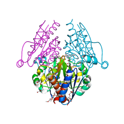
6PT2
 | | Crystal structure of the active delta opioid receptor in complex with the peptide agonist KGCHM07 | | 分子名称: | (2R)-2,3-dihydroxypropyl (9Z)-octadec-9-enoate, CHOLESTEROL, Delta opioid receptor, ... | | 著者 | Claff, T, Yu, J, Blais, V, Patel, N, Martin, C, Wu, L, Han, G.W, Holleran, B.J, Van der Poorten, O, Hanson, M.A, Sarret, P, Gendron, L, Cherezov, V, Katritch, V, Ballet, S, Liu, Z, Muller, C.E, Stevens, R.C. | | 登録日 | 2019-07-14 | | 公開日 | 2019-12-11 | | 最終更新日 | 2023-11-15 | | 実験手法 | X-RAY DIFFRACTION (2.8 Å) | | 主引用文献 | Elucidating the active delta-opioid receptor crystal structure with peptide and small-molecule agonists.
Sci Adv, 5, 2019
|
|
6Q02
 | |
6PZ3
 | |


