Loading
PDBj
MenuPDBj@FacebookPDBj@TwitterPDBj@YouTubewwPDB FoundationwwPDB
RCSB PDBPDBeBMRBAdv. SearchSearch help
PDB: 712 results

3M1Y
DownloadVisualize
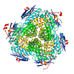
BU of 3m1y by Molmil
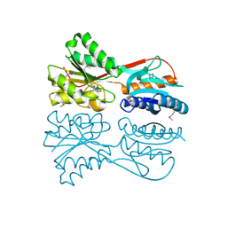
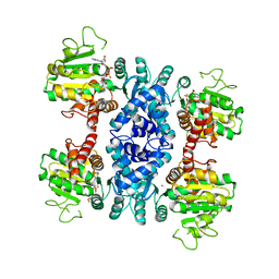
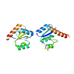
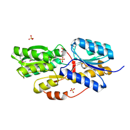
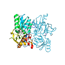
Crystal Structure of a Phosphoserine phosphatase (SerB) from Helicobacter pylori
Descriptor: CHLORIDE ION, MAGNESIUM ION, Phosphoserine phosphatase (SerB)
Authors:Syed Ibrahim, B, Burley, S.K, Swaminathan, S, New York SGX Research Center for Structural Genomics (NYSGXRC)
Deposit date:2010-03-05
Release date:2010-03-23
Last modified:2024-11-06
Method:X-RAY DIFFRACTION (2.4 Å)
Cite:Crystal Structure of a Phosphoserine phosphatase (SerB) from Helicobacter pylori
To be Published
4L0O
DownloadVisualize
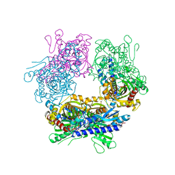
BU of 4l0o by Molmil
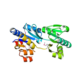
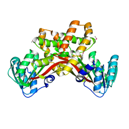
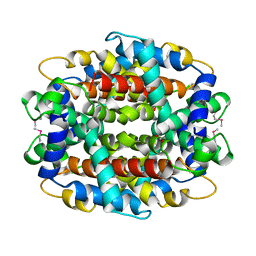
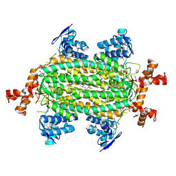
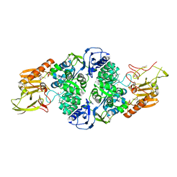
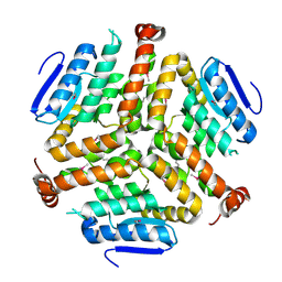
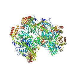
Structure determination of cystathionine gamma-synthase from Helicobacter pylori
Descriptor: Cystathionine gamma-synthase, PHOSPHATE ION, PYRIDOXAL-5'-PHOSPHATE
Authors:Tarique, K.F, Rehman, S.A.A, Gourinath, S.
Deposit date:2013-05-31
Release date:2013-12-25
Last modified:2023-11-08
Method:X-RAY DIFFRACTION (2.76 Å)
Cite:Structure determination of cystathionine gamma-synthase from Helicobacter pylori
To be Published
3MIZ
DownloadVisualize
BU of 3miz by Molmil
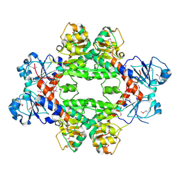
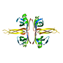
Crystal structure of a putative transcriptional regulator protein, Lacl family from Rhizobium etli
Descriptor: Putative transcriptional regulator protein, LacI family
Authors:Palani, K, Burley, S.K, Swaminathan, S, New York SGX Research Center for Structural Genomics (NYSGXRC)
Deposit date:2010-04-12
Release date:2010-04-21
Last modified:2024-11-06
Method:X-RAY DIFFRACTION (1.91 Å)
Cite:Crystal structure of a putative transcriptional regulator protein, Lacl family from Rhizobium etli
To be Published
3MPO
DownloadVisualize
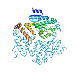
BU of 3mpo by Molmil
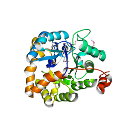
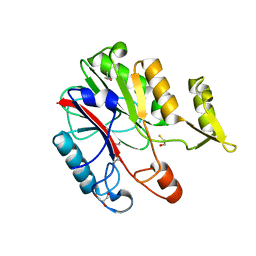
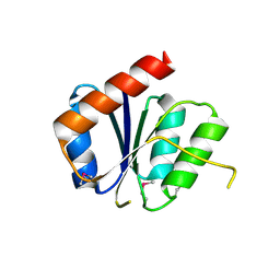
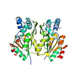
The crystal structure of a hydrolase from Lactobacillus brevis
Descriptor: Predicted hydrolase of the HAD superfamily
Authors:Zhang, Z, Burley, S.K, Swaminathan, S, New York SGX Research Center for Structural Genomics (NYSGXRC)
Deposit date:2010-04-27
Release date:2010-05-12
Last modified:2021-02-10
Method:X-RAY DIFFRACTION (2.9 Å)
Cite:The crystal structure of a hydrolase from Lactobacillus brevis
To be Published
4HY3
DownloadVisualize
BU of 4hy3 by Molmil
Crystal structure of a phosphoglycerate oxidoreductase from rhizobium etli
Descriptor: phosphoglycerate oxidoreductase
Authors:Kumaran, D, Chamala, S, Evans, B, Foti, R, Gizzi, A, Hillerich, B, Kar, A, Lafleur, J, Seidel, R, Villigas, G, Zencheck, W, Almo, S.C, Swaminathan, S, New York Structural Genomics Research Consortium (NYSGRC)
Deposit date:2012-11-12
Release date:2012-12-12
Last modified:2024-10-16
Method:X-RAY DIFFRACTION (2.8 Å)
Cite:Crystal structure of a phosphoglycerate oxidoreductase from rhizobium etli
To be Published
3MSR
DownloadVisualize
BU of 3msr by Molmil
The crystal structure of an amidohydrolase from Mycoplasma synoviae
Descriptor: GLYCEROL, PHOSPHATE ION, amidohydrolases
Authors:Zhang, Z, Burley, S.K, Swaminathan, S, New York SGX Research Center for Structural Genomics (NYSGXRC)
Deposit date:2010-04-29
Release date:2010-05-12
Last modified:2021-02-10
Method:X-RAY DIFFRACTION (2.162 Å)
Cite:The crystal structure of an amidohydrolase from Mycoplasma synoviae
To be Published
2HAE
DownloadVisualize
BU of 2hae by Molmil
Crystal structure of a putative malic enzyme (malate oxidoreductase)
Descriptor: Malate oxidoreductase, NICOTINAMIDE-ADENINE-DINUCLEOTIDE, ZINC ION
Authors:Seetharaman, J, Swaminathan, S, Burley, S.K, New York SGX Research Center for Structural Genomics (NYSGXRC)
Deposit date:2006-06-12
Release date:2006-07-04
Last modified:2023-08-30
Method:X-RAY DIFFRACTION (3.13 Å)
Cite:Crystal structure of a putative malic enzyme (malate oxidoreductase)
To be Published
3MGG
DownloadVisualize
BU of 3mgg by Molmil
Crystal Structure of Methyl Transferase from Methanosarcina mazei
Descriptor: Methyltransferase
Authors:Syed Ibrahim, B, Burley, S.K, Swaminathan, S, New York SGX Research Center for Structural Genomics (NYSGXRC)
Deposit date:2010-04-06
Release date:2010-04-21
Last modified:2021-02-10
Method:X-RAY DIFFRACTION (1.86 Å)
Cite:Crystal Structure of Methyl Transferase from Methanosarcina mazei
To be Published
2QH0
DownloadVisualize
BU of 2qh0 by Molmil
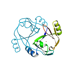
Crystal structure of a glyoxalase from clostridium acetobutylicum
Descriptor: Lactoylglutathione lyase, ZINC ION
Authors:Rao, K.N, Burley, S.K, Swaminathan, S, New York SGX Research Center for Structural Genomics (NYSGXRC)
Deposit date:2007-06-29
Release date:2007-07-17
Last modified:2024-02-21
Method:X-RAY DIFFRACTION (2.45 Å)
Cite:Crystal structure of a glyoxalase from clostridium acetobutylicum.
To be Published
1XFJ
DownloadVisualize
BU of 1xfj by Molmil
Crystal structure of protein CC_0490 from Caulobacter crescentus, Pfam DUF152
Descriptor: ACETATE ION, BETA-MERCAPTOETHANOL, GLYCEROL, ...
Authors:Krishnamurthy, N.R, Kumaran, D, Swaminathan, S, Burley, S.K, New York SGX Research Center for Structural Genomics (NYSGXRC)
Deposit date:2004-09-14
Release date:2004-09-21
Last modified:2021-02-03
Method:X-RAY DIFFRACTION (1.75 Å)
Cite:Crystal structure of a conserved hypothetical protein from Caulobacter crescentus
To be Published
3MSY
DownloadVisualize
BU of 3msy by Molmil
Crystal Structure of Mandelate racemase/muconate lactonizing enzyme from a Marine actinobacterium
Descriptor: Mandelate racemase/muconate lactonizing enzyme
Authors:Satyanarayana, L, Burley, S.K, Swaminathan, S, New York SGX Research Center for Structural Genomics (NYSGXRC)
Deposit date:2010-04-29
Release date:2010-06-30
Last modified:2024-10-30
Method:X-RAY DIFFRACTION (2.5 Å)
Cite:Crystal Structure of Mandelate racemase/muconate lactonizing enzyme from a Marine actinobacterium
To be Published
2GUW
DownloadVisualize
BU of 2guw by Molmil
Crystal structure of AMP Nucleosidase from Salmonella typhimurium LT2
Descriptor: AMP nucleosidase
Authors:Rao, K.N, Swaminathan, S, Burley, S.K, New York SGX Research Center for Structural Genomics (NYSGXRC)
Deposit date:2006-05-01
Release date:2006-06-06
Last modified:2024-02-14
Method:X-RAY DIFFRACTION (2.64 Å)
Cite:Crystal structure of AMP Nucleosidase from Salmonella typhimurium LT2
To be Published
2N3O
DownloadVisualize
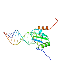
BU of 2n3o by Molmil
Structure of PTB RRM1(41-163) bound to an RNA stemloop containing a structured loop derived from viral internal ribosomal entry site RNA
Descriptor: Polypyrimidine tract-binding protein 1, RNA (5'-R(*GP*GP*GP*AP*CP*CP*UP*GP*GP*UP*CP*UP*UP*UP*CP*CP*AP*GP*GP*UP*CP*CP*C)-3')
Authors:Maris, C, Jayne, S.F, Damberger, F.F, Ravindranathan, S, Allain, F.H.-T.
Deposit date:2015-06-08
Release date:2016-08-10
Last modified:2024-05-01
Method:SOLUTION NMR
Cite:C-terminal helix folding upon pyrimidine-rich hairpin binding to PTB RRM1. Implications for PTB function in Encephalomyocarditis virus IRES activity.
To be Published
4KPK
DownloadVisualize
BU of 4kpk by Molmil
Crystal structure of a enoyl-CoA hydratase from Shewanella pealeana ATCC 700345
Descriptor: 1,2-ETHANEDIOL, Enoyl-CoA hydratase/isomerase
Authors:Kumaran, D, Chamala, S, Evans, B, Foti, R, Gizzi, A, Hillerich, B, Kar, A, Lafleur, J, Seidel, R, Villigas, G, Zencheck, W, Al Obaidi, N, Almo, S.C, Swaminathan, S, New York Structural Genomics Research Consortium (NYSGRC)
Deposit date:2013-05-13
Release date:2013-05-22
Method:X-RAY DIFFRACTION (2.09 Å)
Cite:Crystal structure of a enoyl-CoA hydratase from Shewanella pealeana ATCC 700345
To be Published
3N53
DownloadVisualize
BU of 3n53 by Molmil
Crystal structure of a response regulator receiver modulated diguanylate cyclase from Pelobacter carbinolicus
Descriptor: Response regulator receiver modulated diguanylate cyclase
Authors:Palani, K, Zhang, Z, Burley, S.K, Swaminathan, S, New York SGX Research Center for Structural Genomics (NYSGXRC)
Deposit date:2010-05-24
Release date:2010-07-28
Last modified:2023-11-22
Method:X-RAY DIFFRACTION (2.2 Å)
Cite:Crystal structure of a response regulator receiver modulated diguanylate cyclase from Pelobacter carbinolicus
To be Published
3MHD
DownloadVisualize
BU of 3mhd by Molmil
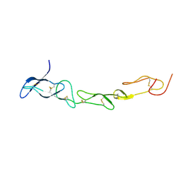
Crystal structure of DCR3
Descriptor: Tumor necrosis factor receptor superfamily member 6B
Authors:Zhan, C, Patskovsky, Y, Yan, Q, Li, Z, Ramagopal, U.A, Nathenson, S.G, Almo, S.C.
Deposit date:2010-04-07
Release date:2011-02-23
Last modified:2024-11-06
Method:X-RAY DIFFRACTION (2.901 Å)
Cite:Decoy Strategies: The Structure of TL1A:DcR3 Complex.
Structure, 19, 2011
1Y9I
DownloadVisualize
BU of 1y9i by Molmil
Crystal structure of low temperature requirement C protein from Listeria monocytogenes
Descriptor: CALCIUM ION, GLYCEROL, MAGNESIUM ION, ...
Authors:Kumaran, D, Swaminathan, S, Burley, S.K, New York SGX Research Center for Structural Genomics (NYSGXRC)
Deposit date:2004-12-15
Release date:2004-12-28
Last modified:2024-10-16
Method:X-RAY DIFFRACTION (1.8 Å)
Cite:Crystal structure of phosphatidylglycerophosphatase (PGPase), a putative membrane-bound lipid phosphatase, reveals a novel binuclear metal binding site and two "proton wires".
Proteins, 64, 2006
4HGV
DownloadVisualize
BU of 4hgv by Molmil
Crystal structure of a fumarate hydratase
Descriptor: Fumarate hydratase class II, SULFATE ION
Authors:Eswaramoorthy, S, Evans, B, Foti, R, Gizzi, A, Hillerich, B, Kar, A, Lafleur, J, Seidel, R, Villigas, G, Zencheck, W, Almo, S.C, Swaminathan, S, New York Structural Genomics Research Consortium (NYSGRC)
Deposit date:2012-10-08
Release date:2012-10-31
Last modified:2024-11-06
Method:X-RAY DIFFRACTION (2.09 Å)
Cite:Crystal structure of a fumarate hydratase
To be Published
3T81
DownloadVisualize
BU of 3t81 by Molmil
Crystal Structure of diiron adenine deaminase
Descriptor: Adenine deaminase 2, FE (III) ION
Authors:Bagaria, A, Kumaran, D, Burley, S.K, Swaminathan, S, New York SGX Research Center for Structural Genomics (NYSGXRC)
Deposit date:2011-08-01
Release date:2011-11-02
Last modified:2023-12-06
Method:X-RAY DIFFRACTION (2.63 Å)
Cite:Crystal Structure of diiron adenine deaminase
TO BE PUBLISHED
2RJK
DownloadVisualize
BU of 2rjk by Molmil
Crystal Structure of Human TL1A Extracellular Domain C95S Mutant
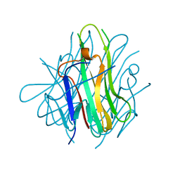
Descriptor: TNF superfamily ligand TL1A
Authors:Zhan, C, Yan, Q, Patskovsky, Y, Shi, W, Ramagopal, U.A, Toro, R, Bonanno, J, Nathenson, S.G, Almo, S.C.
Deposit date:2007-10-15
Release date:2008-08-26
Last modified:2023-08-30
Method:X-RAY DIFFRACTION (2.3 Å)
Cite:Biochemical and structural characterization of the human TL1A ectodomain.
Biochemistry, 48, 2009
2RJO
DownloadVisualize
BU of 2rjo by Molmil
Crystal structure of Twin-arginine translocation pathway signal protein from Burkholderia phytofirmans
Descriptor: SULFATE ION, Twin-arginine translocation pathway signal protein, beta-D-galactopyranose
Authors:Agarwal, R, Burley, S.K, Swaminathan, S, New York SGX Research Center for Structural Genomics (NYSGXRC)
Deposit date:2007-10-15
Release date:2007-10-23
Last modified:2024-11-06
Method:X-RAY DIFFRACTION (2.05 Å)
Cite:Crystal structure of twin-arginine translocation pathway signal protein from Burkholderia phytofirmans.
To be Published
3NHM
DownloadVisualize
BU of 3nhm by Molmil
Crystal structure of a response regulator from Myxococcus xanthus
Descriptor: Response regulator
Authors:Palani, K, Burley, S.K, Swaminathan, S, New York SGX Research Center for Structural Genomics (NYSGXRC)
Deposit date:2010-06-14
Release date:2010-07-21
Last modified:2024-10-30
Method:X-RAY DIFFRACTION (2.19 Å)
Cite:Crystal structure of a response regulator from Myxococcus xanthus
To be Published
3TBH
DownloadVisualize
BU of 3tbh by Molmil
Crystal structure of O-Acetyl Serine Sulfhydrylase in complex with octapeptide derived from Serine Acetyl Transferase of Leishmania donovani
Descriptor: O-acetyl serine sulfhydrylase, SODIUM ION, Serine acetyl transferase derived octapeptide
Authors:Raj, I, Gourinath, S.
Deposit date:2011-08-06
Release date:2012-08-01
Last modified:2023-12-06
Method:X-RAY DIFFRACTION (1.68 Å)
Cite:Crystal structure of O-Acetyl Serine Sulfhydrylase in complex with octapeptide derived from Serine Acetyl Transferase of Leishmania donovani
To be Published
4LK5
DownloadVisualize
BU of 4lk5 by Molmil
Crystal structure of a enoyl-CoA hydratase from Mycobacterium avium subsp. paratuberculosis K-10
Descriptor: enoyl-CoA hydratase
Authors:Kumaran, D, Chamala, S, Evans, B, Foti, R, Gizzi, A, Hillerich, B, Kar, A, Lafleur, J, Seidel, R, Villigas, G, Zencheck, W, Al Obaidi, N, Almo, S.C, Swaminathan, S, New York Structural Genomics Research Consortium (NYSGRC)
Deposit date:2013-07-06
Release date:2013-07-24
Last modified:2024-10-09
Method:X-RAY DIFFRACTION (2.5 Å)
Cite:Crystal structure of a enoyl-CoA hydratase from Mycobacterium avium subsp. paratuberculosis K-10
To be Published
1SRU
DownloadVisualize
BU of 1sru by Molmil
Crystal structure of full length E. coli SSB protein
Descriptor: Single-strand binding protein
Authors:Savvides, S.N, Raghunathan, S, Fuetterer, K, Kozlov, A.G, Lohman, T.M, Waksman, G.
Deposit date:2004-03-23
Release date:2004-08-03
Last modified:2024-02-14
Method:X-RAY DIFFRACTION (3.3 Å)
Cite:The C-terminal domain of full-length E. coli SSB is disordered even when bound to DNA.
Protein Sci., 13, 2004

227111

PDB entries from 2024-11-06

PDB statisticsPDBj update infoContact PDBjnumon