1YIR
 
 | |
5FXT
 | |
3DDA
 | |
5W5A
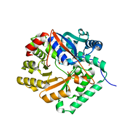
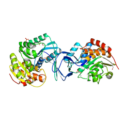
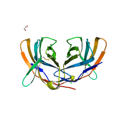
 | | Crystal structure of Mycobacterium tuberculosis CRP-FNR family transcription factor Cmr (Rv1675c) | | 分子名称: | CHLORIDE ION, HTH-type transcriptional regulator Cmr, SULFATE ION | | 著者 | Cheung, J, Cassidy, M, Ginter, C, Ranganathan, S, Pata, D.J, McDonough, K.A. | | 登録日 | 2017-06-14 | | 公開日 | 2017-12-13 | | 最終更新日 | 2019-01-09 | | 実験手法 | X-RAY DIFFRACTION (1.85 Å) | | 主引用文献 | Novel structural features drive DNA binding properties of Cmr, a CRP family protein in TB complex mycobacteria.
Nucleic Acids Res., 46, 2018
|
|
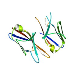
1YYN
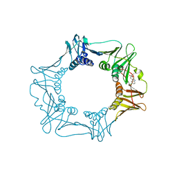
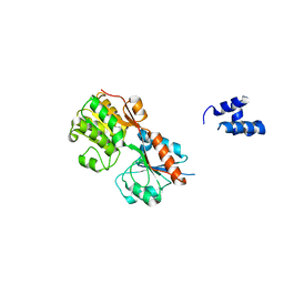
 | | A common binding site for disialyllactose and a tri-peptide in the C-fragment of tetanus neurotoxin | | 分子名称: | N-acetyl-alpha-neuraminic acid-(2-8)-N-acetyl-alpha-neuraminic acid-(2-3)-alpha-D-galactopyranose-(1-4)-beta-D-glucopyranose, Tetanus toxin | | 著者 | Seetharaman, J, Eswaramoorthy, S, Kumaran, D, Swaminathan, S. | | 登録日 | 2005-02-25 | | 公開日 | 2005-03-15 | | 最終更新日 | 2023-10-25 | | 実験手法 | X-RAY DIFFRACTION (2.3 Å) | | 主引用文献 | Common binding site for disialyllactose and tri-peptide in C-fragment of tetanus neurotoxin
Proteins, 61, 2005
|
|
3FFZ
 | | Domain organization in Clostridium butulinum neurotoxin type E is unique: Its implication in faster translocation | | 分子名称: | ACETATE ION, Botulinum neurotoxin type E, SODIUM ION, ... | | 著者 | Kumaran, D, Eswaramoorthy, S, Swaminathan, S. | | 登録日 | 2008-12-04 | | 公開日 | 2008-12-16 | | 最終更新日 | 2023-09-06 | | 実験手法 | X-RAY DIFFRACTION (2.65 Å) | | 主引用文献 | Domain organization in Clostridium botulinum neurotoxin type E is unique: its implication in faster translocation.
J.Mol.Biol., 386, 2009
|
|
3D8K
 | |
2ABQ
 | |
3H5T
 | |
3GRA
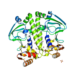
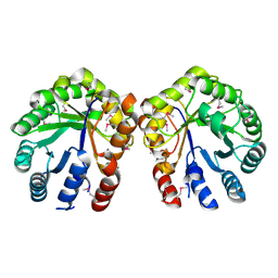
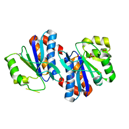 | | Crystal structure of AraC family transcriptional regulator from Pseudomonas putida | | 分子名称: | 1,2-ETHANEDIOL, MAGNESIUM ION, SULFATE ION, ... | | 著者 | Bagaria, A, Kumaran, D, Burley, S.K, Swaminathan, S, New York SGX Research Center for Structural Genomics (NYSGXRC) | | 登録日 | 2009-03-25 | | 公開日 | 2009-04-14 | | 最終更新日 | 2021-02-10 | | 実験手法 | X-RAY DIFFRACTION (2.3 Å) | | 主引用文献 | Crystal structure of AraC family transcriptional regulator from Pseudomonas putida
To be Published
|
|
1DPY
 | | THREE-DIMENSIONAL STRUCTURE OF A NOVEL PHOSPHOLIPASE A2 FROM INDIAN COMMON KRAIT AT 2.45 A RESOLUTION | | 分子名称: | PHOSPHOLIPASE A2, SODIUM ION | | 著者 | Singh, G, Gourinath, S, Sharma, S, Paramasivam, M, Srinivasan, A, Singh, T.P. | | 登録日 | 1999-12-28 | | 公開日 | 2000-06-28 | | 最終更新日 | 2011-07-13 | | 実験手法 | X-RAY DIFFRACTION (2.45 Å) | | 主引用文献 | Sequence and crystal structure determination of a basic phospholipase A2 from common krait (Bungarus caeruleus) at 2.4 A resolution: identification and characterization of its pharmacological sites.
J.Mol.Biol., 307, 2001
|
|
3H5O
 | |
1DQT
 | | THE CRYSTAL STRUCTURE OF MURINE CTLA4 (CD152) | | 分子名称: | 1,2-ETHANEDIOL, CHLORIDE ION, CYTOTOXIC T LYMPHOCYTE ASSOCIATED ANTIGEN 4 | | 著者 | Ostrov, D.A, Shi, W, Schwartz, J.C, Almo, S.C, Nathenson, S.G. | | 登録日 | 2000-01-05 | | 公開日 | 2000-10-27 | | 最終更新日 | 2018-01-31 | | 実験手法 | X-RAY DIFFRACTION (2 Å) | | 主引用文献 | Structure of murine CTLA-4 and its role in modulating T cell responsiveness.
Science, 290, 2000
|
|
3H74
 | | Crystal structure of pyridoxal kinase from Lactobacillus plantarum | | 分子名称: | GLYCEROL, Pyridoxal kinase, SULFATE ION | | 著者 | Bagaria, A, Kumaran, D, Burley, S.K, Swaminathan, S, New York SGX Research Center for Structural Genomics (NYSGXRC) | | 登録日 | 2009-04-24 | | 公開日 | 2009-05-26 | | 最終更新日 | 2021-02-10 | | 実験手法 | X-RAY DIFFRACTION (1.3 Å) | | 主引用文献 | Crystal structure of pyridoxal kinase from Lactobacillus plantarum
To be Published
|
|
3HDG
 | |
3DEC
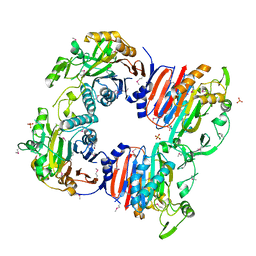
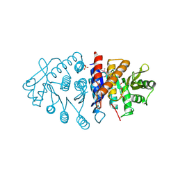
 | | Crystal structure of a glycosyl hydrolases family 2 protein from Bacteroides thetaiotaomicron | | 分子名称: | Beta-galactosidase, POTASSIUM ION | | 著者 | Kumaran, D, Bonanno, J, Romero, R, Burley, S.K, Swaminathan, S, New York SGX Research Center for Structural Genomics (NYSGXRC) | | 登録日 | 2008-06-09 | | 公開日 | 2008-06-17 | | 最終更新日 | 2023-11-15 | | 実験手法 | X-RAY DIFFRACTION (2.8 Å) | | 主引用文献 | Crystal Structure of a Glycosyl Hydrolases Family 2 protein from Bacteroides thetaiotaomicron.
To be Published
|
|
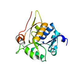
1XFJ
 | | Crystal structure of protein CC_0490 from Caulobacter crescentus, Pfam DUF152 | | 分子名称: | ACETATE ION, BETA-MERCAPTOETHANOL, GLYCEROL, ... | | 著者 | Krishnamurthy, N.R, Kumaran, D, Swaminathan, S, Burley, S.K, New York SGX Research Center for Structural Genomics (NYSGXRC) | | 登録日 | 2004-09-14 | | 公開日 | 2004-09-21 | | 最終更新日 | 2021-02-03 | | 実験手法 | X-RAY DIFFRACTION (1.75 Å) | | 主引用文献 | Crystal structure of a conserved hypothetical protein from Caulobacter crescentus
To be Published
|
|
2ISN
 | | Crystal structure of a phosphatase from a pathogenic strain Toxoplasma gondii | | 分子名称: | NYSGXRC-8828z, phosphatase, PRASEODYMIUM ION, ... | | 著者 | Agarwal, R, Burley, S.K, Swaminathan, S, New York SGX Research Center for Structural Genomics (NYSGXRC) | | 登録日 | 2006-10-18 | | 公開日 | 2006-10-31 | | 最終更新日 | 2021-02-03 | | 実験手法 | X-RAY DIFFRACTION (1.9 Å) | | 主引用文献 | Structural genomics of protein phosphatases.
J.STRUCT.FUNCT.GENOM., 8, 2007
|
|
3EEG
 | |
4HKE
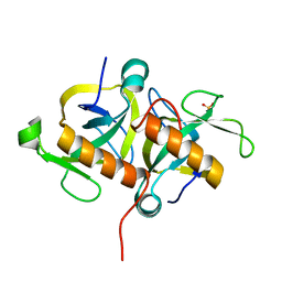 | | Crystal Structure of MoxT of Bacillus anthracis | | 分子名称: | Addiction module toxin component PemK, SULFATE ION | | 著者 | Verma, S, Kumar, S, Gourinath, S, Bhatnagar, R. | | 登録日 | 2012-10-15 | | 公開日 | 2013-11-13 | | 最終更新日 | 2023-11-08 | | 実験手法 | X-RAY DIFFRACTION (1.87 Å) | | 主引用文献 | Structural basis of Bacillus anthracis MoxXT disruption and the modulation of MoxT ribonuclease activity by rationally designed peptides.
J.Biomol.Struct.Dyn., 33, 2015
|
|
4HL9
 | | Crystal structure of antibiotic biosynthesis monooxygenase | | 分子名称: | Antibiotic biosynthesis monooxygenase | | 著者 | Rice, S, Eswaramoorthy, S, Chamala, S, Evans, B, Foti, R, Gizzi, A, Hillerich, B, Kar, A, LaFleur, J, Seidel, R, Villigas, G, Zencheck, W, Almo, S.C, Swaminathan, S, New York Structural Genomics Research Consortium (NYSGRC) | | 登録日 | 2012-10-16 | | 公開日 | 2012-10-31 | | 最終更新日 | 2018-01-24 | | 実験手法 | X-RAY DIFFRACTION (1.93 Å) | | 主引用文献 | Crystal structure of antibiotic biosynthesis monooxygenase
To be Published
|
|
3EUW
 | |
4HWU
 | | Crystal structure of the Ig-C2 type 1 domain from mouse Fibroblast growth factor receptor 2 (FGFR2) [NYSGRC-005912] | | 分子名称: | Fibroblast growth factor receptor 2 | | 著者 | Kumar, P.R, Ahmed, M, Banu, R, Bhosle, R, Calarese, D, Celikigil, A, Chamala, S, Chan, M.K, Chowdhury, S, Fiser, A, Garforth, S, Glenn, A.S, Hillerich, B, Khafizov, K, Love, J, Patel, H, Rubinstein, R, Seidel, R, Stead, M, Toro, R, Nathenson, S.G, Almo, S.C, New York Structural Genomics Research Consortium (NYSGRC), Atoms-to-Animals: The Immune Function Network (IFN) | | 登録日 | 2012-11-08 | | 公開日 | 2012-11-21 | | 実験手法 | X-RAY DIFFRACTION (2.903 Å) | | 主引用文献 | Crystal structure of the Ig-C2 type 1 domain from mouse FGFR2 [NYSGRC-005912]
to be published
|
|
4HUJ
 | | Crystal structure of a hypothetical protein SMa0349 from Sinorhizobium meliloti | | 分子名称: | Uncharacterized protein | | 著者 | Rice, S, Eswaramoorthy, S, Chamala, S, Evans, B, Foti, F, Gizzi, A, Hillerich, B, Kar, A, LaFleur, J, Seidel, R, Villigas, G, Zencheck, W, Almo, S.C, Swaminathan, S, New York Structural Genomics Research Consortium (NYSGRC) | | 登録日 | 2012-11-02 | | 公開日 | 2012-12-12 | | 実験手法 | X-RAY DIFFRACTION (1.77 Å) | | 主引用文献 | Crystal structure of a hypothetical protein SMa0349 from Sinorhizobium meliloti
To be Published
|
|
5ZR2
 | |


