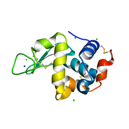
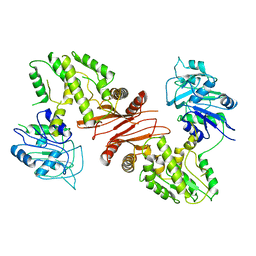
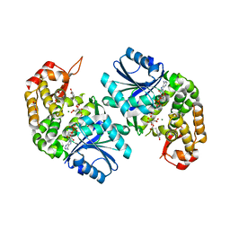
1ESQ
 
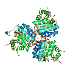 | | CRYSTAL STRUCTURE OF THIAZOLE KINASE MUTANT (C198S) WITH ATP AND THIAZOLE PHOSPHATE. | | 分子名称: | 4-METHYL-5-HYDROXYETHYLTHIAZOLE PHOSPHATE, ADENOSINE-5'-TRIPHOSPHATE, HYDROXYETHYLTHIAZOLE KINASE, ... | | 著者 | Campobasso, N, Mathews, I.I, Begley, T.P, Ealick, S.E. | | 登録日 | 2000-04-10 | | 公開日 | 2000-08-09 | | 最終更新日 | 2024-02-07 | | 実験手法 | X-RAY DIFFRACTION (2.5 Å) | | 主引用文献 | Crystal structure of 4-methyl-5-beta-hydroxyethylthiazole kinase from Bacillus subtilis at 1.5 A resolution.
Biochemistry, 39, 2000
|
|
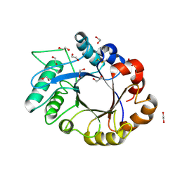
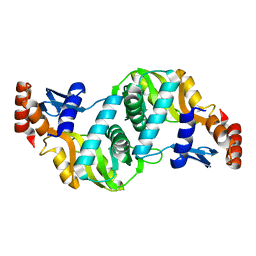
6L8X
 | | Crystal structure of Siraitia grosvenorii ugt transferase mutant2 | | 分子名称: | Glycosyltransferase | | 著者 | Li, J, Shan, N, Yang, J.G, Liu, W.D, Sun, Y.X. | | 登録日 | 2019-11-07 | | 公開日 | 2020-04-08 | | 最終更新日 | 2023-11-22 | | 実験手法 | X-RAY DIFFRACTION (1.55 Å) | | 主引用文献 | Efficient O-Glycosylation of Triterpenes Enabled by Protein Engineering of Plant Glycosyltransferase UGT74AC1
Acs Catalysis, 10, 2020
|
|
1NZ5
 | | The Horse heart myoglobin variant K45E/K63E complexed with Manganese | | 分子名称: | MANGANESE (II) ION, Myoglobin, PROTOPORPHYRIN IX CONTAINING FE | | 著者 | Hunter, C.L, Maurus, R, Mauk, M.R, Lee, H, Raven, E.L, Tong, H, Nguyen, N, Smith, S, Brayer, G.D, Mauk, A.G. | | 登録日 | 2003-02-15 | | 公開日 | 2003-04-08 | | 最終更新日 | 2024-02-14 | | 実験手法 | X-RAY DIFFRACTION (1.7 Å) | | 主引用文献 | Introduction and characterization of a functionally linked metal ion binding site at
the exposed heme edge of myoglobin
Proc.Natl.Acad.Sci.USA, 100, 2003
|
|
1OIS
 | | YEAST DNA TOPOISOMERASE I, N-TERMINAL FRAGMENT | | 分子名称: | DNA TOPOISOMERASE I | | 著者 | Lue, N, Sharma, A, Mondragon, A, Wang, J.C. | | 登録日 | 1996-09-14 | | 公開日 | 1997-03-12 | | 最終更新日 | 2024-02-14 | | 実験手法 | X-RAY DIFFRACTION (1.9 Å) | | 主引用文献 | A 26 kDa yeast DNA topoisomerase I fragment: crystallographic structure and mechanistic implications.
Structure, 3, 1995
|
|
4WM1
 | | High pressure protein crystallography of hen egg white lysozyme at 500 MPa | | 分子名称: | CHLORIDE ION, Lysozyme C, SODIUM ION | | 著者 | Yamada, H, Nagae, T, Watanabe, N. | | 登録日 | 2014-10-08 | | 公開日 | 2015-04-08 | | 最終更新日 | 2020-02-05 | | 実験手法 | X-RAY DIFFRACTION (1.6 Å) | | 主引用文献 | High-pressure protein crystallography of hen egg-white lysozyme
Acta Crystallogr.,Sect.D, 71, 2015
|
|
1OM0
 | | crystal structure of xylanase inhibitor protein (XIP-I) from wheat | | 分子名称: | 1,2-ETHANEDIOL, 2-acetamido-2-deoxy-beta-D-glucopyranose, Xylanase Inhibitor Protein I | | 著者 | Payan, F, Flatman, R, Porciero, S, Williamson, G, Juge, N, Roussel, A. | | 登録日 | 2003-02-24 | | 公開日 | 2003-06-03 | | 最終更新日 | 2023-10-25 | | 実験手法 | X-RAY DIFFRACTION (1.8 Å) | | 主引用文献 | Structural analysis of xylanase inhibitor protein I (XIP-I), a proteinaceous xylanase inhibitor from wheat (Triticum aestivum, var. Soisson).
Biochem.J., 372, 2003
|
|
4WNL
 | | The X-ray structure of a RNA-binding protein complex | | 分子名称: | GLYCEROL, ISOPROPYL ALCOHOL, SULFATE ION, ... | | 著者 | Singh, N, Blobel, G, Shi, H. | | 登録日 | 2014-10-13 | | 公開日 | 2014-12-24 | | 最終更新日 | 2023-12-27 | | 実験手法 | X-RAY DIFFRACTION (2.8 Å) | | 主引用文献 | Hooking She3p onto She2p for myosin-mediated cytoplasmic mRNA transport.
Proc.Natl.Acad.Sci.USA, 112, 2015
|
|
1ONK
 | | Mistletoe lectin I from viscum album | | 分子名称: | 2-acetamido-2-deoxy-beta-D-glucopyranose, AZIDE ION, Beta-galactoside specific lectin I A chain, ... | | 著者 | Gabdoulkhakov, A.G, Savoshkina, Y, Krauspenhaar, R, Stoeva, S, Konareva, N, Kornilov, V, Kornev, A.N, Voelter, W, Nikonov, S.V, Betzel, C, Mikhailov, A.M. | | 登録日 | 2003-02-28 | | 公開日 | 2004-02-28 | | 最終更新日 | 2023-10-25 | | 実験手法 | X-RAY DIFFRACTION (2.1 Å) | | 主引用文献 | Mistletoe lectin I from viscum album
To be Published
|
|
4WIM
 | |
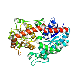
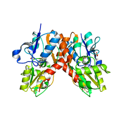
1ORO
 | | A FLEXIBLE LOOP AT THE DIMER INTERFACE IS A PART OF THE ACTIVE SITE OF THE ADJACENT MONOMER OF ESCHERICHIA COLI OROTATE PHOSPHORIBOSYLTRANSFERASE | | 分子名称: | OROTATE PHOSPHORIBOSYLTRANSFERASE, SULFATE ION | | 著者 | Henriksen, A, Aghajari, N, Jensen, K.F, Gajhede, M. | | 登録日 | 1995-09-11 | | 公開日 | 1996-04-03 | | 最終更新日 | 2024-02-14 | | 実験手法 | X-RAY DIFFRACTION (2.4 Å) | | 主引用文献 | A flexible loop at the dimer interface is a part of the active site of the adjacent monomer of Escherichia coli orotate phosphoribosyltransferase.
Biochemistry, 35, 1996
|
|
1ONU
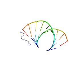
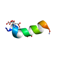 | | NMDA RECEPTOR ANTAGONIST, CONANTOKIN-G, NMR, 17 STRUCTURES | | 分子名称: | CONANTOKIN-G | | 著者 | Skjaerbaek, N, Nielsen, K.J, Lewis, R.J, Alewood, P.F, Craik, D.J. | | 登録日 | 1996-08-27 | | 公開日 | 1997-09-04 | | 最終更新日 | 2022-02-23 | | 実験手法 | SOLUTION NMR | | 主引用文献 | Determination of the solution structures of conantokin-G and conantokin-T by CD and NMR spectroscopy.
J.Biol.Chem., 272, 1997
|
|
1P1W
 | |
6LK2
 | | Crystal structure of Providencia alcalifaciens 3-dehydroquinate synthase (DHQS) in complex with Mg2+, NAD and chlorogenic acid | | 分子名称: | (1R,3R,4S,5R)-3-[3-[3,4-bis(oxidanyl)phenyl]propanoyloxy]-1,4,5-tris(oxidanyl)cyclohexane-1-carboxylic acid, 1,2-ETHANEDIOL, 3-dehydroquinate synthase, ... | | 著者 | Neetu, N, Katiki, M, Kumar, P. | | 登録日 | 2019-12-17 | | 公開日 | 2020-07-29 | | 最終更新日 | 2023-11-22 | | 実験手法 | X-RAY DIFFRACTION (2.503 Å) | | 主引用文献 | Structural and Biochemical Analyses Reveal that Chlorogenic Acid Inhibits the Shikimate Pathway.
J.Bacteriol., 202, 2020
|
|
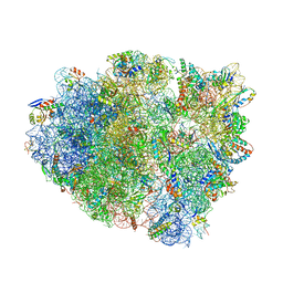
4WRA
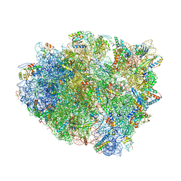 | | Complex of 70S ribosome with tRNA-Tyr and mRNA with A-A mismatch in the first position in the A-site and with antibiotic paromomycin. | | 分子名称: | 16S ribosomal RNA, 23S ribosomal RNA, 30S ribosomal protein S10, ... | | 著者 | Rozov, A, Demeshkina, N, Yusupov, M, Yusupova, G. | | 登録日 | 2014-10-23 | | 公開日 | 2015-06-10 | | 最終更新日 | 2024-01-10 | | 実験手法 | X-RAY DIFFRACTION (3.05 Å) | | 主引用文献 | Structural insights into the translational infidelity mechanism.
Nat Commun, 6, 2015
|
|
4WQ1
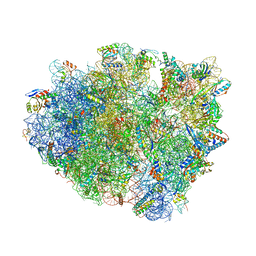 | | Complex of 70S ribosome with tRNA-Tyr and mRNA with C-A mismatch in the first position in the A-site. | | 分子名称: | 16S ribosomal RNA, 23S ribosomal RNA, 30S ribosomal protein S10, ... | | 著者 | Rozov, A, Demeshkina, N, Yusupov, M, Yusupova, G. | | 登録日 | 2014-10-21 | | 公開日 | 2015-06-10 | | 最終更新日 | 2024-01-10 | | 実験手法 | X-RAY DIFFRACTION (3.1 Å) | | 主引用文献 | Structural insights into the translational infidelity mechanism.
Nat Commun, 6, 2015
|
|
1P4L
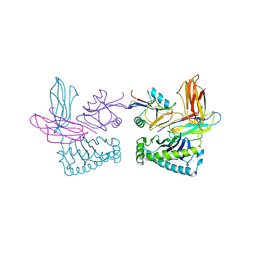 | | Crystal structure of NK receptor Ly49C mutant with its MHC class I ligand H-2Kb | | 分子名称: | Beta-2-microglobulin, LY49-C, MHC CLASS I H-2KB HEAVY CHAIN, ... | | 著者 | Dam, J, Guan, R, Natarajan, K, Dimasi, N, Mariuzza, R.A. | | 登録日 | 2003-04-23 | | 公開日 | 2003-11-11 | | 最終更新日 | 2023-08-16 | | 実験手法 | X-RAY DIFFRACTION (2.9 Å) | | 主引用文献 | Variable MHC class I engagement by Ly49 natural killer cell receptors demonstrated by the crystal structure of Ly49C bound to H-2K(b).
Nat.Immunol., 4, 2003
|
|
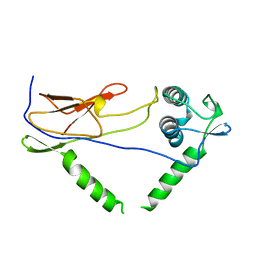
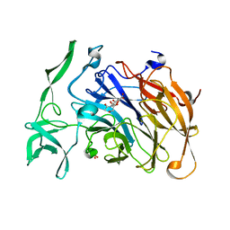
1P5T
 | | Crystal Structure of Dok1 PTB Domain | | 分子名称: | Docking protein 1 | | 著者 | Shi, N, Ye, S, Liu, Y, Zhou, W, Ding, Y, Lou, Z, Qiang, B, Yuan, J, Rao, Z. | | 登録日 | 2003-04-28 | | 公開日 | 2004-02-17 | | 最終更新日 | 2011-07-13 | | 実験手法 | X-RAY DIFFRACTION (2.35 Å) | | 主引用文献 | Structural Basis for the Specific Recognition of RET by the Dok1 Phosphotyrosine Binding Domain
J.BIOL.CHEM., 279, 2004
|
|
1OFX
 | | CRYSTAL STRUCTURE OF AN OKAZAKI FRAGMENT AT 2 ANGSTROMS RESOLUTION | | 分子名称: | DNA (5'-D(*GP*GP*GP*TP*AP*TP*AP*CP*GP*C)-3'), DNA/RNA (5'-R(*GP*CP*GP*)-D(*TP*AP*TP*AP*CP*CP*C)-3'), SPERMINE | | 著者 | Egli, M, Usman, N, Zhang, S, Rich, A. | | 登録日 | 1991-10-17 | | 公開日 | 1993-04-15 | | 最終更新日 | 2024-02-14 | | 実験手法 | X-RAY DIFFRACTION (2 Å) | | 主引用文献 | Crystal structure of an Okazaki fragment at 2-A resolution.
Proc.Natl.Acad.Sci.USA, 89, 1992
|
|
4WL6
 | | Raster-scanning protein crystallography using micro and nano-focused synchrotron beams | | 分子名称: | CHLORIDE ION, Lysozyme C | | 著者 | Coquelle, N, Kapp, U, Shilova, A, Weinhausen, B, Burghammer, M, Colletier, J.P. | | 登録日 | 2014-10-06 | | 公開日 | 2015-05-06 | | 最終更新日 | 2024-01-10 | | 実験手法 | X-RAY DIFFRACTION (1.85 Å) | | 主引用文献 | Raster-scanning serial protein crystallography using micro- and nano-focused synchrotron beams.
Acta Crystallogr.,Sect.D, 71, 2015
|
|
4WLY
 | | High pressure protein crystallography of hen egg white lysozyme at 380 MPa | | 分子名称: | CHLORIDE ION, Lysozyme C, SODIUM ION | | 著者 | Yamada, H, Nagae, T, Watanabe, N. | | 登録日 | 2014-10-08 | | 公開日 | 2015-04-08 | | 最終更新日 | 2020-02-05 | | 実験手法 | X-RAY DIFFRACTION (1.62 Å) | | 主引用文献 | High-pressure protein crystallography of hen egg-white lysozyme
Acta Crystallogr.,Sect.D, 71, 2015
|
|
4WM4
 | | High pressure protein crystallography of hen egg white lysozyme at 800 MPa | | 分子名称: | CHLORIDE ION, Lysozyme C, SODIUM ION | | 著者 | Yamada, H, Nagae, T, Watanabe, N. | | 登録日 | 2014-10-08 | | 公開日 | 2015-04-08 | | 最終更新日 | 2020-02-05 | | 実験手法 | X-RAY DIFFRACTION (1.6 Å) | | 主引用文献 | High-pressure protein crystallography of hen egg-white lysozyme
Acta Crystallogr.,Sect.D, 71, 2015
|
|
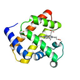
1OXD
 | | Expansion of the Genetic Code Enables Design of a Novel "Gold" Class of Green Fluorescent Proteins | | 分子名称: | cyan fluorescent protein cfp | | 著者 | Hyun Bae, J, Rubini, M, Jung, G, Wiegand, G, Seifert, M.H, Azim, M.K, Kim, J.S, Zumbusch, A, Holak, T.A, Moroder, L, Huber, R, Budisa, N. | | 登録日 | 2003-04-02 | | 公開日 | 2003-12-02 | | 最終更新日 | 2021-10-27 | | 実験手法 | X-RAY DIFFRACTION (1.15 Å) | | 主引用文献 | Expansion of the Genetic Code Enables Design of a Novel "Gold" Class of Green Fluorescent Proteins
J.Mol.Biol., 328, 2003
|
|
1OXE
 | | Expansion of the Genetic Code Enables Design of a Novel "Gold" Class of Green Fluorescent Proteins | | 分子名称: | cyan fluorescent protein cfp | | 著者 | Hyun Bae, J, Rubini, M, Jung, G, Wiegand, G, Seifert, M.H, Azim, M.K, Kim, J.S, Zumbusch, A, Holak, T.A, Moroder, L, Huber, R, Budisa, N. | | 登録日 | 2003-04-02 | | 公開日 | 2003-12-02 | | 最終更新日 | 2021-10-27 | | 実験手法 | X-RAY DIFFRACTION (1.15 Å) | | 主引用文献 | Expansion of the Genetic Code Enables Design of a Novel "Gold" Class of Green Fluorescent Proteins
J.Mol.Biol., 328, 2003
|
|
4WU1
 | | Complex of 70S ribosome with tRNA-Tyr and mRNA with G-U mismatch in the second position in the P-site | | 分子名称: | 16S ribosomal RNA, 23S ribosomal RNA, 30S ribosomal protein S10, ... | | 著者 | Rozov, A, Demeshkina, N, Yusupov, M, Yusupova, G. | | 登録日 | 2014-10-30 | | 公開日 | 2015-06-10 | | 最終更新日 | 2024-01-10 | | 実験手法 | X-RAY DIFFRACTION (3.2 Å) | | 主引用文献 | Structural insights into the translational infidelity mechanism.
Nat Commun, 6, 2015
|
|
4X47
 | | Crystal structure of the intramolecular trans-sialidase from Ruminococcus gnavus in complex with Neu5Ac2en | | 分子名称: | 2-DEOXY-2,3-DEHYDRO-N-ACETYL-NEURAMINIC ACID, Anhydrosialidase, PHOSPHATE ION | | 著者 | Owen, C.D, Tailford, L.E, Taylor, G.L, Juge, N. | | 登録日 | 2014-12-02 | | 公開日 | 2015-07-22 | | 最終更新日 | 2024-01-10 | | 実験手法 | X-RAY DIFFRACTION (2 Å) | | 主引用文献 | Discovery of intramolecular trans-sialidases in human gut microbiota suggests novel mechanisms of mucosal adaptation.
Nat Commun, 6, 2015
|
|


