1G2B
 
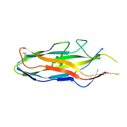
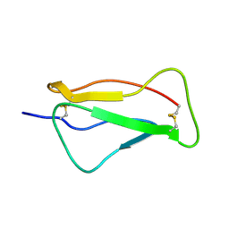
 | | ALPHA-SPECTRIN SRC HOMOLOGY 3 DOMAIN, CIRCULAR PERMUTANT, CUT AT N47-D48 | | 分子名称: | SPECTRIN ALPHA CHAIN, SULFATE ION | | 著者 | Berisio, R, Viguera, A.R, Serrano, L, Wilmanns, M. | | 登録日 | 2000-10-18 | | 公開日 | 2000-11-01 | | 最終更新日 | 2024-02-07 | | 実験手法 | X-RAY DIFFRACTION (1.12 Å) | | 主引用文献 | Atomic resolution structure of a mutant of the spectrin SH3 domain.
Acta Crystallogr.,Sect.D, 57, 2001
|
|
1YE8
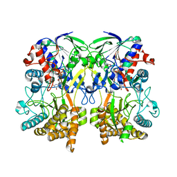
 | | Crystal Structure of THEP1 from the hyperthermophile Aquifex aeolicus | | 分子名称: | Hypothetical UPF0334 kinase-like protein AQ_1292, MAGNESIUM ION, PHOSPHATE ION, ... | | 著者 | Rossbach, M, Daumke, O, Klinger, C, Wittinghofer, A, Kaufmann, M. | | 登録日 | 2004-12-28 | | 公開日 | 2005-03-29 | | 最終更新日 | 2014-11-12 | | 実験手法 | X-RAY DIFFRACTION (1.4 Å) | | 主引用文献 | Crystal structure of THEP1 from the hyperthermophile Aquifex aeolicus: a variation of the RecA fold
BMC Struct.Biol., 5, 2005
|
|
3B6S
 | | Crystal Structure of hla-b*2705 Complexed with the Citrullinated Vasoactive Intestinal Peptide Type 1 Receptor (vipr) Peptide (residues 400-408) | | 分子名称: | Beta-2-microglobulin, HLA class I histocompatibility antigen, B-27 alpha chain, ... | | 著者 | Beltrami, A, Rossmann, M, Fiorillo, M.T, Sorrentino, R, Saenger, W, Ziegler, A, Uchanska-Ziegler, A. | | 登録日 | 2007-10-29 | | 公開日 | 2008-07-22 | | 最終更新日 | 2023-11-15 | | 実験手法 | X-RAY DIFFRACTION (1.8 Å) | | 主引用文献 | Citrullination-dependent Differential Presentation of a Self-peptide by HLA-B27 Subtypes.
J.Biol.Chem., 283, 2008
|
|
1GXB
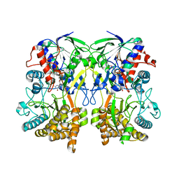
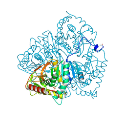
 | | ANTHRANILATE PHOSPHORIBOSYLTRANSFERASE IN COMPLEX WITH PYROPHOSPHATE AND MAGNESIUM | | 分子名称: | ANTHRANILATE PHOSPHORIBOSYLTRANSFERASE, MAGNESIUM ION, PYROPHOSPHATE 2- | | 著者 | Mayans, O, Ivens, A, Nissen, L.J, Kirschner, K, Wilmanns, M. | | 登録日 | 2002-04-02 | | 公開日 | 2003-04-03 | | 最終更新日 | 2024-05-08 | | 実験手法 | X-RAY DIFFRACTION (2.65 Å) | | 主引用文献 | Structural Analysis of Two Enzymes Catalysing Reverse Metabolic Reactions Implies Common Ancestry
Embo J., 21, 2002
|
|
1O9W
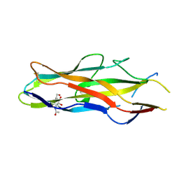
 | | F17-aG lectin domain from Escherichia coli in complex with N-acetyl-glucosamine | | 分子名称: | 2-acetamido-2-deoxy-beta-D-glucopyranose, F17A-G FIMBRIAL ADHESIN | | 著者 | Buts, L, De Genst, E, Loris, R, Oscarson, S, Lahmann, M, Messens, J, Brosens, E, Wyns, L, Bouckaert, J, De Greve, H. | | 登録日 | 2002-12-20 | | 公開日 | 2003-05-29 | | 最終更新日 | 2023-12-13 | | 実験手法 | X-RAY DIFFRACTION (1.65 Å) | | 主引用文献 | The Fimbrial Adhesin F17-G of Enterotoxigenic Escherichia Coli Has an Immunoglobulin-Like Lectin Domain that Binds N-Acetylglucosamine
Mol.Microbiol., 49, 2003
|
|
4Z25
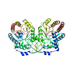
 | | Mimivirus R135 (residues 51-702) | | 分子名称: | FLAVIN-ADENINE DINUCLEOTIDE, Putative GMC-type oxidoreductase R135 | | 著者 | Klose, T, Rossmann, M.G. | | 登録日 | 2015-03-28 | | 公開日 | 2015-06-03 | | 最終更新日 | 2023-09-27 | | 実験手法 | X-RAY DIFFRACTION (3.339 Å) | | 主引用文献 | A Mimivirus Enzyme that Participates in Viral Entry.
Structure, 23, 2015
|
|
1HFB
 | |
1O9V
 | | F17-aG lectin domain from Escherichia coli in complex with a selenium carbohydrate derivative | | 分子名称: | F17-AG LECTIN DOMAIN, methyl 2-acetamido-2-deoxy-1-seleno-beta-D-glucopyranoside | | 著者 | Buts, L, De Genst, E, Loris, R, Oscarson, S, Lahmann, M, Messens, J, Brosens, E, Wyns, L, Bouckaert, J, De Greve, H. | | 登録日 | 2002-12-20 | | 公開日 | 2003-05-29 | | 最終更新日 | 2020-07-29 | | 実験手法 | X-RAY DIFFRACTION (1.75 Å) | | 主引用文献 | The Fimbrial Adhesin F17-G of Enterotoxigenic Escherichia Coli Has an Immunoglobulin-Like Lectin Domain that Binds N-Acetylglucosamine
Mol.Microbiol., 49, 2003
|
|
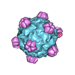
4WM8
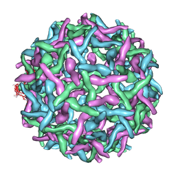
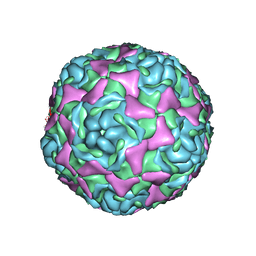 | | Crystal Structure of Human Enterovirus D68 | | 分子名称: | DECANOIC ACID, VP1, VP2, ... | | 著者 | Liu, Y, Sheng, J, Fokine, A, Meng, G, Long, F, Kuhn, R.J, Rossmann, M.G. | | 登録日 | 2014-10-08 | | 公開日 | 2015-01-14 | | 最終更新日 | 2023-12-27 | | 実験手法 | X-RAY DIFFRACTION (2 Å) | | 主引用文献 | Virus structure. Structure and inhibition of EV-D68, a virus that causes respiratory illness in children.
Science, 347, 2015
|
|
1O9Z
 | | F17-aG lectin domain from Escherichia coli (ligand free) | | 分子名称: | F17-AG LECTIN DOMAIN | | 著者 | Buts, L, De Genst, E, Loris, R, Oscarson, S, Lahmann, M, Messens, J, Brosens, E, Wyns, L, Bouckaert, J, De Greve, H. | | 登録日 | 2002-12-23 | | 公開日 | 2003-05-29 | | 最終更新日 | 2023-12-13 | | 実験手法 | X-RAY DIFFRACTION (1.75 Å) | | 主引用文献 | The Fimbrial Adhesin F17-G of Enterotoxigenic Escherichia Coli Has an Immunoglobulin-Like Lectin Domain that Binds N-Acetylglucosamine
Mol.Microbiol., 49, 2003
|
|
4Z26
 | | Mimivirus R135 (residues 51-702) | | 分子名称: | FLAVIN-ADENINE DINUCLEOTIDE, Putative GMC-type oxidoreductase R135 | | 著者 | Klose, T, Rossmann, M.G. | | 登録日 | 2015-03-28 | | 公開日 | 2015-05-13 | | 最終更新日 | 2023-09-27 | | 実験手法 | X-RAY DIFFRACTION (2.915 Å) | | 主引用文献 | A Mimivirus Enzyme that Participates in Viral Entry.
Structure, 23, 2015
|
|
7AC8
 | |
2OF6
 | | Structure of immature West Nile virus | | 分子名称: | envelope glycoprotein E | | 著者 | Zhang, Y, Kaufmann, B, Chipman, P.R, Kuhn, R.J, Rossmann, M.G. | | 登録日 | 2007-01-02 | | 公開日 | 2007-04-03 | | 最終更新日 | 2023-12-27 | | 実験手法 | ELECTRON MICROSCOPY (24 Å) | | 主引用文献 | Structure of immature west nile virus.
J.Virol., 81, 2007
|
|
1Z7Z
 | | Cryo-em structure of human coxsackievirus A21 complexed with five domain icam-1kilifi | | 分子名称: | 2-acetamido-2-deoxy-beta-D-glucopyranose, Intercellular adhesion molecule-1, human coxsackievirus A21 | | 著者 | Xiao, C, Bator-Kelly, C.M, Rieder, E, Chipman, P.R, Craig, A, Kuhn, R.J, Wimmer, E, Rossmann, M.G. | | 登録日 | 2005-03-28 | | 公開日 | 2005-08-02 | | 最終更新日 | 2021-10-20 | | 実験手法 | ELECTRON MICROSCOPY (8 Å) | | 主引用文献 | The crystal structure of coxsackievirus a21 and its interaction with icam-1.
Structure, 13, 2005
|
|
4NAG
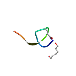 | | Xanthomonins I III are a New Class of Lasso Peptides Featuringa Seven-Membered Macrolactam Ring | | 分子名称: | HEXANE-1,6-DIOL, Xanthomonin I | | 著者 | Hegemann, J.D, Zimmermann, M, Zhu, S, Steuber, H, Harms, K, Xie, X, Marahiel, M.A. | | 登録日 | 2013-10-22 | | 公開日 | 2014-04-30 | | 最終更新日 | 2014-05-21 | | 実験手法 | X-RAY DIFFRACTION (0.81 Å) | | 主引用文献 | Xanthomonins I-III: A New Class of Lasso Peptides with a Seven-Residue Macrolactam Ring.
Angew.Chem.Int.Ed.Engl., 53, 2014
|
|
1M06
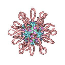
 | | Structural Studies of Bacteriophage alpha3 Assembly, X-Ray Crystallography | | 分子名称: | 5'-D(P*(3DR)P*(3DR)P*(3DR)P*(3DR)P*(3DR)P*(3DR)P*(3DR)P*(3DR)P*(3DR)P*(3DR))-3', Capsid Protein, Major spike protein, ... | | 著者 | Bernal, R.A, Hafenstein, S, Olson, N.H, Bowman, V, Chipman, P.R, Baker, T.S, Fane, B.A, Rossmann, M.G. | | 登録日 | 2002-06-12 | | 公開日 | 2002-12-25 | | 最終更新日 | 2024-04-03 | | 実験手法 | X-RAY DIFFRACTION (3.5 Å) | | 主引用文献 | Structural Studies of Bacteriophage alpha3 Assembly
J.Mol.Biol., 325, 2003
|
|
6LDH
 | |
2QZF
 | | SCR1 of DAF from 1ojv fitted into cryoEM density | | 分子名称: | Complement decay-accelerating factor | | 著者 | Hafenstein, S, Bowman, V.D, Chipman, P.R, Bator Kelly, C.M, Lin, F, Medof, M.E, Rossmann, M.G. | | 登録日 | 2007-08-16 | | 公開日 | 2007-10-30 | | 最終更新日 | 2018-07-18 | | 実験手法 | ELECTRON MICROSCOPY (14 Å) | | 主引用文献 | Interaction of decay-accelerating factor with coxsackievirus b3.
J.Virol., 81, 2007
|
|
2QZH
 | | SCR2/3 of DAF from the NMR structure 1nwv fitted into a cryoEM reconstruction of CVB3-RD complexed with DAF | | 分子名称: | Complement decay-accelerating factor | | 著者 | Hafenstein, S, Bowman, V.D, Chipman, P.R, Bator Kelly, C.M, Lin, F, Medof, M.E, Rossmann, M.G. | | 登録日 | 2007-08-16 | | 公開日 | 2007-10-30 | | 最終更新日 | 2024-02-21 | | 実験手法 | ELECTRON MICROSCOPY (14 Å) | | 主引用文献 | Interaction of decay-accelerating factor with coxsackievirus b3.
J.Virol., 81, 2007
|
|
2R29
 | | Neutralization of dengue virus by a serotype cross-reactive antibody elucidated by cryoelectron microscopy and x-ray crystallography | | 分子名称: | Envelope protein E, Heavy chain of Fab 1A1D-2, Light chain of Fab 1A1D-2 | | 著者 | Lok, S.M, Kostyuchenko, V.K, Nybakken, G.E, Holdaway, H.A, Battisti, A.J, Sukupolvi-petty, S, Sedlak, D, Fremont, D.H, Chipman, P.R, Roehrig, J.T, Diamond, M.S, Kuhn, R.J, Rossmann, M.G. | | 登録日 | 2007-08-24 | | 公開日 | 2007-12-25 | | 最終更新日 | 2018-01-24 | | 実験手法 | X-RAY DIFFRACTION (3 Å) | | 主引用文献 | Binding of a neutralizing antibody to dengue virus alters the arrangement of surface glycoproteins.
Nat.Struct.Mol.Biol., 15, 2008
|
|
1YA5
 | | Crystal structure of the titin domains z1z2 in complex with telethonin | | 分子名称: | N2B-TITIN ISOFORM, SULFATE ION, TELETHONIN | | 著者 | Pinotsis, N, Popov, A, Zou, P, Wilmanns, M. | | 登録日 | 2004-12-17 | | 公開日 | 2005-12-20 | | 最終更新日 | 2024-02-14 | | 実験手法 | X-RAY DIFFRACTION (2.445 Å) | | 主引用文献 | Palindromic assembly of the giant muscle protein titin in the sarcomeric Z-disk
Nature, 439, 2006
|
|
2N5O
 | | Universal Base oligonucleotide structure | | 分子名称: | DNA_(5'-D(*AP*TP*GP*GP*(4EN)P*GP*CP*TP*C)-3'), DNA_(5'-D(*GP*AP*GP*CP*TP*CP*CP*AP*T)-3') | | 著者 | Spring-Connell, A.M, Evich, M.G, Seela, F, Germann, M.W. | | 登録日 | 2015-07-23 | | 公開日 | 2016-09-07 | | 最終更新日 | 2024-05-01 | | 実験手法 | SOLUTION NMR | | 主引用文献 | Using NMR and molecular dynamics to link structure and dynamics effects of the universal base 8-aza, 7-deaza, N8 linked adenosine analog.
Nucleic Acids Res., 44, 2016
|
|
2QZD
 | | Fitted structure of SCR4 of DAF into cryoEM density | | 分子名称: | Complement decay-accelerating factor | | 著者 | Hafenstein, S, Bowman, V.D, Chipman, P.R, Bator Kelly, C.M, Lin, F, Medof, M.E, Rossmann, M.G. | | 登録日 | 2007-08-16 | | 公開日 | 2007-10-30 | | 最終更新日 | 2018-07-18 | | 実験手法 | ELECTRON MICROSCOPY (14 Å) | | 主引用文献 | Interaction of decay-accelerating factor with coxsackievirus b3.
J.Virol., 81, 2007
|
|
2R69
 | | Crystal structure of Fab 1A1D-2 complexed with E-DIII of Dengue virus at 3.8 angstrom resolution | | 分子名称: | Heavy chain of 1A1D-2, Light chain of 1A1D-2, Major envelope protein E | | 著者 | Lok, S.M, Kostyuchenko, V.K, Nybakken, G.E, Holdaway, H.A, Battisti, A.J, Sukupolvi-petty, S, Sedlak, D, Fremont, D.H, Chipman, P.R, Roehrig, J.T, Diamond, M.S, Kuhn, R.J, Rossmann, M.G. | | 登録日 | 2007-09-05 | | 公開日 | 2007-12-25 | | 最終更新日 | 2018-01-24 | | 実験手法 | X-RAY DIFFRACTION (3.8 Å) | | 主引用文献 | Binding of a neutralizing antibody to dengue virus alters the arrangement of surface glycoproteins.
Nat.Struct.Mol.Biol., 15, 2008
|
|
2IDF
 | | P. aeruginosa azurin N42C/M64E double mutant, BMME-linked dimer | | 分子名称: | 1-[PYRROL-1-YL-2,5-DIONE-METHOXYMETHYL]-PYRROLE-2,5-DIONE, Azurin, COPPER (II) ION, ... | | 著者 | Einsle, O, de Jongh, T.E, Hoffmann, M, Cavazzini, D, Rossi, G.L, Ubbink, M, Canters, G.W. | | 登録日 | 2006-09-15 | | 公開日 | 2008-03-18 | | 最終更新日 | 2023-08-30 | | 実験手法 | X-RAY DIFFRACTION (2.25 Å) | | 主引用文献 | Electron transfer in a crosslinked protein dimer mediated by a hydrogen-bonded network across the dimer interface
To be Published
|
|


