4GGC
 
 | |
3D3C
 | |
3D3B
 | |
4ZUZ
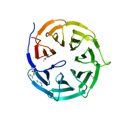
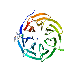
 | | SidC 1-871 | | 分子名称: | SidC | | 著者 | Luo, X, Wasilko, D.J, Liu, Y, Sun, J, Wu, X, Luo, Z.-Q, Mao, Y. | | 登録日 | 2015-05-18 | | 公開日 | 2015-07-29 | | 最終更新日 | 2023-09-27 | | 実験手法 | X-RAY DIFFRACTION (2.86 Å) | | 主引用文献 | Structure of the Legionella Virulence Factor, SidC Reveals a Unique PI(4)P-Specific Binding Domain Essential for Its Targeting to the Bacterial Phagosome.
Plos Pathog., 11, 2015
|
|
1KLQ
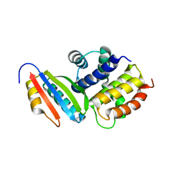
 | | The Mad2 Spindle Checkpoint Protein Undergoes Similar Major Conformational Changes upon Binding to Either Mad1 or Cdc20 | | 分子名称: | MITOTIC SPINDLE ASSEMBLY CHECKPOINT PROTEIN MAD2A, Mad2-binding peptide | | 著者 | Luo, X, Tang, Z, Rizo, J, Yu, H. | | 登録日 | 2001-12-12 | | 公開日 | 2002-01-25 | | 最終更新日 | 2024-05-22 | | 実験手法 | SOLUTION NMR | | 主引用文献 | The Mad2 spindle checkpoint protein undergoes similar major conformational changes upon binding to either Mad1 or Cdc20.
Mol.Cell, 9, 2002
|
|
5HGU
 | |
1DUJ
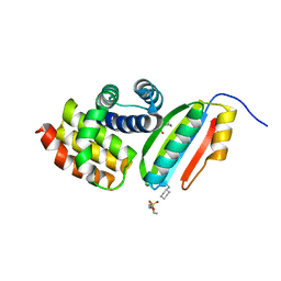
 | | SOLUTION STRUCTURE OF THE SPINDLE ASSEMBLY CHECKPOINT PROTEIN HUMAN MAD2 | | 分子名称: | SPINDLE ASSEMBLY CHECKPOINT PROTEIN | | 著者 | Luo, X, Fang, G, Coldiron, M, Lin, Y, Yu, H. | | 登録日 | 2000-01-17 | | 公開日 | 2000-03-08 | | 最終更新日 | 2024-05-22 | | 実験手法 | SOLUTION NMR | | 主引用文献 | Structure of the Mad2 spindle assembly checkpoint protein and its interaction with Cdc20.
Nat.Struct.Biol., 7, 2000
|
|
5XPI
 | | Structure of UHRF1 TTD in complex with NV01 | | 分子名称: | E3 ubiquitin-protein ligase UHRF1, N-[3-(diethylamino)propyl]-2-(12-methyl-9-oxidanylidene-5-thia-1,10,11-triazatricyclo[6.4.0.0^2,6]dodeca-2(6),3,7,11-tetraen-10-yl)ethanamide | | 著者 | Luo, X, Zhao, K. | | 登録日 | 2017-06-02 | | 公開日 | 2018-04-25 | | 最終更新日 | 2023-11-22 | | 実験手法 | X-RAY DIFFRACTION (2.2 Å) | | 主引用文献 | Discovery of Small-Molecule Antagonists of the H3K9me3 Binding to UHRF1 Tandem Tudor Domain
SLAS Discov, 23, 2018
|
|
1S2H
 | | The Mad2 spindle checkpoint protein possesses two distinct natively folded states | | 分子名称: | Mitotic spindle assembly checkpoint protein MAD2A | | 著者 | Luo, X, Tang, Z, Xia, G, Wassmann, K, Matsumoto, T, Rizo, J, Yu, H. | | 登録日 | 2004-01-08 | | 公開日 | 2004-03-30 | | 最終更新日 | 2024-05-22 | | 実験手法 | SOLUTION NMR | | 主引用文献 | The Mad2 spindle checkpoint protein has two distinct natively folded states.
Nat.Struct.Mol.Biol., 11, 2004
|
|
5BRM
 | |
4LGD
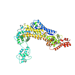
 | | Structural Basis for Autoactivation of Human Mst2 Kinase and Its Regulation by RASSF5 | | 分子名称: | MAGNESIUM ION, PHOSPHOAMINOPHOSPHONIC ACID-ADENYLATE ESTER, Ras association domain family member 5, ... | | 著者 | Luo, X, Ni, L, Tomchick, D.R. | | 登録日 | 2013-06-27 | | 公開日 | 2013-09-18 | | 最終更新日 | 2023-09-20 | | 実験手法 | X-RAY DIFFRACTION (3.05 Å) | | 主引用文献 | Structural Basis for Autoactivation of Human Mst2 Kinase and Its Regulation by RASSF5.
Structure, 21, 2013
|
|
4LG4
 | |
4N14
 | | Crystal structure of Cdc20 and apcin complex | | 分子名称: | 2-(2-methyl-5-nitro-1H-imidazol-1-yl)ethyl [(1R)-2,2,2-trichloro-1-(pyrimidin-2-ylamino)ethyl]carbamate, Cell division cycle protein 20 homolog | | 著者 | Luo, X, Tian, W, Yu, H. | | 登録日 | 2013-10-03 | | 公開日 | 2014-08-20 | | 最終更新日 | 2024-02-28 | | 実験手法 | X-RAY DIFFRACTION (2.1 Å) | | 主引用文献 | Synergistic blockade of mitotic exit by two chemical inhibitors of the APC/C.
Nature, 514, 2014
|
|
4O98
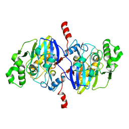 | | Crystal structure of Pseudomonas oleovorans PoOPH mutant H250I/I263W | | 分子名称: | ZINC ION, organophosphorus hydrolase | | 著者 | Luo, X.J, Kong, X.D, Zhao, J, Chen, Q, Zhou, J.H, Xu, J.H. | | 登録日 | 2014-01-02 | | 公開日 | 2014-12-03 | | 最終更新日 | 2017-11-22 | | 実験手法 | X-RAY DIFFRACTION (2.251 Å) | | 主引用文献 | Switching a newly discovered lactonase into an efficient and thermostable phosphotriesterase by simple double mutations His250Ile/Ile263Trp
Biotechnol.Bioeng., 111, 2014
|
|
1TBD
 | | SOLUTION STRUCTURE OF THE ORIGIN DNA BINDING DOMAIN OF SV40 T-ANTIGEN, NMR, MINIMIZED AVERAGE STRUCTURE | | 分子名称: | SV40 T-ANTIGEN | | 著者 | Luo, X, Sanford, D.G, Bullock, P.A, Bachovchin, W.W. | | 登録日 | 1996-11-04 | | 公開日 | 1997-03-12 | | 最終更新日 | 2024-05-22 | | 実験手法 | SOLUTION NMR | | 主引用文献 | Solution structure of the origin DNA-binding domain of SV40 T-antigen.
Nat.Struct.Biol., 3, 1996
|
|
3IMQ
 | | Crystal structure of the NusB101-S10(delta loop) complex | | 分子名称: | 30S ribosomal protein S10, N utilization substance protein B, POTASSIUM ION | | 著者 | Luo, X, Wahl, M.C. | | 登録日 | 2009-08-11 | | 公開日 | 2009-11-10 | | 最終更新日 | 2023-09-06 | | 実験手法 | X-RAY DIFFRACTION (2.5 Å) | | 主引用文献 | Fine tuning of the E. coli NusB:NusE complex affinity to BoxA RNA is required for processive antitermination.
Nucleic Acids Res., 38, 2010
|
|
6L1W
 | | Zinc-finger Antiviral Protein (ZAP) bound to RNA | | 分子名称: | RNA (5'-R(*CP*GP*UP*CP*GP*U)-3'), ZINC ION, Zinc finger CCCH-type antiviral protein 1 | | 著者 | Luo, X, Wang, X, Gao, Y, Zhu, J, Liu, S, Gao, G, Gao, P. | | 登録日 | 2019-09-30 | | 公開日 | 2020-01-01 | | 最終更新日 | 2023-11-22 | | 実験手法 | X-RAY DIFFRACTION (2.194 Å) | | 主引用文献 | Molecular Mechanism of RNA Recognition by Zinc-Finger Antiviral Protein.
Cell Rep, 30, 2020
|
|
4DZO
 | |
5U36
 | | Crystal Structure Of A Mutant M. Jannashii Tyrosyl-tRNA Synthetase | | 分子名称: | Tyrosine--tRNA ligase | | 著者 | Luo, X, Fu, G, Zhu, X, Wilson, I.A, Wang, F. | | 登録日 | 2016-12-01 | | 公開日 | 2017-06-07 | | 最終更新日 | 2023-10-04 | | 実験手法 | X-RAY DIFFRACTION (3.03 Å) | | 主引用文献 | Genetically encoding phosphotyrosine and its nonhydrolyzable analog in bacteria.
Nat. Chem. Biol., 13, 2017
|
|
4GGD
 | |
4GGA
 | |
2TBD
 | | SV40 T ANTIGEN DNA-BINDING DOMAIN, NMR, 30 STRUCTURES | | 分子名称: | SV40 T ANTIGEN | | 著者 | Luo, X, Sanford, D.G, Bullock, P.A, Bachovchin, W.W. | | 登録日 | 1997-01-09 | | 公開日 | 1997-04-01 | | 最終更新日 | 2024-05-22 | | 実験手法 | SOLUTION NMR | | 主引用文献 | Solution structure of the origin DNA-binding domain of SV40 T-antigen.
Nat.Struct.Biol., 3, 1996
|
|
2QYF
 | |
5WC2
 | | Crystal Structure of ADP-bound human TRIP13 | | 分子名称: | ADENOSINE-5'-DIPHOSPHATE, Pachytene checkpoint protein 2 homolog | | 著者 | Jeong, B.-C, Luo, X. | | 登録日 | 2017-06-29 | | 公開日 | 2018-04-25 | | 最終更新日 | 2023-10-04 | | 実験手法 | X-RAY DIFFRACTION (2.5 Å) | | 主引用文献 | Mechanistic insight into TRIP13-catalyzed Mad2 structural transition and spindle checkpoint silencing.
Nat Commun, 8, 2017
|
|
6OCG
 | | Crystal structure of VASH1-SVBP complex bound with EpoY | | 分子名称: | CHLORIDE ION, GLYCEROL, N-[(3R)-4-ethoxy-3-hydroxy-4-oxobutanoyl]-L-tyrosine, ... | | 著者 | Li, F, Luo, X, Yu, H. | | 登録日 | 2019-03-23 | | 公開日 | 2019-06-26 | | 最終更新日 | 2023-10-11 | | 実験手法 | X-RAY DIFFRACTION (1.833 Å) | | 主引用文献 | Structural basis of tubulin detyrosination by vasohibins.
Nat.Struct.Mol.Biol., 26, 2019
|
|


