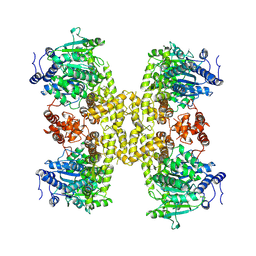
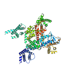
5Z96
 
 | | Structure of the mouse TRPC4 ion channel | | 分子名称: | 2-(HEXADECANOYLOXY)-1-[(PHOSPHONOOXY)METHYL]ETHYL HEXADECANOATE, CHOLESTEROL HEMISUCCINATE, SODIUM ION, ... | | 著者 | Duan, J, Li, Z, Li, J, Zhang, J. | | 登録日 | 2018-02-02 | | 公開日 | 2018-04-18 | | 最終更新日 | 2018-08-29 | | 実験手法 | ELECTRON MICROSCOPY (3.28 Å) | | 主引用文献 | Structure of the mouse TRPC4 ion channel.
Nat Commun, 9, 2018
|
|
8HC5
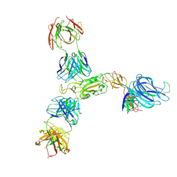 | | SARS-CoV-2 wildtype S1 in complex with YB9-258 Fab and R1-32 Fab | | 分子名称: | 2-acetamido-2-deoxy-beta-D-glucopyranose, Heavy chain of R1-32 Fab, Heavy chain of YB9-258 Fab, ... | | 著者 | Liu, B, Gao, X, Chen, Q, Li, Z, Su, M, He, J, Xiong, X. | | 登録日 | 2022-11-01 | | 公開日 | 2023-01-25 | | 最終更新日 | 2023-05-03 | | 実験手法 | ELECTRON MICROSCOPY (3.43 Å) | | 主引用文献 | Somatically hypermutated antibodies isolated from SARS-CoV-2 Delta infected patients cross-neutralize heterologous variants.
Nat Commun, 14, 2023
|
|
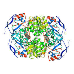
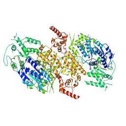
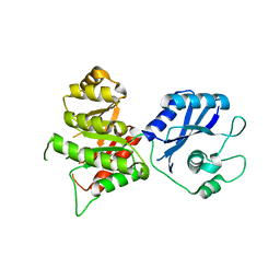
5Z9T
 | | a new PL6 alginate lyase complex with trisaccharide | | 分子名称: | GLYCEROL, MALONATE ION, SODIUM ION, ... | | 著者 | Liu, W.Z, Lyu, Q.Q, Zhang, K.K, Li, Z.J. | | 登録日 | 2018-02-05 | | 公開日 | 2019-03-27 | | 最終更新日 | 2023-11-22 | | 実験手法 | X-RAY DIFFRACTION (1.8 Å) | | 主引用文献 | Structural insights into a novel Ca2+-independent PL-6 alginate lyase from Vibrio OU02 identify the possible subsites responsible for product distribution.
Biochim Biophys Acta Gen Subj, 1863, 2019
|
|
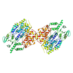
7KJY
 | | Symmetry in Yeast Alcohol Dehydrogenase 1 - Open Form with NADH | | 分子名称: | Alcohol dehydrogenase, NICOTINAMIDE-ADENINE-DINUCLEOTIDE, ZINC ION | | 著者 | Subramanian, R, Chang, L, Li, Z, Plapp, B.V. | | 登録日 | 2020-10-26 | | 公開日 | 2021-03-31 | | 実験手法 | ELECTRON MICROSCOPY (3.2 Å) | | 主引用文献 | Cryo-Electron Microscopy Structures of Yeast Alcohol Dehydrogenase.
Biochemistry, 60, 2021
|
|
5X3P
 | |
9ASL
 | |
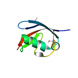
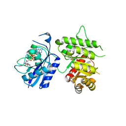
5X4L
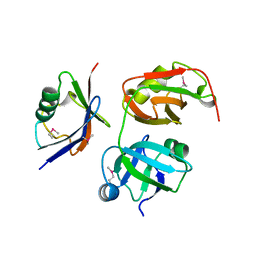 | | Crystal structure of the UBX domain of human UBXD7 in complex with p97 N domain | | 分子名称: | Transitional endoplasmic reticulum ATPase, UBX domain-containing protein 7 | | 著者 | Jiang, T, Li, Z, Wang, Y, Xu, M. | | 登録日 | 2017-02-13 | | 公開日 | 2017-03-22 | | 最終更新日 | 2023-11-22 | | 実験手法 | X-RAY DIFFRACTION (2.402 Å) | | 主引用文献 | Crystal structures of the UBX domain of human UBXD7 and its complex with p97 ATPase
Biochem. Biophys. Res. Commun., 486, 2017
|
|
9C5Q
 | |
8W0A
 | |
9ASK
 | |
9ASJ
 | |
7V55
 | |
7V53
 | |
3MGM
 | | Crystal structure of human NUDT16 | | 分子名称: | U8 snoRNA-decapping enzyme | | 著者 | Yan, J, Lu, G, Zhang, J, Qi, J, Li, Z, Gao, F. | | 登録日 | 2010-04-07 | | 公開日 | 2011-04-20 | | 最終更新日 | 2023-11-01 | | 実験手法 | X-RAY DIFFRACTION (1.801 Å) | | 主引用文献 | Crystal structure and the template mRNA decapping activity of human NUDT16
To be Published
|
|
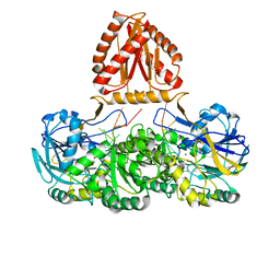
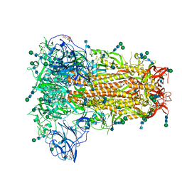
6LQA
 | | voltage-gated sodium channel Nav1.5 with quinidine | | 分子名称: | 2-acetamido-2-deoxy-beta-D-glucopyranose, Quinidine, Sodium channel protein type 5 subunit alpha | | 著者 | Yan, N, Li, Z, Pan, X, Huang, G. | | 登録日 | 2020-01-13 | | 公開日 | 2021-03-24 | | 最終更新日 | 2021-05-19 | | 実験手法 | ELECTRON MICROSCOPY (3.3 Å) | | 主引用文献 | Structural Basis for Pore Blockade of the Human Cardiac Sodium Channel Na v 1.5 by the Antiarrhythmic Drug Quinidine*.
Angew.Chem.Int.Ed.Engl., 60, 2021
|
|
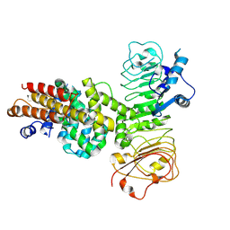
3MCN
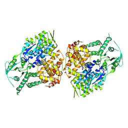
 | | Crystal Structure of the 6-hyroxymethyl-7,8-dihydropterin pyrophosphokinase dihydropteroate synthase bifunctional enzyme from Francisella tularensis | | 分子名称: | 2,6-diamino-5-nitropyrimidin-4(3H)-one, 2-amino-4-hydroxy-6-hydroxymethyldihydropteridine pyrophosphokinase/dihydropteroate synthase, MAGNESIUM ION | | 著者 | Pemble IV, C.W, Mehta, P.K, Mehra, S, Li, Z, Lee, R.E, White, S.W. | | 登録日 | 2010-03-29 | | 公開日 | 2010-12-22 | | 最終更新日 | 2023-09-06 | | 実験手法 | X-RAY DIFFRACTION (2.2 Å) | | 主引用文献 | Crystal structure of the 6-hydroxymethyl-7,8-dihydropterin pyrophosphokinase.dihydropteroate synthase bifunctional enzyme from Francisella tularensis.
Plos One, 5, 2010
|
|
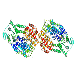
3MCM
 | | Crystal Structure of the 6-hyroxymethyl-7,8-dihydropterin pyrophosphokinase dihydropteroate synthase bifunctional enzyme from Francisella tularensis | | 分子名称: | 2-amino-4-hydroxy-6-hydroxymethyldihydropteridine pyrophosphokinase/dihydropteroate synthase, MAGNESIUM ION | | 著者 | Pemble IV, C.W, Mehta, P.K, Mehra, S, Li, Z, Lee, R.E, White, S.W. | | 登録日 | 2010-03-29 | | 公開日 | 2010-12-22 | | 最終更新日 | 2023-09-06 | | 実験手法 | X-RAY DIFFRACTION (2.2 Å) | | 主引用文献 | Crystal structure of the 6-hydroxymethyl-7,8-dihydropterin pyrophosphokinase.dihydropteroate synthase bifunctional enzyme from Francisella tularensis.
Plos One, 5, 2010
|
|
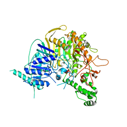
3MCO
 | | Crystal Structure of the 6-hyroxymethyl-7,8-dihydropterin pyrophosphokinase dihydropteroate synthase bifunctional enzyme from Francisella tularensis | | 分子名称: | 2-AMINO-6-HYDROXYMETHYL-7,8-DIHYDRO-3H-PTERIDIN-4-ONE, 2-amino-4-hydroxy-6-hydroxymethyldihydropteridine pyrophosphokinase/dihydropteroate synthase, DIPHOSPHOMETHYLPHOSPHONIC ACID ADENOSYL ESTER, ... | | 著者 | Pemble IV, C.W, Mehta, P.K, Mehra, S, Li, Z, Lee, R.E, White, S.W. | | 登録日 | 2010-03-29 | | 公開日 | 2010-12-22 | | 最終更新日 | 2023-09-06 | | 実験手法 | X-RAY DIFFRACTION (2.3 Å) | | 主引用文献 | Crystal structure of the 6-hydroxymethyl-7,8-dihydropterin pyrophosphokinase.dihydropteroate synthase bifunctional enzyme from Francisella tularensis.
Plos One, 5, 2010
|
|
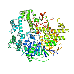
3MHD
 | | Crystal structure of DCR3 | | 分子名称: | Tumor necrosis factor receptor superfamily member 6B | | 著者 | Zhan, C, Patskovsky, Y, Yan, Q, Li, Z, Ramagopal, U.A, Nathenson, S.G, Almo, S.C. | | 登録日 | 2010-04-07 | | 公開日 | 2011-02-23 | | 最終更新日 | 2023-09-06 | | 実験手法 | X-RAY DIFFRACTION (2.901 Å) | | 主引用文献 | Decoy Strategies: The Structure of TL1A:DcR3 Complex.
Structure, 19, 2011
|
|
7WNU
 | | Mycobacterium tuberculosis Rnase J complex with 7nt RNA | | 分子名称: | RNA (5'-R(P*AP*AP*AP*AP*AP*AP*A)-3'), Ribonuclease J, ZINC ION | | 著者 | Li, J, Hu, J, Bao, L, Zhan, B, Li, Z. | | 登録日 | 2022-01-19 | | 公開日 | 2023-01-25 | | 最終更新日 | 2023-11-29 | | 実験手法 | X-RAY DIFFRACTION (3.2 Å) | | 主引用文献 | Structural ans functional study of RNase J from Mycobacterium tuberculosis
To Be Published
|
|
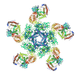
8JAY
 | | CrtSPARTA Octamer bound with guide-target | | 分子名称: | DNA (25-MER), MAGNESIUM ION, Piwi domain-containing protein, ... | | 著者 | Guo, L.J, Huang, P.P, Li, Z.X, Xiao, Y.B, Chen, M.R. | | 登録日 | 2023-05-07 | | 公開日 | 2024-03-20 | | 最終更新日 | 2024-04-10 | | 実験手法 | ELECTRON MICROSCOPY (4.2 Å) | | 主引用文献 | Auto-inhibition and activation of a short Argonaute-associated TIR-APAZ defense system.
Nat.Chem.Biol., 20, 2024
|
|
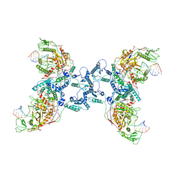
7XE0
 | | Cryo-EM structure of plant NLR Sr35 resistosome | | 分子名称: | ADENOSINE-5'-TRIPHOSPHATE, AvrSr35, Sr35 | | 著者 | Ouyang, S.Y, Zhao, Y.B, Li, Z.K, Liu, M.X. | | 登録日 | 2022-03-29 | | 公開日 | 2022-09-28 | | 最終更新日 | 2024-06-26 | | 実験手法 | ELECTRON MICROSCOPY (3.33 Å) | | 主引用文献 | Pathogen effector AvrSr35 triggers Sr35 resistosome assembly via a direct recognition mechanism.
Sci Adv, 8, 2022
|
|
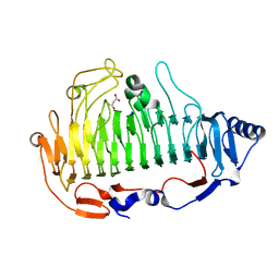
3OW8
 | | Crystal Structure of the WD repeat-containing protein 61 | | 分子名称: | UNKNOWN ATOM OR ION, WD repeat-containing protein 61 | | 著者 | Tempel, W, Li, Z, Chao, X, Lam, R, Wernimont, A.K, He, H, Seitova, A, Pan, P.W, Li, Y, Bountra, C, Weigelt, J, Arrowsmith, C.H, Edwards, A.M, Bochkarev, A, Min, J, Structural Genomics Consortium (SGC) | | 登録日 | 2010-09-17 | | 公開日 | 2010-09-29 | | 最終更新日 | 2023-09-06 | | 実験手法 | X-RAY DIFFRACTION (2.3 Å) | | 主引用文献 | Structure and function of WD40 domain proteins.
Protein Cell, 2, 2011
|
|
6NZK
 | | Structural basis for human coronavirus attachment to sialic acid receptors | | 分子名称: | 2-acetamido-2-deoxy-beta-D-glucopyranose, 2-acetamido-2-deoxy-beta-D-glucopyranose-(1-4)-2-acetamido-2-deoxy-beta-D-glucopyranose, Spike surface glycoprotein, ... | | 著者 | Tortorici, M.A, Walls, A.C, Lang, Y, Wang, C, Li, Z, Koerhuis, D, Boons, G.J, Bosch, B.J, Rey, F.A, de Groot, R, Veesler, D. | | 登録日 | 2019-02-13 | | 公開日 | 2019-06-05 | | 最終更新日 | 2020-07-29 | | 実験手法 | ELECTRON MICROSCOPY (2.8 Å) | | 主引用文献 | Structural basis for human coronavirus attachment to sialic acid receptors.
Nat.Struct.Mol.Biol., 26, 2019
|
|
7XVG
 | | Cryo-EM structure of binary complex of plant NLR Sr35 and effector AvrSr35 | | 分子名称: | AvrSr35, Sr35 | | 著者 | Ouyang, S.Y, Zhao, Y.B, Li, Z.K, Liu, M.X. | | 登録日 | 2022-05-23 | | 公開日 | 2022-09-28 | | 最終更新日 | 2024-07-03 | | 実験手法 | ELECTRON MICROSCOPY (3.6 Å) | | 主引用文献 | Pathogen effector AvrSr35 triggers Sr35 resistosome assembly via a direct recognition mechanism.
Sci Adv, 8, 2022
|
|


