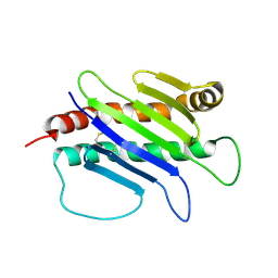
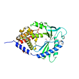
6O8B
 
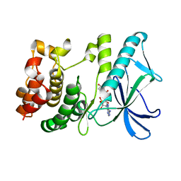
 | | Crystal structure of STING CTD in complex with TBK1 | | 分子名称: | N-(3-{[5-iodo-4-({3-[(thiophen-2-ylcarbonyl)amino]propyl}amino)pyrimidin-2-yl]amino}phenyl)pyrrolidine-1-carboxamide, Serine/threonine-protein kinase TBK1, Stimulator of interferon genes protein | | 著者 | Li, P, Zhao, B, Du, F. | | 登録日 | 2019-03-09 | | 公開日 | 2019-05-22 | | 最終更新日 | 2023-10-11 | | 実験手法 | X-RAY DIFFRACTION (3.4 Å) | | 主引用文献 | A conserved PLPLRT/SD motif of STING mediates the recruitment and activation of TBK1.
Nature, 569, 2019
|
|
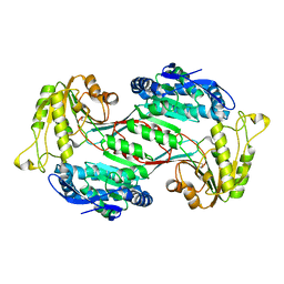
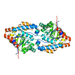
4LEY
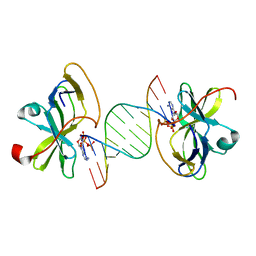
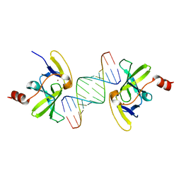
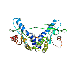
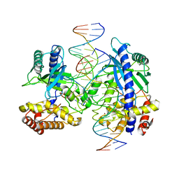 | | Structure of mouse cGAS bound to 18 bp DNA | | 分子名称: | 18 bp dsDNA, Cyclic GMP-AMP synthase, ZINC ION | | 著者 | Li, P. | | 登録日 | 2013-06-26 | | 公開日 | 2013-12-25 | | 最終更新日 | 2024-04-03 | | 実験手法 | X-RAY DIFFRACTION (2.5 Å) | | 主引用文献 | Cyclic GMP-AMP Synthase Is Activated by Double-Stranded DNA-Induced Oligomerization.
Immunity, 39, 2013
|
|
3GA3
 | |
5JEJ
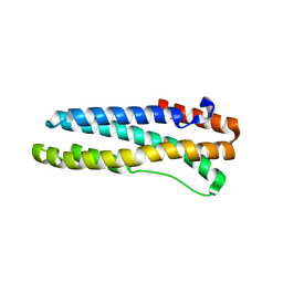
 | | Phosphorylated STING in complex with IRF-3 CTD | | 分子名称: | Interferon regulatory factor 3, Stimulator of interferon genes protein | | 著者 | Li, P, Shu, C. | | 登録日 | 2016-04-18 | | 公開日 | 2016-06-15 | | 最終更新日 | 2016-06-29 | | 実験手法 | X-RAY DIFFRACTION (2 Å) | | 主引用文献 | Structural basis for concerted recruitment and activation of IRF-3 by innate immune adaptor proteins.
Proc.Natl.Acad.Sci.USA, 113, 2016
|
|
2OTU
 | | Crystal structure of Fv polyglutamine complex | | 分子名称: | Fv heavy chain variable domain, Fv light chain variable domain, peptide antigen | | 著者 | Li, P. | | 登録日 | 2007-02-09 | | 公開日 | 2007-04-24 | | 最終更新日 | 2023-08-30 | | 実験手法 | X-RAY DIFFRACTION (1.68 Å) | | 主引用文献 | Implications of the structure of a poly-Gln/anti-poly-Gln complex for disease progression and therapy
To be Published
|
|
3LRR
 | |
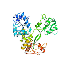
6J76
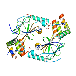
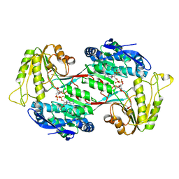 | | Structure of 3,6-anhydro-L-galactose Dehydrogenase in Complex with NAP | | 分子名称: | Aldehyde dehydrogenase A, NADP NICOTINAMIDE-ADENINE-DINUCLEOTIDE PHOSPHATE | | 著者 | Li, P.Y, Wang, Y, Chen, X.L, Zhang, Y.Z. | | 登録日 | 2019-01-17 | | 公開日 | 2020-01-22 | | 最終更新日 | 2023-11-22 | | 実験手法 | X-RAY DIFFRACTION (2.368 Å) | | 主引用文献 | 3,6-Anhydro-L-Galactose Dehydrogenase VvAHGD is a Member of a New Aldehyde Dehydrogenase Family and Catalyzes by a Novel Mechanism with Conformational Switch of Two Catalytic Residues Cysteine 282 and Glutamate 248.
J.Mol.Biol., 432, 2020
|
|
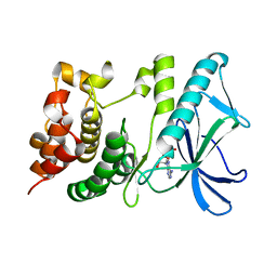
4LEW
 | | Structure of human cGAS | | 分子名称: | Cyclic GMP-AMP synthase, ZINC ION | | 著者 | Li, P. | | 登録日 | 2013-06-26 | | 公開日 | 2013-12-25 | | 最終更新日 | 2014-01-08 | | 実験手法 | X-RAY DIFFRACTION (2.04 Å) | | 主引用文献 | Cyclic GMP-AMP Synthase Is Activated by Double-Stranded DNA-Induced Oligomerization.
Immunity, 39, 2013
|
|
2OTW
 | | Crystal structure of Fv polyglutamine complex | | 分子名称: | Fv heavy chain variable domain VH, Fv light chain avriable domain VL, poly-Gln peptide antigen | | 著者 | Li, P. | | 登録日 | 2007-02-09 | | 公開日 | 2007-04-24 | | 最終更新日 | 2023-08-30 | | 実験手法 | X-RAY DIFFRACTION (2.35 Å) | | 主引用文献 | Implications of the structure of a poly-Gln/anti-poly-Gln complex for disease progression and therapy
To be Published
|
|
1JFM
 | |
6J75
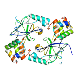
 | | Structure of 3,6-anhydro-L-galactose Dehydrogenase | | 分子名称: | Aldehyde dehydrogenase A | | 著者 | Li, P.Y, Wang, Y, Chen, X.L, Zhang, Y.Z. | | 登録日 | 2019-01-17 | | 公開日 | 2020-01-22 | | 最終更新日 | 2023-11-22 | | 実験手法 | X-RAY DIFFRACTION (2.695 Å) | | 主引用文献 | 3,6-Anhydro-L-Galactose Dehydrogenase VvAHGD is a Member of a New Aldehyde Dehydrogenase Family and Catalyzes by a Novel Mechanism with Conformational Switch of Two Catalytic Residues Cysteine 282 and Glutamate 248.
J.Mol.Biol., 432, 2020
|
|
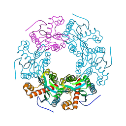
5X1T
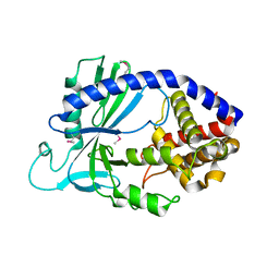
 | | PpkA-294 | | 分子名称: | ADENOSINE-5'-DIPHOSPHATE, PpkA-294 | | 著者 | Li, P.P, Ran, T.T, Xu, D.Q, Wang, W.W. | | 登録日 | 2017-01-26 | | 公開日 | 2018-01-31 | | 最終更新日 | 2024-03-20 | | 実験手法 | X-RAY DIFFRACTION (1.55 Å) | | 主引用文献 | Crystal structures of the kinase domain of PpkA, a key regulatory component of T6SS, reveal a general inhibitory mechanism.
Biochem.J., 475, 2018
|
|
5X1Q
 | | PpkA-294 with ATP and MnCl2 | | 分子名称: | ADENOSINE-5'-TRIPHOSPHATE, GLYCEROL, MANGANESE (II) ION, ... | | 著者 | Li, P.P, Ran, T.T, Xu, D.Q, Wang, W.W. | | 登録日 | 2017-01-26 | | 公開日 | 2018-01-31 | | 最終更新日 | 2024-03-20 | | 実験手法 | X-RAY DIFFRACTION (1.602 Å) | | 主引用文献 | Crystal structures of the kinase domain of PpkA, a key regulatory component of T6SS, reveal a general inhibitory mechanism.
Biochem.J., 475, 2018
|
|
5X1S
 | | PpkA-294 with Amppcp | | 分子名称: | GLYCEROL, PHOSPHOMETHYLPHOSPHONIC ACID ADENYLATE ESTER, PpkA | | 著者 | Li, P.P, Ran, T.T, Xu, D.Q, Wang, W.W. | | 登録日 | 2017-01-26 | | 公開日 | 2018-01-31 | | 最終更新日 | 2024-03-20 | | 実験手法 | X-RAY DIFFRACTION (1.45 Å) | | 主引用文献 | Crystal structures of the kinase domain of PpkA, a key regulatory component of T6SS, reveal a general inhibitory mechanism.
Biochem.J., 475, 2018
|
|
7JFM
 | |
7JFL
 | |
3OG8
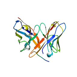
 | | Crystal structure of human RIG-I CTD bound to a 14-bp blunt-ended dsRNA | | 分子名称: | Antiviral innate immune response receptor RIG-I, RNA (5'-R(*GP*GP*CP*GP*CP*GP*CP*GP*CP*GP*CP*GP*CP*C)-3'), ZINC ION | | 著者 | Li, P. | | 登録日 | 2010-08-16 | | 公開日 | 2010-11-03 | | 最終更新日 | 2024-02-21 | | 実験手法 | X-RAY DIFFRACTION (2.4 Å) | | 主引用文献 | Crystal structure of RIG-I C-terminal domain bound to blunt-ended double-strand RNA without 5' triphosphate.
Nucleic Acids Res., 39, 2011
|
|
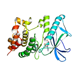
4LEV
 | | Structure of human cGAS | | 分子名称: | Cyclic GMP-AMP synthase, ZINC ION | | 著者 | Li, P. | | 登録日 | 2013-06-26 | | 公開日 | 2013-12-25 | | 最終更新日 | 2014-01-08 | | 実験手法 | X-RAY DIFFRACTION (1.952 Å) | | 主引用文献 | Cyclic GMP-AMP Synthase Is Activated by Double-Stranded DNA-Induced Oligomerization.
Immunity, 39, 2013
|
|
3E3H
 | | Crystal structure of the OP hydrolase mutant from Brevundimonas diminuta | | 分子名称: | COBALT (II) ION, DIETHYL 4-METHYLBENZYLPHOSPHONATE, Parathion hydrolase | | 著者 | Li, P, Reeves, T.E, Grimsley, J.K, Wild, J.R. | | 登録日 | 2008-08-07 | | 公開日 | 2008-10-07 | | 最終更新日 | 2023-11-15 | | 実験手法 | X-RAY DIFFRACTION (2.15 Å) | | 主引用文献 | Balancing the stability and the catalytic specificities of OP hydrolases with enhanced V-agent activities.
Protein Eng.Des.Sel., 21, 2008
|
|
5WSO
 | |
1B3J
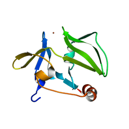
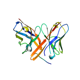
 | | STRUCTURE OF THE MHC CLASS I HOMOLOG MIC-A, A GAMMADELTA T CELL LIGAND | | 分子名称: | 2-acetamido-2-deoxy-beta-D-glucopyranose-(1-4)-2-acetamido-2-deoxy-beta-D-glucopyranose, MHC CLASS I HOMOLOG MIC-A | | 著者 | Li, P, Willie, S, Bauer, S, Morris, D, Spies, T, Strong, R. | | 登録日 | 1998-12-11 | | 公開日 | 1999-07-09 | | 最終更新日 | 2023-12-27 | | 実験手法 | X-RAY DIFFRACTION (3 Å) | | 主引用文献 | Crystal structure of the MHC class I homolog MIC-A, a gammadelta T cell ligand.
Immunity, 10, 1999
|
|
5V5F
 | | Crystal structure of RICE1 (PNT2) | | 分子名称: | At3g11770 | | 著者 | Li, P. | | 登録日 | 2017-03-14 | | 公開日 | 2017-09-13 | | 最終更新日 | 2024-03-06 | | 実験手法 | X-RAY DIFFRACTION (2.945 Å) | | 主引用文献 | RISC-interacting clearing 3'- 5' exoribonucleases (RICEs) degrade uridylated cleavage fragments to maintain functional RISC in Arabidopsis thaliana.
Elife, 6, 2017
|
|
3S2X
 | |
3U9M
 | |
4EMT
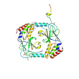
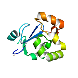
 | | Crystal Structure of human STING bound to c-di-GMP | | 分子名称: | 9,9'-[(2R,3R,3aS,5S,7aR,9R,10R,10aS,12S,14aR)-3,5,10,12-tetrahydroxy-5,12-dioxidooctahydro-2H,7H-difuro[3,2-d:3',2'-j][1,3,7,9,2,8]tetraoxadiphosphacyclododecine-2,9-diyl]bis(2-amino-1,9-dihydro-6H-purin-6-one), CALCIUM ION, Transmembrane protein 173 | | 著者 | Li, P. | | 登録日 | 2012-04-12 | | 公開日 | 2012-06-13 | | 最終更新日 | 2012-07-25 | | 実験手法 | X-RAY DIFFRACTION (1.5 Å) | | 主引用文献 | Structure of STING bound to cyclic di-GMP reveals the mechanism of cyclic dinucleotide recognition by the immune system.
Nat.Struct.Mol.Biol., 19, 2012
|
|


