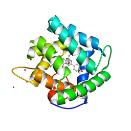
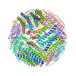
8X40
 
 | |
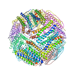
8IKQ
 | |
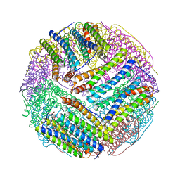
1XGD
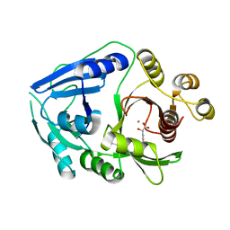
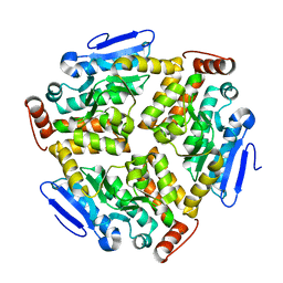
 | | Apo R268A human aldose reductase | | 分子名称: | Aldose reductase | | 著者 | Brownlee, J.M, Bohren, K.M, Milne, A.C, Gabbay, K.H, Harrison, D.H.T. | | 登録日 | 2004-09-16 | | 公開日 | 2005-03-29 | | 最終更新日 | 2023-08-23 | | 実験手法 | X-RAY DIFFRACTION (2.1 Å) | | 主引用文献 | The structure of Apo R268A human aldose reductase: Hinges and latches that control the kinetic mechanism
Biochim.Biophys.Acta, 1748, 2005
|
|
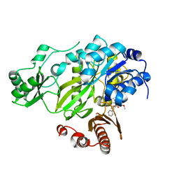
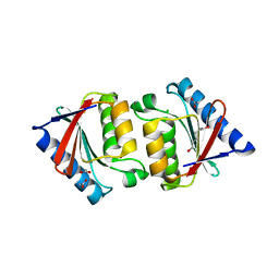
1T47
 | | Structure of fe2-HPPD bound to NTBC | | 分子名称: | 2-{HYDROXY[2-NITRO-4-(TRIFLUOROMETHYL)PHENYL]METHYLENE}CYCLOHEXANE-1,3-DIONE, 4-hydroxyphenylpyruvate dioxygenase, FE (II) ION | | 著者 | Brownlee, J, Johnson-Winters, K, Harrison, D.H.T, Moran, G.R. | | 登録日 | 2004-04-28 | | 公開日 | 2004-06-15 | | 最終更新日 | 2024-02-14 | | 実験手法 | X-RAY DIFFRACTION (2.5 Å) | | 主引用文献 | Structure of the Ferrous Form of (4-Hydroxyphenyl)pyruvate Dioxygenase from Streptomyces avermitilis in Complex with the Therapeutic Herbicide, NTBC
Biochemistry, 43, 2004
|
|
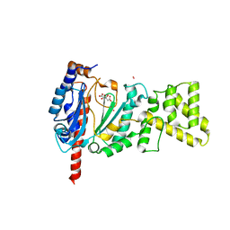
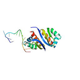
2R5V
 | | Hydroxymandelate Synthase Crystal Structure | | 分子名称: | (2S)-hydroxy(4-hydroxyphenyl)ethanoic acid, COBALT (II) ION, PCZA361.1, ... | | 著者 | Brownlee, J.M, He, P, Moran, G.R, Harrison, D.H.T. | | 登録日 | 2007-09-04 | | 公開日 | 2008-03-18 | | 最終更新日 | 2024-02-21 | | 実験手法 | X-RAY DIFFRACTION (2.3 Å) | | 主引用文献 | Two roads diverged: the structure of hydroxymandelate synthase from Amycolatopsis orientalis in complex with 4-hydroxymandelate.
Biochemistry, 47, 2008
|
|
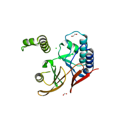
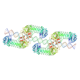
4V46
 | | Crystal structure of the BAFF-BAFF-R complex | | 分子名称: | MAGNESIUM ION, Tumor necrosis factor ligand superfamily member 13B, Tumor necrosis factor receptor superfamily member 13C | | 著者 | Kim, H.M, Yu, K.S, Lee, M.E, Shin, D.R, Kim, Y.S, Paik, S.G, Yoo, O.J, Lee, H, Lee, J.-O. | | 登録日 | 2003-03-23 | | 公開日 | 2014-07-09 | | 最終更新日 | 2024-10-09 | | 実験手法 | X-RAY DIFFRACTION (3.3 Å) | | 主引用文献 | Crystal structure of the BAFF-BAFF-R complex and its implications for receptor activation
NAT.STRUCT.BIOL., 10, 2003
|
|
3JRX
 | | Crystal structure of the BC domain of ACC2 in complex with soraphen A | | 分子名称: | Acetyl-CoA carboxylase 2, SORAPHEN A | | 著者 | Cho, Y.S, Lee, J.I, Shin, D, Kim, H.T, Lee, T.G, Heo, Y.S. | | 登録日 | 2009-09-09 | | 公開日 | 2010-01-12 | | 最終更新日 | 2023-11-01 | | 実験手法 | X-RAY DIFFRACTION (2.5 Å) | | 主引用文献 | Molecular mechanism for the regulation of human ACC2 through phosphorylation by AMPK.
Biochem.Biophys.Res.Commun., 391, 2010
|
|
4MM2
 | | Crystal structure of yeast primase catalytic subunit | | 分子名称: | CADMIUM ION, CITRIC ACID, DNA primase small subunit | | 著者 | Park, K.R, An, J.Y, Lee, Y, Youn, H.S, Lee, J.G, Kang, J.Y, Kim, T.G, Lim, J.J, Eom, S.H, Wang, J. | | 登録日 | 2013-09-07 | | 公開日 | 2014-09-10 | | 最終更新日 | 2023-11-08 | | 実験手法 | X-RAY DIFFRACTION (1.6 Å) | | 主引用文献 | Crystal structure of yeast primase catalytic subunit
To be Published
|
|
1QV0
 | | Atomic resolution structure of obelin from Obelia longissima | | 分子名称: | C2-HYDROPEROXY-COELENTERAZINE, COBALT (II) ION, GLYCEROL, ... | | 著者 | Liu, Z.J, Vysotski, E.S, Deng, L, Lee, J, Rose, J, Wang, B.C. | | 登録日 | 2003-08-26 | | 公開日 | 2003-11-11 | | 最終更新日 | 2023-08-16 | | 実験手法 | X-RAY DIFFRACTION (1.1 Å) | | 主引用文献 | Atomic resolution structure of obelin: soaking with calcium enhances electron density of the second oxygen atom substituted at the C2-position of coelenterazine.
Biochem.Biophys.Res.Commun., 311, 2003
|
|
1HOX
 | | CRYSTAL STRUCTURE OF RABBIT PHOSPHOGLUCOSE ISOMERASE COMPLEXED WITH FRUCTOSE-6-PHOSPHATE | | 分子名称: | 6-O-phosphono-beta-D-fructofuranose, PHOSPHOGLUCOSE ISOMERASE | | 著者 | Jeffrey, C.J, Lee, J.H, Chang, K.Z, Patel, V. | | 登録日 | 2000-12-11 | | 公開日 | 2001-07-20 | | 最終更新日 | 2023-08-09 | | 実験手法 | X-RAY DIFFRACTION (2.1 Å) | | 主引用文献 | Crystal structure of rabbit phosphoglucose isomerase complexed with its substrate D-fructose 6-phosphate.
Biochemistry, 40, 2001
|
|
6ZIF
 | |
2PQU
 | |
2PNM
 | |
6LVP
 | |
5JFY
 | |
2PY9
 | |
5WLY
 | | E. coli LpxH- 8 mutations | | 分子名称: | 1,2-ETHANEDIOL, CHLORIDE ION, FORMIC ACID, ... | | 著者 | Bohl, T.E, Aihara, H, Shi, K, Lee, J.K. | | 登録日 | 2017-07-28 | | 公開日 | 2018-04-11 | | 最終更新日 | 2023-10-04 | | 実験手法 | X-RAY DIFFRACTION (2 Å) | | 主引用文献 | The substrate-binding cap of the UDP-diacylglucosamine pyrophosphatase LpxH is highly flexible, enabling facile substrate binding and product release.
J. Biol. Chem., 293, 2018
|
|
3BVK
 | | Structural basis for the iron uptake mechanism of Helicobacter pylori ferritin | | 分子名称: | FE (III) ION, Ferritin, GLYCEROL | | 著者 | Kim, K.H, Cho, K.J, Lee, J.H, Shin, H.J, Yang, I.S. | | 登録日 | 2008-01-07 | | 公開日 | 2009-01-13 | | 最終更新日 | 2024-03-13 | | 実験手法 | X-RAY DIFFRACTION (1.5 Å) | | 主引用文献 | Structural basis for the iron uptake mechanism of Helicobacter pylori ferritin
To be Published
|
|
3BVL
 | | Structural basis for the iron uptake mechanism of Helicobacter pylori ferritin | | 分子名称: | FE (III) ION, Ferritin, GLYCEROL | | 著者 | Kim, K.H, Cho, K.J, Lee, J.H, Shin, H.J, Yang, I.S. | | 登録日 | 2008-01-07 | | 公開日 | 2009-01-13 | | 最終更新日 | 2024-03-13 | | 実験手法 | X-RAY DIFFRACTION (1.8 Å) | | 主引用文献 | Structural basis for the iron uptake mechanism of Helicobacter pylori ferritin
To be Published
|
|
3BVF
 | | Structural basis for the iron uptake mechanism of Helicobacter pylori ferritin | | 分子名称: | FE (III) ION, Ferritin, GLYCEROL, ... | | 著者 | Kim, K.H, Cho, K.J, Lee, J.H, Shin, H.J, Yang, I.S. | | 登録日 | 2008-01-07 | | 公開日 | 2009-01-13 | | 最終更新日 | 2024-03-13 | | 実験手法 | X-RAY DIFFRACTION (1.5 Å) | | 主引用文献 | Structural basis for the iron uptake mechanism of Helicobacter pylori ferritin
To be Published
|
|
3BVI
 | | Structural basis for the iron uptake mechanism of Helicobacter pylori ferritin | | 分子名称: | FE (III) ION, Ferritin, GLYCEROL | | 著者 | Kim, K.H, Cho, K.J, Lee, J.H, Shin, H.J, Yang, I.S. | | 登録日 | 2008-01-07 | | 公開日 | 2009-01-13 | | 最終更新日 | 2024-03-13 | | 実験手法 | X-RAY DIFFRACTION (2 Å) | | 主引用文献 | Structural basis for the iron uptake mechanism of Helicobacter pylori ferritin
To be Published
|
|
7WV5
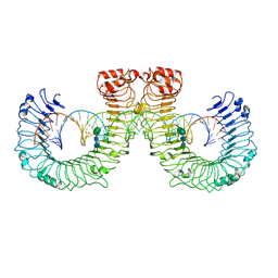 | | ectoTLR3-poly(I:C) | | 分子名称: | 2-acetamido-2-deoxy-beta-D-glucopyranose, 2-acetamido-2-deoxy-beta-D-glucopyranose-(1-4)-2-acetamido-2-deoxy-beta-D-glucopyranose, RNA (46-MER), ... | | 著者 | Lim, C.S, Jang, Y.H, Lee, G.Y, Han, G.M, Lee, J.O. | | 登録日 | 2022-02-09 | | 公開日 | 2022-11-16 | | 最終更新日 | 2024-10-16 | | 実験手法 | ELECTRON MICROSCOPY (3.1 Å) | | 主引用文献 | TLR3 forms a highly organized cluster when bound to a poly(I:C) RNA ligand.
Nat Commun, 13, 2022
|
|
7WV3
 | | Toll-like receptor3 linear cluster | | 分子名称: | 2-acetamido-2-deoxy-beta-D-glucopyranose, 2-acetamido-2-deoxy-beta-D-glucopyranose-(1-4)-2-acetamido-2-deoxy-beta-D-glucopyranose, RNA (80-MER), ... | | 著者 | Lim, C.S, Jang, Y.H, Lee, G.Y, Han, G.M, Lee, J.O. | | 登録日 | 2022-02-09 | | 公開日 | 2022-11-16 | | 最終更新日 | 2024-10-30 | | 実験手法 | ELECTRON MICROSCOPY (2.26 Å) | | 主引用文献 | TLR3 forms a highly organized cluster when bound to a poly(I:C) RNA ligand.
Nat Commun, 13, 2022
|
|
3BPM
 | | Crystal Structure of Falcipain-3 with Its inhibitor, Leupeptin | | 分子名称: | Cysteine protease falcipain-3, Leupeptin, SULFATE ION | | 著者 | kerr, I.D, Lee, J.H, Brinen, L.S. | | 登録日 | 2007-12-18 | | 公開日 | 2008-12-30 | | 最終更新日 | 2023-08-30 | | 実験手法 | X-RAY DIFFRACTION (2.5 Å) | | 主引用文献 | Structures of falcipain-2 and falcipain-3 bound to small molecule inhibitors: implications for substrate specificity.
J.Med.Chem., 52, 2009
|
|
3BPF
 | | Crystal Structure of Falcipain-2 with Its inhibitor, E64 | | 分子名称: | Cysteine protease falcipain-2, GLYCEROL, N-[N-[1-HYDROXYCARBOXYETHYL-CARBONYL]LEUCYLAMINO-BUTYL]-GUANIDINE | | 著者 | kerr, I.D, Lee, J.H, Brinen, L.S. | | 登録日 | 2007-12-18 | | 公開日 | 2008-12-30 | | 最終更新日 | 2023-08-30 | | 実験手法 | X-RAY DIFFRACTION (2.9 Å) | | 主引用文献 | Structures of falcipain-2 and falcipain-3 bound to small molecule inhibitors: implications for substrate specificity.
J.Med.Chem., 52, 2009
|
|


