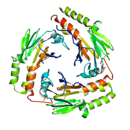
1U7H
 
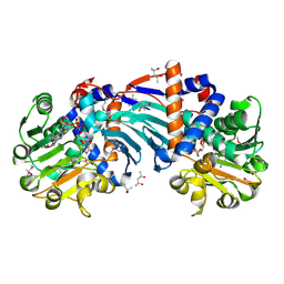 | | Structure and a Proposed Mechanism for Ornithine Cyclodeaminase from Pseudomonas putida | | 分子名称: | (4S)-2-METHYL-2,4-PENTANEDIOL, NICOTINAMIDE-ADENINE-DINUCLEOTIDE, SODIUM ION, ... | | 著者 | Alam, S, Goodman, J.L, Wang, S, Ruzicka, F.J, Frey, P.A, Wedekind, J.E. | | 登録日 | 2004-08-03 | | 公開日 | 2004-11-09 | | 最終更新日 | 2024-04-03 | | 実験手法 | X-RAY DIFFRACTION (1.8 Å) | | 主引用文献 | Ornithine Cyclodeaminase: Structure, Mechanism of Action, and Implications for
the u-Crystallin Family;
Biochemistry, 43, 2004
|
|
3CWK
 | |
3D96
 | |
2JQ4
 | | Complete resonance assignments and solution structure calculation of ATC2521 (NESG ID: AtT6) from Agrobacterium tumefaciens | | 分子名称: | Hypothetical protein Atu2571 | | 著者 | Srisailam, S, Lemak, A, Yee, A, Karra, M.D, Lukin, J.A, Arrowsmith, C.H, Northeast Structural Genomics Consortium (NESG) | | 登録日 | 2007-05-29 | | 公開日 | 2007-06-12 | | 最終更新日 | 2024-05-08 | | 実験手法 | SOLUTION NMR | | 主引用文献 | Solution Structure of a protein (ATC2521)of unknown function from Agrobacterium tumefaciens
To be Published
|
|
3D95
 | |
1X7D
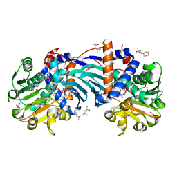 | | Crystal Structure Analysis of Ornithine Cyclodeaminase Complexed with NAD and ornithine to 1.6 Angstroms | | 分子名称: | (4S)-2-METHYL-2,4-PENTANEDIOL, 2-(N-MORPHOLINO)-ETHANESULFONIC ACID, L-ornithine, ... | | 著者 | Alam, S, Goodman, J.L, Wang, S, Ruzicka, F.J, Frey, P.A, Wedekind, J.E. | | 登録日 | 2004-08-13 | | 公開日 | 2004-11-09 | | 最終更新日 | 2023-11-15 | | 実験手法 | X-RAY DIFFRACTION (1.6 Å) | | 主引用文献 | Ornithine Cyclodeaminase: Structure, Mechanism of Action, and Implications for
the u-Crystallin Family
Biochemistry, 43, 2004
|
|
3D97
 | |
2FRS
 | |
2FS6
 | |
2FR3
 | |
2FS7
 | |
2G79
 | |
2G78
 | |
2G7B
 | |
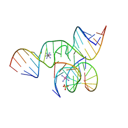
1X9K
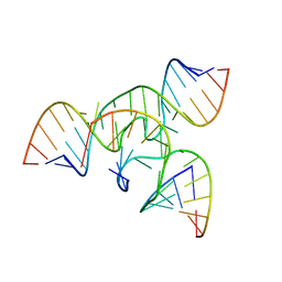 | | An all-RNA Hairpin Ribozyme with mutation U39C | | 分子名称: | 5'-R(*AP*AP*UP*AP*GP*AP*GP*AP*AP*GP*CP*GP*A)-3', 5'-R(*GP*GP*CP*AP*GP*AP*GP*AP*AP*AP*CP*AP*CP*AP*CP*GP*A)-3', 5'-R(*UP*CP*GP*CP*AP*GP*UP*CP*CP*UP*AP*UP*U)-3', ... | | 著者 | Alam, S, Grum-Tokars, V, Krucinska, J, Kundracik, M.L, Wedekind, J.E. | | 登録日 | 2004-08-21 | | 公開日 | 2005-11-22 | | 最終更新日 | 2023-08-23 | | 実験手法 | X-RAY DIFFRACTION (3.17 Å) | | 主引用文献 | Conformational Heterogeneity at Position U37 of an All-RNA Hairpin Ribozyme with Implications for Metal Binding and the Catalytic Structure of the S-Turn.
Biochemistry, 44, 2005
|
|
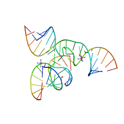
1X9C
 | | An all-RNA Hairpin Ribozyme with mutation U39C | | 分子名称: | 5'-R(*CP*GP*GP*UP*GP*AP*GP*AP*AP*GP*GP*G)-3', 5'-R(*GP*GP*CP*AP*GP*AP*GP*AP*AP*AP*CP*AP*CP*AP*CP*GP*A)-3', 5'-R(*UP*CP*CP*CP*(A2M)P*GP*UP*CP*CP*AP*CP*CP*G)-3', ... | | 著者 | Alam, S, Grum-Tokars, V, Krucinska, J, Kundracik, M.L, Wedekind, J.E. | | 登録日 | 2004-08-20 | | 公開日 | 2005-11-22 | | 最終更新日 | 2023-08-23 | | 実験手法 | X-RAY DIFFRACTION (2.19 Å) | | 主引用文献 | Conformational Heterogeneity at Position U37 of an All-RNA Hairpin Ribozyme with Implications for Metal Binding and the Catalytic Structure of the S-Turn.
Biochemistry, 44, 2005
|
|
2GQB
 | | Solution Structure of a conserved unknown protein RPA2825 from Rhodopseudomonas palustris; (Northeast Structural Genomics Consortium Target RpT4; Ontario Centre for Structural Proteomics Target rp2812 ) | | 分子名称: | conserved hypothetical protein | | 著者 | Srisailam, S, Lukin, J.A, Yee, A, Lemak, A, Arrowsmith, C.H, Northeast Structural Genomics Consortium (NESG) | | 登録日 | 2006-04-20 | | 公開日 | 2006-10-24 | | 最終更新日 | 2024-05-29 | | 実験手法 | SOLUTION NMR | | 主引用文献 | Solution Structure of a conserved unknown protein RPA2825 from Rhodopseudomonas palustris
To be Published
|
|
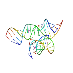
2D2K
 | | Crystal Structure of a minimal, native (U39) all-RNA hairpin ribozyme | | 分子名称: | 5'-R(*CP*GP*GP*UP*GP*AP*GP*AP*AP*GP*GP*G)-3', 5'-R(*GP*GP*CP*AP*GP*AP*GP*AP*AP*AP*CP*AP*CP*AP*CP*GP*A)-3', 5'-R(*UP*CP*CP*CP*(A2M)P*GP*UP*CP*CP*AP*CP*CP*G)-3', ... | | 著者 | Alam, S, Grum-Tokars, V, Krucinska, J, Kundracik, M.L, Wedekind, J.E. | | 登録日 | 2005-09-11 | | 公開日 | 2005-11-01 | | 最終更新日 | 2023-10-25 | | 実験手法 | X-RAY DIFFRACTION (2.65 Å) | | 主引用文献 | Conformational heterogeneity at position U37 of an all-RNA hairpin ribozyme with implications for metal binding and the catalytic structure of the S-turn
Biochemistry, 44, 2005
|
|
2D2L
 | | Crystal Structure of a minimal, all-RNA hairpin ribozyme with a propyl linker (C3) at position U39 | | 分子名称: | 5'-R(*CP*GP*GP*UP*GP*AP*GP*AP*AP*GP*GP*G)-3', 5'-R(*GP*GP*CP*AP*GP*AP*GP*AP*AP*AP*CP*AP*CP*AP*CP*GP*A)-3', 5'-R(*UP*CP*CP*CP*(A2M)P*GP*UP*CP*CP*AP*CP*CP*G)-3', ... | | 著者 | Alam, S, Grum-Tokars, V, Krucinska, J, Kundracik, M.L, Wedekind, J.E. | | 登録日 | 2005-09-11 | | 公開日 | 2005-11-01 | | 最終更新日 | 2023-10-25 | | 実験手法 | X-RAY DIFFRACTION (2.5 Å) | | 主引用文献 | Conformational heterogeneity at position U37 of an all-RNA hairpin ribozyme with implications for metal binding and the catalytic structure of the S-turn
Biochemistry, 44, 2005
|
|
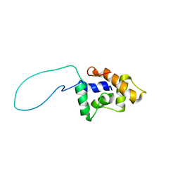
2AKL
 | | Solution structure for phn-A like protein PA0128 from Pseudomonas aeruginosa | | 分子名称: | ZINC ION, phnA-like protein pa0128 | | 著者 | Srisailam, S, Yee, A, Lemak, A, Lukin, J.A, Bansal, S, Prestegard, J.H, Arrowsmith, C.H, Northeast Structural Genomics Consortium (NESG), Ontario Centre for Structural Proteomics (OCSP) | | 登録日 | 2005-08-03 | | 公開日 | 2006-07-18 | | 最終更新日 | 2024-05-22 | | 実験手法 | SOLUTION NMR | | 主引用文献 | Sequence Specific Resonance Assignment of a Hypothetical Protein PA0128 from Pseudomonas Aeruginosa
J.Biomol.Nmr, 36, 2006
|
|
1FJK
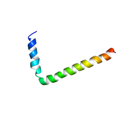 | | NMR Solution Structure of Phospholamban (C41F) | | 分子名称: | CARDIAC PHOSPHOLAMBAN | | 著者 | Lamberth, S, Griesinger, C, Schmid, H, Carafoli, E, Muenchbach, M, Vorherr, T, Krebs, J. | | 登録日 | 2000-08-08 | | 公開日 | 2000-09-06 | | 最終更新日 | 2024-05-22 | | 実験手法 | SOLUTION NMR | | 主引用文献 | NMR Solution Structure of Phospholamban
HELV.CHIM.ACTA, 83, 2000
|
|
1FJP
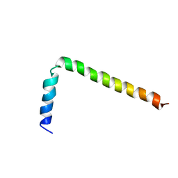 | | NMR Solution Structure of Phospholamban (C41F) | | 分子名称: | CARDIAC PHOSPHOLAMBAN | | 著者 | Lamberth, S, Griesinger, C, Schmid, H, Carafoli, E, Muenchbach, M, Vorherr, T, Krebs, J. | | 登録日 | 2000-08-08 | | 公開日 | 2000-09-06 | | 最終更新日 | 2024-05-22 | | 実験手法 | SOLUTION NMR | | 主引用文献 | NMR Solution Structure of Phospholamban
HELV.CHIM.ACTA, 83, 2000
|
|
1U7V
 | | Crystal Structure of the phosphorylated Smad2/Smad4 heterotrimeric complex | | 分子名称: | Mothers against decapentaplegic homolog 2, Mothers against decapentaplegic homolog 4 | | 著者 | Chacko, B.M, Qin, B.Y, Tiwari, A, Shi, G, Lam, S, Hayward, L.J, de Caestecker, M, Lin, K. | | 登録日 | 2004-08-04 | | 公開日 | 2004-09-28 | | 最終更新日 | 2023-08-23 | | 実験手法 | X-RAY DIFFRACTION (2.7 Å) | | 主引用文献 | Structural basis of heteromeric smad protein assembly in tgf-Beta signaling
Mol.Cell, 15, 2004
|
|
1U7F
 | | Crystal Structure of the phosphorylated Smad3/Smad4 heterotrimeric complex | | 分子名称: | Mothers against decapentaplegic homolog 3, Mothers against decapentaplegic homolog 4 | | 著者 | Chacko, B.M, Qin, B.Y, Tiwari, A, Shi, G, Lam, S, Hayward, L.J, de Caestecker, M, Lin, K. | | 登録日 | 2004-08-03 | | 公開日 | 2004-09-28 | | 最終更新日 | 2023-08-23 | | 実験手法 | X-RAY DIFFRACTION (2.6 Å) | | 主引用文献 | Structural basis of heteromeric smad protein assembly in tgf-Beta signaling
Mol.Cell, 15, 2004
|
|
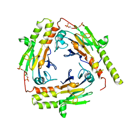
3DSH
 | | Crystal structure of dimeric interferon regulatory factor 5 (IRF-5) transactivation domain | | 分子名称: | Interferon regulatory factor 5 | | 著者 | Chen, W, Lam, S.S, Srinath, H, Jiang, Z, Correia, J.J, Schiffer, C, Fitzgerald, K.A, Lin, K, Royer Jr, W.E. | | 登録日 | 2008-07-12 | | 公開日 | 2008-10-07 | | 最終更新日 | 2024-02-21 | | 実験手法 | X-RAY DIFFRACTION (2 Å) | | 主引用文献 | Insights into interferon regulatory factor activation from the crystal structure of dimeric IRF5.
Nat.Struct.Mol.Biol., 15, 2008
|
|


