Loading
PDBj
メニューPDBj@FacebookPDBj@TwitterPDBj@YouTubewwPDB FoundationwwPDB
RCSB PDBPDBeBMRBAdv. SearchSearch help
PDB: 122 件

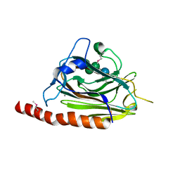
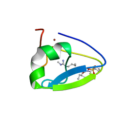
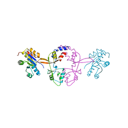
5K24
DownloadVisualize
BU of 5k24 by Molmil
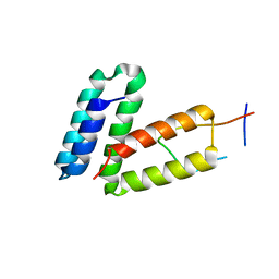
Crystal structure of the complex between phosphatase PRL-2 in the oxidized state with the Bateman domain of murine magnesium transporter CNNM3
分子名称: CITRATE ANION, Metal transporter CNNM3, Protein tyrosine phosphatase type IVA 2
著者Kozlov, G, Gulerez, I, Gehring, K.
登録日2016-05-18
公開日2016-10-12
最終更新日2018-10-03
実験手法X-RAY DIFFRACTION (3.1 Å)
主引用文献PRL3 phosphatase active site is required for binding the putative magnesium transporter CNNM3.
Sci Rep, 7, 2017
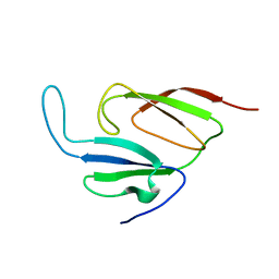
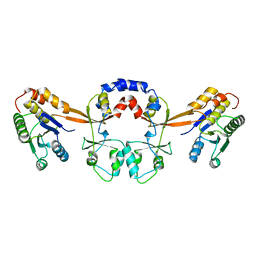
5K22
DownloadVisualize
BU of 5k22 by Molmil
Crystal structure of the complex between human PRL-2 phosphatase in reduced state and Bateman domain of human CNNM3
分子名称: Metal transporter CNNM3, Protein tyrosine phosphatase type IVA 2
著者Kozlov, G, Wu, H, Gehring, K.
登録日2016-05-18
公開日2016-10-12
最終更新日2024-03-06
実験手法X-RAY DIFFRACTION (3 Å)
主引用文献Phosphocysteine in the PRL-CNNM pathway mediates magnesium homeostasis.
EMBO Rep., 17, 2016
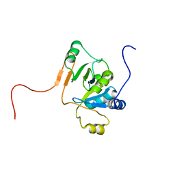
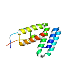
1JGN
DownloadVisualize
BU of 1jgn by Molmil
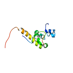
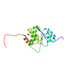
Solution structure of the C-terminal PABC domain of human poly(A)-binding protein in complex with the peptide from Paip2
分子名称: polyadenylate-binding protein 1, polyadenylate-binding protein-interacting protein 2
著者Kozlov, G, Siddiqui, N, Coillet-Matillon, S, Ekiel, I, Gehring, K.
登録日2001-06-26
公開日2003-06-24
最終更新日2024-05-22
実験手法SOLUTION NMR
主引用文献Structural basis of ligand recognition by PABC, a highly specific peptide-binding domain found in poly(A)-binding protein and a HECT ubiquitin ligase
EMBO J., 23, 2004
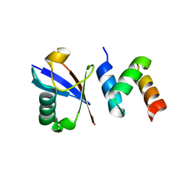
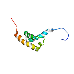
1JH4
DownloadVisualize
BU of 1jh4 by Molmil
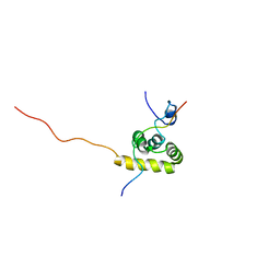
Solution structure of the C-terminal PABC domain of human poly(A)-binding protein in complex with the peptide from Paip1
分子名称: polyadenylate-binding protein 1, polyadenylate-binding protein-interacting protein-1
著者Kozlov, G, Siddiqui, N, Coillet-Matillon, S, Ekiel, I, Gehring, K.
登録日2001-06-27
公開日2003-06-24
最終更新日2024-05-22
実験手法SOLUTION NMR
主引用文献Structural basis of ligand recognition by PABC, a highly specific peptide-binding domain found in poly(A)-binding protein and a HECT ubiquitin ligase
EMBO J., 23, 2004
2H8L
DownloadVisualize
BU of 2h8l by Molmil
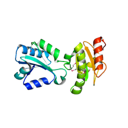
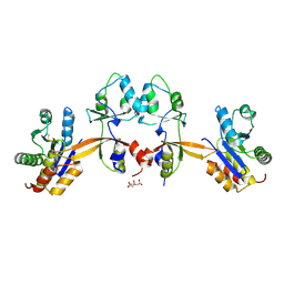
Crystal structure of the bb' fragment of ERp57
分子名称: Protein disulfide-isomerase A3
著者Kozlov, G, Schrag, J.D, Cygler, M, Gehring, K.
登録日2006-06-07
公開日2006-08-29
最終更新日2011-07-13
実験手法X-RAY DIFFRACTION (2 Å)
主引用文献Crystal Structure of the bb' Domains of the Protein Disulfide Isomerase ERp57.
Structure, 14, 2006
3O10
DownloadVisualize
BU of 3o10 by Molmil
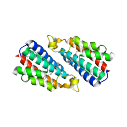
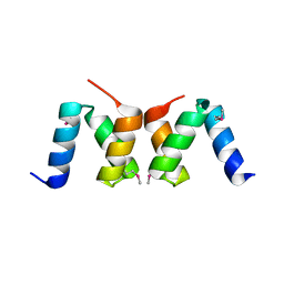
Crystal structure of the HEPN domain from human sacsin
分子名称: MALONATE ION, Sacsin
著者Kozlov, G, Gehring, K.
登録日2010-07-20
公開日2011-03-30
最終更新日2014-07-30
実験手法X-RAY DIFFRACTION (1.9 Å)
主引用文献Structural basis of defects in the sacsin HEPN domain responsible for autosomal recessive spastic ataxia of Charlevoix-Saguenay (ARSACS).
J.Biol.Chem., 286, 2011
3O0X
DownloadVisualize
BU of 3o0x by Molmil
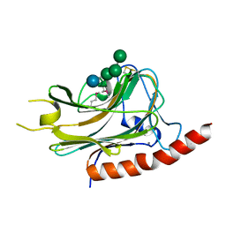
Structural basis of carbohydrate recognition by calreticulin
分子名称: CALCIUM ION, Calreticulin, alpha-D-glucopyranose-(1-3)-alpha-D-mannopyranose-(1-2)-alpha-D-mannopyranose-(1-2)-alpha-D-mannopyranose
著者Kozlov, G, Gehring, K.
登録日2010-07-20
公開日2010-09-29
最終更新日2023-12-06
実験手法X-RAY DIFFRACTION (2.01 Å)
主引用文献Structural basis of carbohydrate recognition by calreticulin.
J.Biol.Chem., 285, 2010
1Y7X
DownloadVisualize
BU of 1y7x by Molmil
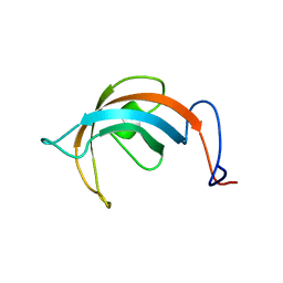
Solution structure of a two-repeat fragment of major vault protein
分子名称: Major vault protein
著者Kozlov, G, Vavelyuk, O, Minailiuc, O, Banville, D, Gehring, K, Ekiel, I.
登録日2004-12-10
公開日2005-12-20
最終更新日2024-05-22
実験手法SOLUTION NMR
主引用文献Solution structure of a two-repeat fragment of major vault protein.
J.Mol.Biol., 356, 2006
1JCU
DownloadVisualize
BU of 1jcu by Molmil
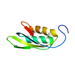
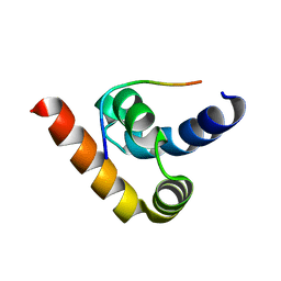
Solution Structure of MTH1692 Protein from Methanobacterium thermoautotrophicum
分子名称: conserved protein MTH1692
著者Kozlov, G, Ekiel, I, Gehring, K.
登録日2001-06-11
公開日2002-07-24
最終更新日2024-05-22
実験手法SOLUTION NMR
主引用文献An NMR approach to structural proteomics.
Proc.Natl.Acad.Sci.USA, 99, 2002
2OOB
DownloadVisualize
BU of 2oob by Molmil
crystal structure of the UBA domain from Cbl-b ubiquitin ligase in complex with ubiquitin
分子名称: E3 ubiquitin-protein ligase CBL-B, Ubiquitin
著者Kozlov, G, Gehring, K.
登録日2007-01-25
公開日2007-02-06
最終更新日2023-08-30
実験手法X-RAY DIFFRACTION (1.9 Å)
主引用文献Structural basis for ubiquitin-mediated dimerization and activation of the ubiquitin protein ligase Cbl-b.
Mol.Cell, 27, 2007
1R6H
DownloadVisualize
BU of 1r6h by Molmil
Solution Structure of human PRL-3
分子名称: protein tyrosine phosphatase type IVA, member 3 isoform 1
著者Kozlov, G, Gehring, K, Ekiel, I.
登録日2003-10-15
公開日2004-01-13
最終更新日2024-05-22
実験手法SOLUTION NMR
主引用文献Structural Insights into Molecular Function of the Metastasis-associated Phosphatase PRL-3.
J.Biol.Chem., 279, 2004
1RWU
DownloadVisualize
BU of 1rwu by Molmil
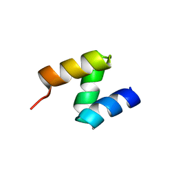
Solution structure of conserved protein YbeD from E. coli
分子名称: Hypothetical UPF0250 protein ybeD
著者Kozlov, G, Arrowsmith, C.H, Gehring, K, Northeast Structural Genomics Consortium (NESG)
登録日2003-12-17
公開日2004-12-21
最終更新日2024-05-22
実験手法SOLUTION NMR
主引用文献Structural similarity of YbeD protein from Escherichia coli to allosteric regulatory domains
J.Bacteriol., 186, 2004
1SSL
DownloadVisualize
BU of 1ssl by Molmil
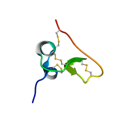
Solution structure of the PSI domain from the Met receptor
分子名称: Hepatocyte growth factor receptor
著者Kozlov, G, Perreault, A, Schrag, J.D, Cygler, M, Gehring, K, Ekiel, I.
登録日2004-03-24
公開日2004-10-12
最終更新日2022-03-02
実験手法SOLUTION NMR
主引用文献Insights into function of PSI domains from structure of the Met receptor PSI domain.
Biochem.Biophys.Res.Commun., 321, 2004
3O0V
DownloadVisualize
BU of 3o0v by Molmil
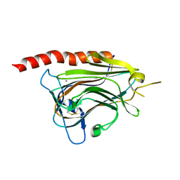
Crystal structure of the calreticulin lectin domain
分子名称: CALCIUM ION, Calreticulin
著者Kozlov, G, Gehring, K.
登録日2010-07-20
公開日2010-09-29
最終更新日2023-09-06
実験手法X-RAY DIFFRACTION (2.3 Å)
主引用文献Structural basis of carbohydrate recognition by calreticulin.
J.Biol.Chem., 285, 2010
3NTW
DownloadVisualize
BU of 3ntw by Molmil
Structure of the MLLE domain of EDD in complex with a PAM2 peptide from Paip1
分子名称: E3 ubiquitin-protein ligase UBR5, Polyadenylate-binding protein-interacting protein 1
著者Kozlov, G, Gehring, K.
登録日2010-07-05
公開日2011-07-06
最終更新日2023-09-06
実験手法X-RAY DIFFRACTION (2.6 Å)
主引用文献The MLLE domain of the ubiquitin ligase UBR5 binds to its catalytic domain to regulate substrate binding.
J. Biol. Chem., 290, 2015
3O0W
DownloadVisualize
BU of 3o0w by Molmil
Structural basis of carbohydrate recognition by calreticulin
分子名称: CALCIUM ION, Calreticulin, alpha-D-glucopyranose-(1-3)-alpha-D-mannopyranose-(1-2)-alpha-D-mannopyranose-(1-2)-alpha-D-mannopyranose
著者Kozlov, G, Gehring, K.
登録日2010-07-20
公開日2010-09-29
最終更新日2020-07-29
実験手法X-RAY DIFFRACTION (1.95 Å)
主引用文献Structural basis of carbohydrate recognition by calreticulin.
J.Biol.Chem., 285, 2010
2OO9
DownloadVisualize
BU of 2oo9 by Molmil
crystal structure of the UBA domain from human c-Cbl ubiquitin ligase
分子名称: E3 ubiquitin-protein ligase CBL
著者Kozlov, G, Gehring, K.
登録日2007-01-25
公開日2007-02-06
最終更新日2023-12-27
実験手法X-RAY DIFFRACTION (2.1 Å)
主引用文献Structural basis for UBA-mediated dimerization of c-Cbl ubiquitin ligase.
J.Biol.Chem., 282, 2007
2OOA
DownloadVisualize
BU of 2ooa by Molmil
crystal structure of the UBA domain from Cbl-b ubiquitin ligase
分子名称: E3 ubiquitin-protein ligase CBL-B
著者Kozlov, G, Gehring, K.
登録日2007-01-25
公開日2007-02-06
最終更新日2023-12-27
実験手法X-RAY DIFFRACTION (1.56 Å)
主引用文献Structural basis for ubiquitin-mediated dimerization and activation of the ubiquitin protein ligase Cbl-b.
Mol.Cell, 27, 2007
5TDC
DownloadVisualize
BU of 5tdc by Molmil
Crystal structure of the human UBR-box domain from UBR1 in complex with monomethylated arginine peptide.
分子名称: E3 ubiquitin-protein ligase UBR1, NMM-ILE-PHE-SER peptide, SULFATE ION, ...
著者Kozlov, G, Munoz-Escobar, J, Matta-Camacho, E, Gehring, K.
登録日2016-09-19
公開日2017-03-22
最終更新日2023-10-04
実験手法X-RAY DIFFRACTION (1.607 Å)
主引用文献Bound Waters Mediate Binding of Diverse Substrates to a Ubiquitin Ligase.
Structure, 25, 2017
5TSR
DownloadVisualize
BU of 5tsr by Molmil
Crystal structure of PRL-3 phosphatase in complex with the Bateman domain of CNNM3 magnesium transporter
分子名称: Metal transporter CNNM3, Protein tyrosine phosphatase type IVA 3
著者Kozlov, G, Zhang, H, Gehring, K.
登録日2016-10-31
公開日2017-02-08
最終更新日2023-10-04
実験手法X-RAY DIFFRACTION (3.188 Å)
主引用文献PRL3 phosphatase active site is required for binding the putative magnesium transporter CNNM3.
Sci Rep, 7, 2017
5V8Z
DownloadVisualize
BU of 5v8z by Molmil
Crystal structure of ERp29 D-domain in complex with the P-domain of calmegin
分子名称: Calmegin, Endoplasmic reticulum resident protein 29
著者Kozlov, G, Munoz-Escobar, J, Gehring, K.
登録日2017-03-22
公開日2017-06-21
最終更新日2023-10-04
実験手法X-RAY DIFFRACTION (2.105 Å)
主引用文献Mapping the ER Interactome: The P Domains of Calnexin and Calreticulin as Plurivalent Adapters for Foldases and Chaperones.
Structure, 25, 2017
1IFW
DownloadVisualize
BU of 1ifw by Molmil
SOLUTION STRUCTURE OF C-TERMINAL DOMAIN OF POLY(A) BINDING PROTEIN FROM SACCHAROMYCES CEREVISIAE
分子名称: POLYADENYLATE-BINDING PROTEIN, CYTOPLASMIC AND NUCLEAR
著者Kozlov, G, Siddiqui, N, Coillet-Matillon, S, Sprules, T, Ekiel, I, Gehring, K.
登録日2001-04-13
公開日2002-07-24
最終更新日2024-05-22
実験手法SOLUTION NMR
主引用文献Solution structure of the orphan PABC domain from Saccharomyces cerevisiae poly(A)-binding protein.
J.Biol.Chem., 277, 2002
1L1P
DownloadVisualize
BU of 1l1p by Molmil
Solution Structure of the PPIase Domain from E. coli Trigger Factor
分子名称: trigger factor
著者Kozlov, G, Trempe, J.-F, Perreault, A, Wong, M, Denisov, A, Ghandi, S, Gehring, K, Ekiel, I, Montreal-Kingston Bacterial Structural Genomics Initiative (BSGI)
登録日2002-02-19
公開日2003-06-24
最終更新日2024-05-22
実験手法SOLUTION NMR
主引用文献Solution Structure of the Closed Form of a Peptidyl-Prolyl Isomerase Reveals the Mechanism of Protein Folding
To be Published
5V90
DownloadVisualize
BU of 5v90 by Molmil
Crystal structure of ERp29 D-domain in complex with the P-domain of calreticulin
分子名称: Calreticulin, Endoplasmic reticulum resident protein 29, GLYCEROL
著者Kozlov, G, Munoz-Escobar, J, Gehring, K.
登録日2017-03-22
公開日2017-06-21
最終更新日2023-10-04
実験手法X-RAY DIFFRACTION (3.255 Å)
主引用文献Mapping the ER Interactome: The P Domains of Calnexin and Calreticulin as Plurivalent Adapters for Foldases and Chaperones.
Structure, 25, 2017
5VRQ
DownloadVisualize
BU of 5vrq by Molmil
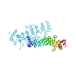
Crystal structure of Legionella pneumophila effector AnkC
分子名称: Ankyrin repeat-containing protein
著者Kozlov, G, Wong, K, Wang, W, Skubak, P, Munoz-Escobar, J, Liu, Y, Pannu, N.S, Gehring, K, Montreal-Kingston Bacterial Structural Genomics Initiative (BSGI)
登録日2017-05-11
公開日2017-11-29
最終更新日2024-03-13
実験手法X-RAY DIFFRACTION (3.205 Å)
主引用文献Ankyrin repeats as a dimerization module.
Biochem. Biophys. Res. Commun., 495, 2018

221051

件を2024-06-12に公開中

PDB statisticsPDBj update infoContact PDBjnumon