1ZFA
 
 | | GGA Duplex A-DNA | | 分子名称: | 5'-D(*CP*CP*TP*CP*CP*GP*GP*AP*GP*G)-3', CALCIUM ION, SODIUM ION | | 著者 | Hays, F.A, Teegarden, A.T, Jones, Z.J.R, Harms, M, Raup, D, Watson, J, Cavaliere, E, Ho, P.S. | | 登録日 | 2005-04-20 | | 公開日 | 2005-05-10 | | 最終更新日 | 2023-08-23 | | 実験手法 | X-RAY DIFFRACTION (1.56 Å) | | 主引用文献 | How sequence defines structure: a crystallographic map of DNA structure and conformation.
Proc.Natl.Acad.Sci.Usa, 102, 2005
|
|
3PFW
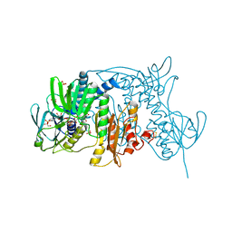
 | | Crystal structure of human sperm-specific glyceraldehyde-3-phosphate dehydrogenase (GAPDS) complex with NAD, a binary form | | 分子名称: | GLYCEROL, Glyceraldehyde-3-phosphate dehydrogenase, testis-specific, ... | | 著者 | Chaikuad, A, Shafqat, N, Yue, W.W, Cocking, R, Bray, J.E, von Delft, F, Arrowsmith, C.H, Edwards, A.M, Weigelt, J, Bountra, C, Oppermann, U, Structural Genomics Consortium (SGC) | | 登録日 | 2010-10-29 | | 公開日 | 2010-12-15 | | 最終更新日 | 2023-09-06 | | 実験手法 | X-RAY DIFFRACTION (2.15 Å) | | 主引用文献 | Structure and kinetic characterization of human sperm-specific glyceraldehyde-3-phosphate dehydrogenase, GAPDS.
Biochem.J., 435, 2011
|
|
1Z6I
 | | Crystal structure of the ectodomain of Drosophila transmembrane receptor PGRP-LCa | | 分子名称: | 2-acetamido-2-deoxy-beta-D-glucopyranose, Peptidoglycan-recognition protein-LC, SULFATE ION | | 著者 | Chang, C.-I, Ihara, K, Chelliah, Y, Mengin-Lecreulx, D, Wakatsuki, S, Deisenhofer, J. | | 登録日 | 2005-03-22 | | 公開日 | 2005-07-19 | | 最終更新日 | 2020-07-29 | | 実験手法 | X-RAY DIFFRACTION (2.5 Å) | | 主引用文献 | Structure of the ectodomain of Drosophila peptidoglycan-recognition protein LCa suggests a molecular mechanism for pattern recognition
Proc.Natl.Acad.Sci.Usa, 102, 2005
|
|
1ZJH
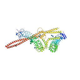
 | | Structure of human muscle pyruvate kinase (PKM2) | | 分子名称: | Pyruvate kinase, isozymes M1/M2 | | 著者 | Choe, J, Atanassova, A, Arrowsmith, C, Edwards, A, Sundstrom, M, Bochkarev, A, Park, H, Structural Genomics Consortium (SGC) | | 登録日 | 2005-04-28 | | 公開日 | 2005-05-17 | | 最終更新日 | 2023-08-23 | | 実験手法 | X-RAY DIFFRACTION (2.2 Å) | | 主引用文献 | Structure of human muscle pyruvate kinase (PKM2).
TO BE PUBLISHED
|
|
1ZJR
 | | Crystal Structure of A. aeolicus TrmH/SpoU tRNA modifying enzyme | | 分子名称: | GLYCEROL, SULFATE ION, tRNA (Guanosine-2'-O-)-methyltransferase | | 著者 | Pleshe, E, Truesdell, J, Batey, R.T. | | 登録日 | 2005-04-30 | | 公開日 | 2005-08-09 | | 最終更新日 | 2023-08-23 | | 実験手法 | X-RAY DIFFRACTION (1.85 Å) | | 主引用文献 | Structure of a class II TrmH tRNA-modifying enzyme from Aquifex aeolicus.
Acta Crystallogr.,Sect.F, 61, 2005
|
|
1ZK7
 | | Crystal Structure of Tn501 MerA | | 分子名称: | FLAVIN-ADENINE DINUCLEOTIDE, GLYCEROL, Mercuric reductase, ... | | 著者 | Dong, A, Ledwidge, R, Patel, B, Fiedler, D, Falkowski, M, Zelikova, J, Summers, A.O, Pai, E.F, Miller, S.M. | | 登録日 | 2005-05-02 | | 公開日 | 2005-07-05 | | 最終更新日 | 2023-08-23 | | 実験手法 | X-RAY DIFFRACTION (1.6 Å) | | 主引用文献 | NmerA, the Metal Binding Domain of Mercuric Ion Reductase, Removes Hg(2+) from Proteins, Delivers It to the Catalytic Core, and Protects Cells under Glutathione-Depleted Conditions
Biochemistry, 44, 2005
|
|
1ZNZ
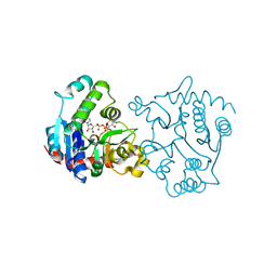
 | | Crystal Structure Of The Reduced Form Of Mycobacterium tuberculosis Guanylate Kinase In Complex With GDP | | 分子名称: | GUANOSINE-5'-DIPHOSPHATE, Guanylate kinase | | 著者 | Hible, G, Christova, P, Renault, L, Seclaman, E, Thompson, A, Girard, E, Munier-Lehmann, H, Cherfils, J. | | 登録日 | 2005-05-12 | | 公開日 | 2005-11-29 | | 最終更新日 | 2023-10-25 | | 実験手法 | X-RAY DIFFRACTION (2.5 Å) | | 主引用文献 | Unique GMP-binding site in Mycobacterium tuberculosis guanosine monophosphate kinase
Proteins, 62, 2006
|
|
1ZBQ
 | | Crystal Structure Of Human 17-Beta-Hydroxysteroid Dehydrogenase Type 4 In Complex With NAD | | 分子名称: | 17-beta-hydroxysteroid dehydrogenase 4, NICOTINAMIDE-ADENINE-DINUCLEOTIDE | | 著者 | Lukacik, P, Shafqat, N, Kavanagh, K, Bray, J, von Delft, F, Edwards, A, Arrowsmith, C, Sundstrom, M, Oppermann, U, Structural Genomics Consortium (SGC) | | 登録日 | 2005-04-08 | | 公開日 | 2005-04-26 | | 最終更新日 | 2023-08-23 | | 実験手法 | X-RAY DIFFRACTION (2.19 Å) | | 主引用文献 | Crystal Structure Of Human 17-Beta-Hydroxysteroid Dehydrogenase Type 4 In Complex With NAD
To be Published
|
|
1ZDG
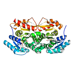
 | | Ser159 mutant of glycogenin complexed with UDP-glucose and manganese | | 分子名称: | Glycogenin-1, MANGANESE (II) ION, SULFATE ION, ... | | 著者 | Hurley, T.D, Stout, S.L, Miner, E, Zhou, J, Roach, P.J. | | 登録日 | 2005-04-14 | | 公開日 | 2005-04-26 | | 最終更新日 | 2023-08-23 | | 実験手法 | X-RAY DIFFRACTION (2.3 Å) | | 主引用文献 | Requirements for catalysis in mammalian glycogenin.
J.Biol.Chem., 280, 2005
|
|
1YV9
 | |
1ZF8
 | | GGT Duplex A-DNA | | 分子名称: | 5'-D(*CP*CP*AP*CP*CP*GP*GP*TP*GP*G)-3', CALCIUM ION | | 著者 | Hays, F.A, Teegarden, A.T, Jones, Z.J.R, Harms, M, Raup, D, Watson, J, Cavaliere, E, Ho, P.S. | | 登録日 | 2005-04-20 | | 公開日 | 2005-05-10 | | 最終更新日 | 2024-04-03 | | 実験手法 | X-RAY DIFFRACTION (1.48 Å) | | 主引用文献 | How sequence defines structure: a crystallographic map of DNA structure and conformation.
Proc.Natl.Acad.Sci.Usa, 102, 2005
|
|
1YZG
 | | Structure of Human ADP-ribosylation factor-like 8 | | 分子名称: | ADP-ribosylation factor-like 8, GUANOSINE-5'-DIPHOSPHATE | | 著者 | Choe, J, Atanassova, A, Arrowsmith, C, Edwards, A, Sundstrom, M, Bochkarev, A, Park, H, Structural Genomics Consortium (SGC) | | 登録日 | 2005-02-28 | | 公開日 | 2005-03-22 | | 最終更新日 | 2023-08-23 | | 実験手法 | X-RAY DIFFRACTION (2 Å) | | 主引用文献 | Structure of Human ADP-ribosylation factor-like 8
To be Published, 2005
|
|
1ZGX
 | |
1YZZ
 | | Humanized caban33 at room temperature | | 分子名称: | anti-VSG immunoglobulin heavy chain variable domain cAbAn33 | | 著者 | Conrath, K, Vincke, C, Stijlemans, B, Schymkowitz, J, Decanniere, K, Muyldermans, S, Loris, R. | | 登録日 | 2005-03-01 | | 公開日 | 2005-06-14 | | 最終更新日 | 2023-08-23 | | 実験手法 | X-RAY DIFFRACTION (2.7 Å) | | 主引用文献 | Antigen binding and solubility effects upon the veneering of a camel VHH in framework-2 to mimic a VH.
J.Mol.Biol., 350, 2005
|
|
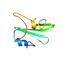
1Z09
 | | Solution structure of km23 | | 分子名称: | Dynein light chain 2A, cytoplasmic | | 著者 | Ilangovan, U, Ding, W, Mulder, K, Hinck, A.P, Zuniga, J, Trbovich, J.A, Demeler, B, Groppe, J.C. | | 登録日 | 2005-03-01 | | 公開日 | 2005-08-02 | | 最終更新日 | 2024-05-22 | | 実験手法 | SOLUTION NMR | | 主引用文献 | Structure and Dynamics of the Homodimeric Dynein Light Chain km23.
J.Mol.Biol., 352 (2), 2005
|
|
3U32
 | | ATP synthase c10 ring reacted with DCCD at pH 5.5 | | 分子名称: | ATP synthase subunit C, mitochondrial, DICYCLOHEXYLUREA | | 著者 | Symersky, J, Pagadala, V, Osowski, D, Krah, A, Meier, T, Faraldo-Gomez, J, Mueller, D.M. | | 登録日 | 2011-10-04 | | 公開日 | 2012-02-08 | | 最終更新日 | 2023-09-13 | | 実験手法 | X-RAY DIFFRACTION (2 Å) | | 主引用文献 | Structure of the c(10) ring of the yeast mitochondrial ATP synthase in the open conformation.
Nat.Struct.Mol.Biol., 19, 2012
|
|
1Z15
 | | Crystal structure analysis of periplasmic Leu/Ile/Val-binding protein in superopen form | | 分子名称: | Leu/Ile/Val-binding protein | | 著者 | Trakhanov, S.D, Vyas, N.K, Kristensen, D.M, Ma, J, Quiocho, F.A. | | 登録日 | 2005-03-03 | | 公開日 | 2005-10-04 | | 最終更新日 | 2023-08-23 | | 実験手法 | X-RAY DIFFRACTION (1.7 Å) | | 主引用文献 | Ligand-free and -bound structures of the binding protein (LivJ) of the Escherichia coli ABC leucine/isoleucine/valine transport system: trajectory and dynamics of the interdomain rotation and ligand specificity.
Biochemistry, 44, 2005
|
|
3OJA
 | | Crystal structure of LRIM1/APL1C complex | | 分子名称: | 2-acetamido-2-deoxy-beta-D-glucopyranose, 2-acetamido-2-deoxy-beta-D-glucopyranose-(1-4)-2-acetamido-2-deoxy-beta-D-glucopyranose, Anopheles Plasmodium-responsive Leucine-rich repeat protein 1, ... | | 著者 | Baxter, R.H.G, Deisenhofer, J. | | 登録日 | 2010-08-20 | | 公開日 | 2010-09-22 | | 最終更新日 | 2020-07-29 | | 実験手法 | X-RAY DIFFRACTION (2.7 Å) | | 主引用文献 | A heterodimeric complex of the LRR proteins LRIM1 and APL1C regulates complement-like immunity in Anopheles gambiae.
Proc.Natl.Acad.Sci.USA, 107, 2010
|
|
1ZIW
 | | Human Toll-like Receptor 3 extracellular domain structure | | 分子名称: | 2-acetamido-2-deoxy-beta-D-glucopyranose, 2-acetamido-2-deoxy-beta-D-glucopyranose-(1-4)-2-acetamido-2-deoxy-beta-D-glucopyranose, GLYCEROL, ... | | 著者 | Wilson, I.A, Choe, J. | | 登録日 | 2005-04-27 | | 公開日 | 2005-06-28 | | 最終更新日 | 2020-07-29 | | 実験手法 | X-RAY DIFFRACTION (2.1 Å) | | 主引用文献 | Crystal structure of human toll-like receptor 3 (TLR3) ectodomain.
Science, 309, 2005
|
|
1Z3F
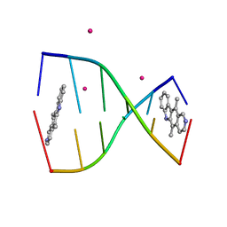 | | Structure of ellipticine in complex with a 6-bp DNA | | 分子名称: | 5'-D(*CP*GP*AP*TP*CP*G)-3', COBALT (II) ION, ELLIPTICINE | | 著者 | Canals, A, Purciolas, M, Aymami, J, Coll, M. | | 登録日 | 2005-03-12 | | 公開日 | 2005-07-19 | | 最終更新日 | 2024-02-14 | | 実験手法 | X-RAY DIFFRACTION (1.5 Å) | | 主引用文献 | The anticancer agent ellipticine unwinds DNA by intercalative binding in an orientation parallel to base pairs.
Acta Crystallogr.,Sect.D, 61, 2005
|
|
1Z3K
 | |
1Z6Y
 | | Structure Of Human ADP-Ribosylation Factor-Like 5 | | 分子名称: | ADP-ribosylation factor-like protein 5, GUANOSINE-5'-DIPHOSPHATE | | 著者 | Choe, J, Atanassova, A, Arrowsmith, C, Edwards, A, Sundstrom, M, Bochkarev, A, Park, H, Structural Genomics Consortium (SGC) | | 登録日 | 2005-03-23 | | 公開日 | 2005-04-05 | | 最終更新日 | 2023-08-23 | | 実験手法 | X-RAY DIFFRACTION (2.4 Å) | | 主引用文献 | Structure Of Human ADP-Ribosylation Factor-Like 5
To be Published
|
|
3U2F
 | | ATP synthase c10 ring in proton-unlocked conformation at PH 8.3 | | 分子名称: | ATP synthase subunit C, mitochondrial | | 著者 | Symersky, J, Pagadala, V, Osowski, D, Krah, A, Meier, T, Faraldo-Gomez, J, Mueller, D.M. | | 登録日 | 2011-10-03 | | 公開日 | 2012-02-08 | | 最終更新日 | 2023-09-13 | | 実験手法 | X-RAY DIFFRACTION (2 Å) | | 主引用文献 | Structure of the c(10) ring of the yeast mitochondrial ATP synthase in the open conformation.
Nat.Struct.Mol.Biol., 19, 2012
|
|
3U2Z
 | | Activator-Bound Structure of Human Pyruvate Kinase M2 | | 分子名称: | 1,6-di-O-phosphono-beta-D-fructofuranose, 6-(3-aminobenzyl)-4-methyl-2-methylsulfinyl-4,6-dihydro-5H-thieno[2',3':4,5]pyrrolo[2,3-d]pyridazin-5-one, Pyruvate kinase isozymes M1/M2, ... | | 著者 | Hong, B, Dimov, S, Tempel, W, Auld, D, Thomas, C, Boxer, M, Jianq, J.-K, Skoumbourdis, A, Min, S, Southall, N, Arrowsmith, C.H, Edwards, A.M, Bountra, C, Weigelt, J, Inglese, J, Park, H, Structural Genomics Consortium (SGC) | | 登録日 | 2011-10-04 | | 公開日 | 2012-09-12 | | 最終更新日 | 2023-09-13 | | 実験手法 | X-RAY DIFFRACTION (2.1 Å) | | 主引用文献 | Pyruvate kinase M2 activators promote tetramer formation and suppress tumorigenesis.
Nat.Chem.Biol., 8, 2012
|
|
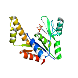
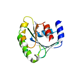
2A0T
 | | NMR structure of the FHA1 domain of Rad53 in complex with a biological relevant phosphopeptide derived from Madt1 | | 分子名称: | Hypothetical 73.8 kDa protein in SAS3-SEC17 intergenic region, residues 301-310, Serine/threonine-protein kinase RAD53 | | 著者 | Mahajan, A, Yuan, C, Pike, B.L, Heierhorst, J, Chang, C.-F, Tsai, M.-D. | | 登録日 | 2005-06-16 | | 公開日 | 2005-11-08 | | 最終更新日 | 2022-03-09 | | 実験手法 | SOLUTION NMR | | 主引用文献 | FHA Domain-Ligand Interactions: Importance of Integrating Chemical and Biological Approaches
J.Am.Chem.Soc., 127, 2005
|
|


