6U38
 
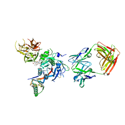 | | PCSK9 in complex with a Fab and compound 8 | | 分子名称: | 2-fluoro-4-{[(1R)-1-methyl-6-{[(2S)-oxan-2-yl]methoxy}-1-{2-oxo-2-[(1,3-thiazol-2-yl)amino]ethyl}-1,2,3,4-tetrahydroisoquinolin-7-yl]oxy}benzoic acid, Fab Heavy Chain, Fab Light Chain, ... | | 著者 | Lu, J, Soisson, S. | | 登録日 | 2019-08-21 | | 公開日 | 2019-11-06 | | 最終更新日 | 2020-01-29 | | 実験手法 | X-RAY DIFFRACTION (2.73 Å) | | 主引用文献 | From Screening to Targeted Degradation: Strategies for the Discovery and Optimization of Small Molecule Ligands for PCSK9.
Cell Chem Biol, 27, 2020
|
|
2EAL
 | | Crystal structure of human galectin-9 N-terminal CRD in complex with Forssman pentasaccharide | | 分子名称: | 2-acetamido-2-deoxy-alpha-D-galactopyranose-(1-3)-2-acetamido-2-deoxy-beta-D-galactopyranose-(1-3)-beta-D-galactopyranose, Galectin-9 | | 著者 | Nagae, M, Nakamura-Tsuruta, S, Nishi, N, Nakamura, T, Hirabayashi, J, Wakatsuki, S, Kato, R. | | 登録日 | 2007-01-31 | | 公開日 | 2007-09-25 | | 最終更新日 | 2024-03-13 | | 実験手法 | X-RAY DIFFRACTION (1.85 Å) | | 主引用文献 | Structural analysis of the human galectin-9 N-terminal carbohydrate recognition domain reveals unexpected properties that differ from the mouse orthologue.
J.Mol.Biol., 375, 2008
|
|
2EJN
 | | Structural characterization of the tetrameric form of the major cat allergen fel D 1 | | 分子名称: | CALCIUM ION, Major allergen I polypeptide chain 1, chain 2 | | 著者 | Kaiser, L, Velickovic, T.C, Badia-Martinez, D, Adedoyin, J, Thunberg, S, Hallen, D, Berndt, K, Gronlund, H, Gafvelin, G, van Hage, M, Achour, A. | | 登録日 | 2007-03-19 | | 公開日 | 2007-04-17 | | 最終更新日 | 2023-10-25 | | 実験手法 | X-RAY DIFFRACTION (1.64 Å) | | 主引用文献 | Structural characterization of the tetrameric form of the major cat allergen Fel d 1
J.Mol.Biol., 370, 2007
|
|
2ELA
 | | Crystal Structure of the PTB domain of human APPL1 | | 分子名称: | Adapter protein containing PH domain, PTB domain and leucine zipper motif 1 | | 著者 | Li, J, Mao, X, Dong, L.Q, Liu, F, Tong, L. | | 登録日 | 2007-03-27 | | 公開日 | 2007-05-29 | | 最終更新日 | 2024-03-13 | | 実験手法 | X-RAY DIFFRACTION (2 Å) | | 主引用文献 | Crystal Structures of the BAR-PH and PTB Domains of Human APPL1
Structure, 15, 2007
|
|
2ELB
 | | Crystal Structure of the BAR-PH domain of human APPL1 | | 分子名称: | Adapter protein containing PH domain, PTB domain and leucine zipper motif 1 | | 著者 | Li, J, Mao, X, Dong, L.Q, Liu, F, Tong, L. | | 登録日 | 2007-03-27 | | 公開日 | 2007-05-29 | | 最終更新日 | 2011-07-13 | | 実験手法 | X-RAY DIFFRACTION (2.6 Å) | | 主引用文献 | Crystal Structures of the BAR-PH and PTB Domains of Human APPL1
Structure, 15, 2007
|
|
2EAK
 | | Crystal structure of human galectin-9 N-terminal CRD in complex with lactose | | 分子名称: | (2S,3S)-1,4-DIMERCAPTOBUTANE-2,3-DIOL, GLYCEROL, Galectin-9, ... | | 著者 | Nagae, M, Nakamura-Tsuruta, S, Nishi, N, Nakamura, T, Hirabayashi, J, Wakatsuki, S, Kato, R. | | 登録日 | 2007-01-31 | | 公開日 | 2007-09-25 | | 最終更新日 | 2020-07-29 | | 実験手法 | X-RAY DIFFRACTION (1.97 Å) | | 主引用文献 | Structural analysis of the human galectin-9 N-terminal carbohydrate recognition domain reveals unexpected properties that differ from the mouse orthologue.
J.Mol.Biol., 375, 2008
|
|
2EPH
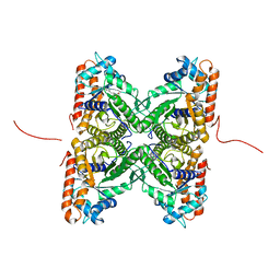 | | Crystal structure of fructose-bisphosphate aldolase from Plasmodium falciparum in complex with TRAP-tail determined at 2.7 angstrom resolution | | 分子名称: | Fructose-bisphosphate aldolase, PbTRAP | | 著者 | Bosch, J, Buscaglia, C.A, Krumm, B, Cardozo, T, Nussenzweig, V, Hol, W.G.J, Structural Genomics of Pathogenic Protozoa Consortium (SGPP) | | 登録日 | 2007-03-30 | | 公開日 | 2007-04-17 | | 最終更新日 | 2023-08-23 | | 実験手法 | X-RAY DIFFRACTION (2.7 Å) | | 主引用文献 | Aldolase provides an unusual binding site for thrombospondin-related anonymous protein in the invasion machinery of the malaria parasite.
Proc.Natl.Acad.Sci.Usa, 104, 2007
|
|
2ES9
 | | Crystal structure of Q8ZRJ2 from salmonella typhimurium. NESG TARGET STR65 | | 分子名称: | putative cytoplasmic protein | | 著者 | Benach, J, Abashidze, M, Jayaraman, S, Janjua, H, Cooper, B, Rong, X, Acton, T.B, Montelione, G.T, Tong, L, Hunt, J.F, Northeast Structural Genomics Consortium (NESG) | | 登録日 | 2005-10-25 | | 公開日 | 2005-11-01 | | 最終更新日 | 2024-03-06 | | 実験手法 | X-RAY DIFFRACTION (2 Å) | | 主引用文献 | Crystal structure of Q8ZRJ2 from Salmonella typhimurium NESG TARGET STR65.
To be Published
|
|
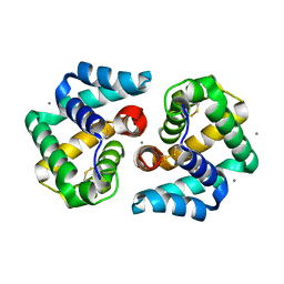
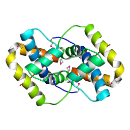
2EUD
 | | Structures of Yeast Ribonucleotide Reductase I complexed with Ligands and Subunit Peptides | | 分子名称: | GEMCITABINE DIPHOSPHATE, MAGNESIUM ION, PHOSPHOAMINOPHOSPHONIC ACID-ADENYLATE ESTER, ... | | 著者 | Dealwis, C, Xu, H, Faber, C, Uchiki, T, Fairman, J.W, Racca, J. | | 登録日 | 2005-10-28 | | 公開日 | 2006-03-07 | | 最終更新日 | 2024-02-14 | | 実験手法 | X-RAY DIFFRACTION (2.3 Å) | | 主引用文献 | Structures of eukaryotic ribonucleotide reductase I define gemcitabine diphosphate binding and subunit assembly.
Proc.Natl.Acad.Sci.Usa, 103, 2006
|
|
2EY2
 | |
2FJ9
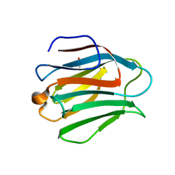
 | | High resolution crystal structure of the unliganded human ACBP | | 分子名称: | Acyl-CoA-binding protein, CHLORIDE ION, LEAD (II) ION, ... | | 著者 | Taskinen, J.P, van Aalten, D.M, Knudsen, J, Wierenga, R.K. | | 登録日 | 2006-01-02 | | 公開日 | 2006-10-31 | | 最終更新日 | 2024-02-14 | | 実験手法 | X-RAY DIFFRACTION (1.6 Å) | | 主引用文献 | High resolution crystal structures of unliganded and liganded human liver ACBP reveal a new mode of binding for the acyl-CoA ligand.
Proteins, 66, 2006
|
|
2EUC
 | | Crystal structure of YfmB from Bacillus subtilis. NESG TARGET SR324 | | 分子名称: | Hypothetical protein yfmB | | 著者 | Benach, J, Abashidze, M, Jayaraman, S, Janjua, H, Cooper, B, Rong, X, Acton, T.B, Montelione, G.T, Hunt, J.F, Tong, L, Northeast Structural Genomics Consortium (NESG) | | 登録日 | 2005-10-28 | | 公開日 | 2005-11-08 | | 最終更新日 | 2011-07-13 | | 実験手法 | X-RAY DIFFRACTION (2.5 Å) | | 主引用文献 | Crystal structure of YfmB from bacillus subtilis NESG TARGET SR324.
To be Published
|
|
6TUB
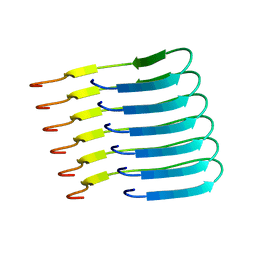 | | Beta-endorphin amyloid fibril | | 分子名称: | Beta-endorphin | | 著者 | Verasdonck, J, Seuring, C, Gath, J, Ghosh, D, Nespovitaya, N, Waelti, M.A, Maji, S, Cadalbert, R, Boeckmann, A, Guentert, P, Meier, B.H, Riek, R. | | 登録日 | 2020-01-05 | | 公開日 | 2020-10-28 | | 最終更新日 | 2024-05-15 | | 実験手法 | SOLID-STATE NMR | | 主引用文献 | The three-dimensional structure of human beta-endorphin amyloid fibrils.
Nat.Struct.Mol.Biol., 27, 2020
|
|
8RGS
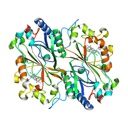 | | Serial synchrotron in plate room temperature structure of Dye Type Peroxidase Aa | | 分子名称: | Deferrochelatase, PROTOPORPHYRIN IX CONTAINING FE | | 著者 | Thompson, A.J, Hough, M.A, Williams, L.J, Worrall, J.A.R, Sanchez-Weatherby, J, Mikolajek, H, Sandy, J. | | 登録日 | 2023-12-14 | | 公開日 | 2023-12-27 | | 最終更新日 | 2024-04-17 | | 実験手法 | X-RAY DIFFRACTION (1.79 Å) | | 主引用文献 | Efficient in situ screening of and data collection from microcrystals in crystallization plates.
Acta Crystallogr D Struct Biol, 80, 2024
|
|
8RGW
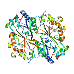 | | Serial synchrotron in plate room temperature structure of Dye Type Peroxidase Aa, 12 drops merged | | 分子名称: | Deferrochelatase, PROTOPORPHYRIN IX CONTAINING FE | | 著者 | Thompson, A.J, Hough, M.A, Williams, L.J, Worrall, J.A.R, Sanchez-Weatherby, J, Mikolajek, H, Sandy, J. | | 登録日 | 2023-12-14 | | 公開日 | 2023-12-27 | | 最終更新日 | 2024-04-17 | | 実験手法 | X-RAY DIFFRACTION (1.88 Å) | | 主引用文献 | Efficient in situ screening of and data collection from microcrystals in crystallization plates.
Acta Crystallogr D Struct Biol, 80, 2024
|
|
8RGY
 | | Serial synchrotron in plate room temperature structure of Dye Type Peroxidase Aa, 8 drops merged | | 分子名称: | Deferrochelatase, PROTOPORPHYRIN IX CONTAINING FE | | 著者 | Thompson, A.J, Hough, M.A, Williams, L.J, Worrall, J.A.R, Sanchez-Weatherby, J, Mikolajek, H, Sandy, J. | | 登録日 | 2023-12-14 | | 公開日 | 2023-12-27 | | 最終更新日 | 2024-04-17 | | 実験手法 | X-RAY DIFFRACTION (2.07 Å) | | 主引用文献 | Efficient in situ screening of and data collection from microcrystals in crystallization plates.
Acta Crystallogr D Struct Biol, 80, 2024
|
|
8RGE
 | | Serial synchrotron in plate room temperature structure of Lysozyme. | | 分子名称: | CHLORIDE ION, Lysozyme C, SODIUM ION | | 著者 | Thompson, A.J, Hough, M.A, Sanchez-Weatherby, J, Williams, L.J, Sandy, J, Worrall, J.A.R. | | 登録日 | 2023-12-13 | | 公開日 | 2023-12-27 | | 最終更新日 | 2024-04-17 | | 実験手法 | X-RAY DIFFRACTION (1.88 Å) | | 主引用文献 | Efficient in situ screening of and data collection from microcrystals in crystallization plates.
Acta Crystallogr D Struct Biol, 80, 2024
|
|
4YY0
 | | The structure of hemagglutinin from a H6N1 influenza virus (A/chicken/Taiwan/A2837/2013) | | 分子名称: | 2-acetamido-2-deoxy-beta-D-glucopyranose, HA1, HA2 | | 著者 | Wang, F, Qi, J, Bi, Y, Zhang, W, Wang, M, Wang, M, Liu, J, Yan, J, Shi, Y, Gao, G.F. | | 登録日 | 2015-03-23 | | 公開日 | 2016-03-23 | | 最終更新日 | 2020-07-29 | | 実験手法 | X-RAY DIFFRACTION (2.59 Å) | | 主引用文献 | Structure of hemagglutinin from a H6N1 influenza virus (A/chicken/Taiwan/A2837/2013)
To Be Published
|
|
7RCO
 | |
2PKT
 | | Crystal structure of the human CLP-36 (PDLIM1) bound to the C-terminal peptide of human alpha-actinin-1 | | 分子名称: | ACETATE ION, CALCIUM ION, CHLORIDE ION, ... | | 著者 | Uppenberg, J, Gileadi, C, Elkins, J, Bray, J, Burgess-Brown, N, Salah, E, Gileadi, O, Bunkoczi, G, Ugochukwu, E, Umeano, C, von Delft, F, Weigelt, J, Arrowsmith, C.H, Edwards, A, Sundstrom, M, Doyle, D.A, Structural Genomics Consortium (SGC) | | 登録日 | 2007-04-18 | | 公開日 | 2007-05-08 | | 最終更新日 | 2024-04-03 | | 実験手法 | X-RAY DIFFRACTION (1.5 Å) | | 主引用文献 | Unusual binding interactions in PDZ domain crystal structures help explain binding mechanisms
Protein Sci., 19, 2010
|
|
4NGS
 | |
4Z8Q
 | | CRYSTAL STRUCTURE OF AvrRxo1-ORF1:AvrRxo1-ORF2 COMPLEX, SELENOMETHIONINE SUBSTITUTED. | | 分子名称: | AvrRxo1-ORF1, AvrRxo1-ORF2, PHOSPHATE ION | | 著者 | Han, Q, Zhou, C, Wu, S, Liu, Y, Yang, Z, Miao, J, Triplett, L, Cheng, Q, Tokuhisa, J, Deblais, L, Robinson, H, Leach, J.E, Li, J, Zhao, B. | | 登録日 | 2015-04-09 | | 公開日 | 2015-09-23 | | 最終更新日 | 2022-03-16 | | 実験手法 | X-RAY DIFFRACTION (1.89 Å) | | 主引用文献 | Crystal Structure of Xanthomonas AvrRxo1-ORF1, a Type III Effector with a Polynucleotide Kinase Domain, and Its Interactor AvrRxo1-ORF2.
Structure, 23, 2015
|
|
2Q0Z
 | | Crystal structure of Q9P172/Sec63 from Homo sapiens. Northeast Structural Genomics Target HR1979. | | 分子名称: | Protein PRO2281 | | 著者 | Benach, J, Neely, H, Abashidze, M, Edstrom, W.C, Seetharaman, J, Zhao, L, Fang, Y, Cunningham, K, Owens, L, Ma, L.-C, Xiao, R, Liu, J, Baran, M.C, Acton, T.B, Rost, B, Montelione, G.T, Tong, L, Hunt, J.F, Northeast Structural Genomics Consortium (NESG) | | 登録日 | 2007-05-23 | | 公開日 | 2007-06-05 | | 最終更新日 | 2018-01-24 | | 実験手法 | X-RAY DIFFRACTION (2 Å) | | 主引用文献 | Crystal structure of Q9P172/Sec63 from Homo sapiens.
To be Published
|
|
4ZJP
 | | Structure of an ABC-Transporter Solute Binding Protein (SBP_IPR025997) from Actinobacillus Succinogenes (Asuc_0197, TARGET EFI-511067) with bound beta-D-ribopyranose | | 分子名称: | 1,2-ETHANEDIOL, Monosaccharide-transporting ATPase, beta-D-ribopyranose | | 著者 | Yadava, U, Vetting, M.W, Al Obaidi, N.F, Toro, R, Morisco, L.L, Benach, J, Wasserman, S.R, Attonito, J.D, Glenn, A.S, Chamala, S, Chowdhury, S, Lafleur, J, Love, J, Seidel, R.D, Whalen, K.L, Gerlt, J.A, Almo, S.C, Enzyme Function Initiative (EFI) | | 登録日 | 2015-04-29 | | 公開日 | 2015-05-20 | | 最終更新日 | 2023-11-15 | | 実験手法 | X-RAY DIFFRACTION (1.63 Å) | | 主引用文献 | Structure of an ABC-Transporter Solute Binding Protein (SBP_IPR025997) from Actinobacillus Succinogenes (Asuc_0197, TARGET EFI-511067) with bound beta-D-ribopyranose
To be published
|
|
8B24
 | | Time-resolved structure of K+-dependent Na+-PPase from Thermotoga maritima 3600-seconds post reaction initiation with Na+ | | 分子名称: | DIPHOSPHATE, K(+)-stimulated pyrophosphate-energized sodium pump, MAGNESIUM ION, ... | | 著者 | Strauss, J, Vidilaseris, K, Goldman, A. | | 登録日 | 2022-09-12 | | 公開日 | 2024-01-17 | | 最終更新日 | 2024-04-10 | | 実験手法 | X-RAY DIFFRACTION (4.53 Å) | | 主引用文献 | Functional and structural asymmetry suggest a unifying principle for catalysis in membrane-bound pyrophosphatases.
Embo Rep., 25, 2024
|
|


