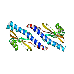
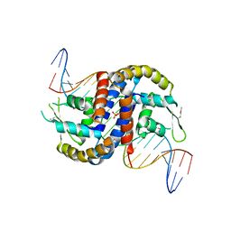
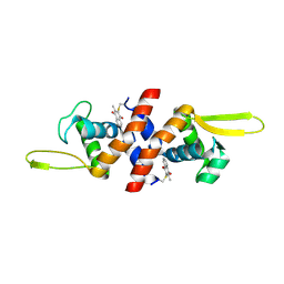
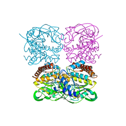
3ORR
 
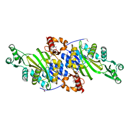 | | Crystal Structure of N5-Carboxyaminoimidazole synthetase from Staphylococcus aureus | | 分子名称: | N5-carboxyaminoimidazole ribonucleotide synthetase | | 著者 | Brugarolas, P, Duguid, E.M, Zhang, W, Poor, C.B, He, C. | | 登録日 | 2010-09-07 | | 公開日 | 2011-07-20 | | 最終更新日 | 2013-04-24 | | 実験手法 | X-RAY DIFFRACTION (2.23 Å) | | 主引用文献 | Structural and biochemical characterization of N5-carboxyaminoimidazole ribonucleotide synthetase and N5-carboxyaminoimidazole ribonucleotide mutase from Staphylococcus aureus.
Acta Crystallogr.,Sect.D, 67, 2011
|
|
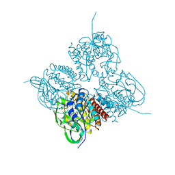
3ORS
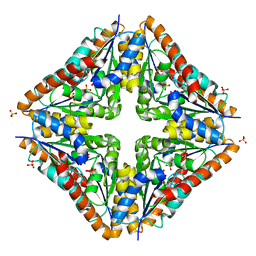 | | Crystal Structure of N5-Carboxyaminoimidazole Ribonucleotide Mutase from Staphylococcus aureus | | 分子名称: | N5-Carboxyaminoimidazole Ribonucleotide Mutase, SULFATE ION | | 著者 | Brugarolas, P, Duguid, E.M, Zhang, W, Poor, C.B, He, C. | | 登録日 | 2010-09-07 | | 公開日 | 2011-07-20 | | 最終更新日 | 2023-09-06 | | 実験手法 | X-RAY DIFFRACTION (1.45 Å) | | 主引用文献 | Structural and biochemical characterization of N5-carboxyaminoimidazole ribonucleotide synthetase and N5-carboxyaminoimidazole ribonucleotide mutase from Staphylococcus aureus.
Acta Crystallogr.,Sect.D, 67, 2011
|
|
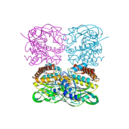
3ORQ
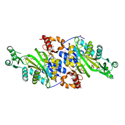 | | Crystal Structure of N5-Carboxyaminoimidazole synthetase from Staphylococcus aureus complexed with ADP | | 分子名称: | ADENOSINE-5'-DIPHOSPHATE, GLYCEROL, MAGNESIUM ION, ... | | 著者 | Brugarolas, P, Duguid, E.M, Zhang, W, Poor, C.B, He, C. | | 登録日 | 2010-09-07 | | 公開日 | 2011-07-20 | | 最終更新日 | 2012-03-28 | | 実験手法 | X-RAY DIFFRACTION (2.23 Å) | | 主引用文献 | Structural and biochemical characterization of N5-carboxyaminoimidazole ribonucleotide synthetase and N5-carboxyaminoimidazole ribonucleotide mutase from Staphylococcus aureus.
Acta Crystallogr.,Sect.D, 67, 2011
|
|
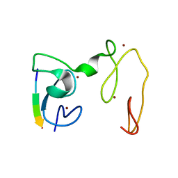
3R26
 | |
3RKP
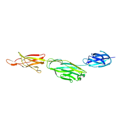 | | Crystal structure of BcpA*(D312A), the major pilin subunit of Bacillus cereus | | 分子名称: | Collagen adhesion protein | | 著者 | Hendrickx, A.P, Poor, C.B, Jureller, J.E, Budzik, J.M, He, C, Schneewind, O. | | 登録日 | 2011-04-18 | | 公開日 | 2012-05-30 | | 最終更新日 | 2023-09-13 | | 実験手法 | X-RAY DIFFRACTION (2.243 Å) | | 主引用文献 | Isopeptide bonds of the major pilin protein BcpA influence pilus structure and bundle formation on the surface of Bacillus cereus.
Mol.Microbiol., 85, 2012
|
|
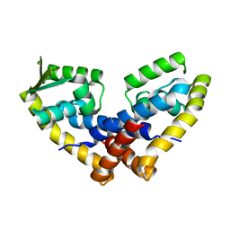
3HSR
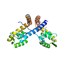 | | Crystal structure of Staphylococcus aureus protein SarZ in mixed disulfide form | | 分子名称: | ACETATE ION, GLYCEROL, HTH-type transcriptional regulator sarZ, ... | | 著者 | Poor, C.B, Duguid, E, Rice, P.A, He, C. | | 登録日 | 2009-06-10 | | 公開日 | 2009-07-07 | | 最終更新日 | 2023-09-06 | | 実験手法 | X-RAY DIFFRACTION (1.9 Å) | | 主引用文献 | Crystal structures of the reduced, sulfenic acid, and mixed disulfide forms of SarZ, a redox active global regulator in Staphylococcus aureus.
J.Biol.Chem., 284, 2009
|
|
3HSE
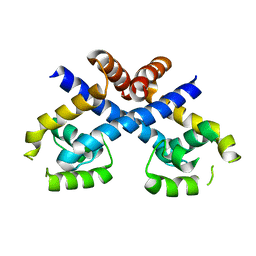 | | Crystal structure of Staphylococcus aureus protein SarZ in reduced form | | 分子名称: | HTH-type transcriptional regulator sarZ | | 著者 | Poor, C.B, Duguid, E, Rice, P.A, He, C. | | 登録日 | 2009-06-10 | | 公開日 | 2009-07-07 | | 最終更新日 | 2023-09-06 | | 実験手法 | X-RAY DIFFRACTION (2.9 Å) | | 主引用文献 | Crystal structures of the reduced, sulfenic acid, and mixed disulfide forms of SarZ, a redox active global regulator in Staphylococcus aureus.
J.Biol.Chem., 284, 2009
|
|
3HRM
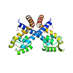 | | Crystal structure of Staphylococcus aureus protein SarZ in sulfenic acid form | | 分子名称: | HTH-type transcriptional regulator sarZ | | 著者 | Poor, C.B, Duguid, E, Rice, P.A, He, C. | | 登録日 | 2009-06-09 | | 公開日 | 2009-07-07 | | 最終更新日 | 2017-11-01 | | 実験手法 | X-RAY DIFFRACTION (2.3 Å) | | 主引用文献 | Crystal structures of the reduced, sulfenic acid, and mixed disulfide forms of SarZ, a redox active global regulator in Staphylococcus aureus.
J.Biol.Chem., 284, 2009
|
|
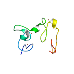
4YWZ
 | |
7RFP
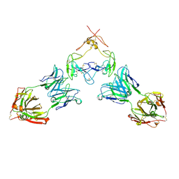 | | Mouse GITR (mGITR) with DTA-1 Fab fragment | | 分子名称: | DTA-1 (heavy chain), DTA-1 (light chain), Tumor necrosis factor receptor superfamily member 18,Enhanced green fluorescent protein | | 著者 | Meyerson, J.R, He, C. | | 登録日 | 2021-07-14 | | 公開日 | 2022-03-09 | | 実験手法 | ELECTRON MICROSCOPY (4.4 Å) | | 主引用文献 | Therapeutic antibody activation of the glucocorticoid-induced TNF receptor by a clustering mechanism.
Sci Adv, 8, 2022
|
|
3KPT
 | | Crystal structure of BcpA, the major pilin subunit of Bacillus cereus | | 分子名称: | CALCIUM ION, Collagen adhesion protein | | 著者 | Poor, C.B, Budzik, J.M, Schneewind, O, He, C. | | 登録日 | 2009-11-16 | | 公開日 | 2009-11-24 | | 最終更新日 | 2021-10-13 | | 実験手法 | X-RAY DIFFRACTION (2.102 Å) | | 主引用文献 | Intramolecular amide bonds stabilize pili on the surface of bacilli.
Proc.Natl.Acad.Sci.USA, 106, 2009
|
|
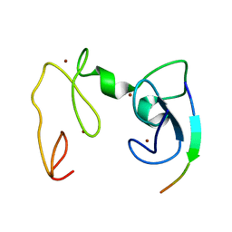
4FX0
 | |
4FX4
 | |
4GNE
 | | Crystal Structure of NSD3 tandem PHD5-C5HCH domains complexed with H3 peptide 1-7 | | 分子名称: | Histone H3.3, Histone-lysine N-methyltransferase NSD3, ZINC ION | | 著者 | Li, F, He, C, Wu, J, Shi, Y. | | 登録日 | 2012-08-17 | | 公開日 | 2013-01-02 | | 最終更新日 | 2024-03-20 | | 実験手法 | X-RAY DIFFRACTION (1.47 Å) | | 主引用文献 | The methyltransferase NSD3 has chromatin-binding motifs, PHD5-C5HCH, that are distinct from other NSD (nuclear receptor SET domain) family members in their histone H3 recognition.
J.Biol.Chem., 288, 2013
|
|
4GND
 | | Crystal Structure of NSD3 tandem PHD5-C5HCH domains | | 分子名称: | Histone-lysine N-methyltransferase NSD3, ZINC ION | | 著者 | Li, F, He, C, Wu, J, Shi, Y. | | 登録日 | 2012-08-17 | | 公開日 | 2013-01-02 | | 最終更新日 | 2023-11-08 | | 実験手法 | X-RAY DIFFRACTION (2.27 Å) | | 主引用文献 | The methyltransferase NSD3 has chromatin-binding motifs, PHD5-C5HCH, that are distinct from other NSD (nuclear receptor SET domain) family members in their histone H3 recognition.
J.Biol.Chem., 288, 2013
|
|
4GNF
 | | Crystal Structure of NSD3 tandem PHD5-C5HCH domains complexed with H3 peptide 1-15 | | 分子名称: | Histone H3.3, Histone-lysine N-methyltransferase NSD3, ZINC ION | | 著者 | Li, F, He, C, Wu, J, Shi, Y. | | 登録日 | 2012-08-17 | | 公開日 | 2013-01-02 | | 最終更新日 | 2023-11-08 | | 実験手法 | X-RAY DIFFRACTION (1.55 Å) | | 主引用文献 | The methyltransferase NSD3 has chromatin-binding motifs, PHD5-C5HCH, that are distinct from other NSD (nuclear receptor SET domain) family members in their histone H3 recognition.
J.Biol.Chem., 288, 2013
|
|
4GNG
 | | Crystal Structure of NSD3 tandem PHD5-C5HCH domains complexed with H3K9me3 peptide | | 分子名称: | GLYCEROL, Histone H3.3, Histone-lysine N-methyltransferase NSD3, ... | | 著者 | Li, F, He, C, Wu, J, Shi, Y. | | 登録日 | 2012-08-17 | | 公開日 | 2013-01-02 | | 最終更新日 | 2023-11-08 | | 実験手法 | X-RAY DIFFRACTION (1.73 Å) | | 主引用文献 | The methyltransferase NSD3 has chromatin-binding motifs, PHD5-C5HCH, that are distinct from other NSD (nuclear receptor SET domain) family members in their histone H3 recognition.
J.Biol.Chem., 288, 2013
|
|
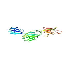
4GXL
 | | The crystal structure of Galectin-8 C-CRD in complex with NDP52 | | 分子名称: | GLYCEROL, Galectin-8, Peptide from Calcium-binding and coiled-coil domain-containing protein 2 | | 著者 | Li, S, Wandel, M.P, Li, F, Liu, Z, He, C, Wu, J, Shi, Y, Randow, F. | | 登録日 | 2012-09-04 | | 公開日 | 2013-05-08 | | 最終更新日 | 2024-03-20 | | 実験手法 | X-RAY DIFFRACTION (2.023 Å) | | 主引用文献 | Sterical hindrance promotes selectivity of the autophagy cargo receptor NDP52 for the danger receptor galectin-8 in antibacterial autophagy
Sci.Signal., 6, 2013
|
|
4HQM
 | | The crystal structure of QsrR-menadione complex | | 分子名称: | 2-methylnaphthalene-1,4-diol, QsrR protein | | 著者 | Ji, Q, Zhang, L, Jones, M.B, Sun, F, Deng, X, Liang, H, Brugarolas, P, Gao, N, Peterson, S.N, Lan, L, Bae, T, He, C. | | 登録日 | 2012-10-25 | | 公開日 | 2013-03-06 | | 最終更新日 | 2013-05-22 | | 実験手法 | X-RAY DIFFRACTION (2.55 Å) | | 主引用文献 | Molecular mechanism of quinone signaling mediated through S-quinonization of a YodB family repressor QsrR.
Proc.Natl.Acad.Sci.USA, 110, 2013
|
|
4LMG
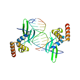 | | Crystal structure of AFT2 in complex with DNA | | 分子名称: | 5'-D(*AP*AP*GP*TP*GP*CP*AP*CP*CP*CP*AP*TP*T)-3', 5'-D(*TP*AP*AP*TP*GP*GP*GP*TP*GP*CP*AP*CP*T)-3', Iron-regulated transcriptional activator AFT2, ... | | 著者 | Poor, C.B, Sanishvili, R, Schuermann, J.P, He, C. | | 登録日 | 2013-07-10 | | 公開日 | 2014-03-05 | | 最終更新日 | 2024-02-28 | | 実験手法 | X-RAY DIFFRACTION (2.2 Å) | | 主引用文献 | Molecular mechanism and structure of the Saccharomyces cerevisiae iron regulator Aft2.
Proc.Natl.Acad.Sci.USA, 111, 2014
|
|
8HL8
 | | Crystal structrue of MtdL R257K mutant | | 分子名称: | MANGANESE (II) ION, Transglycosylse | | 著者 | Li, F.D, He, C. | | 登録日 | 2022-11-29 | | 公開日 | 2023-03-29 | | 最終更新日 | 2024-05-29 | | 実験手法 | X-RAY DIFFRACTION (2.5 Å) | | 主引用文献 | Structures of the NDP-pyranose mutase belonging to glycosyltransferase family 75 reveal residues important for Mn 2+ coordination and substrate binding.
J.Biol.Chem., 299, 2023
|
|
7XPR
 | | Crystal structrue of SeMet-MtdL:GDP | | 分子名称: | GUANOSINE-5'-DIPHOSPHATE, Transglycosylse | | 著者 | Li, F.D, He, C. | | 登録日 | 2022-05-05 | | 公開日 | 2023-03-29 | | 実験手法 | X-RAY DIFFRACTION (2.1 Å) | | 主引用文献 | Structures of the NDP-pyranose mutase belonging to glycosyltransferase family 75 reveal residues important for Mn 2+ coordination and substrate binding.
J.Biol.Chem., 299, 2023
|
|
7XPS
 | | Crystal structrue of MtdL:GDP:Mn | | 分子名称: | GUANOSINE-5'-DIPHOSPHATE, MANGANESE (II) ION, Transglycosylse | | 著者 | Li, F.D, He, C. | | 登録日 | 2022-05-05 | | 公開日 | 2023-03-29 | | 最終更新日 | 2023-11-29 | | 実験手法 | X-RAY DIFFRACTION (2.1 Å) | | 主引用文献 | Structures of the NDP-pyranose mutase belonging to glycosyltransferase family 75 reveal residues important for Mn 2+ coordination and substrate binding.
J.Biol.Chem., 299, 2023
|
|
7XPU
 | | crystal structure of MtdL-S228A-His soaked GDP-Fucp and Mn | | 分子名称: | GLYCEROL, GUANOSINE-5'-DIPHOSPHATE-BETA-L-FUCOPYRANOSE, MANGANESE (II) ION, ... | | 著者 | Li, F.D, He, C. | | 登録日 | 2022-05-05 | | 公開日 | 2023-03-29 | | 最終更新日 | 2023-11-29 | | 実験手法 | X-RAY DIFFRACTION (2.2 Å) | | 主引用文献 | Structures of the NDP-pyranose mutase belonging to glycosyltransferase family 75 reveal residues important for Mn 2+ coordination and substrate binding.
J.Biol.Chem., 299, 2023
|
|
7XPT
 | | Crystal structrue of MtdL:GDP:Mn soaked with GDP-Glc | | 分子名称: | GDP-ALPHA-D-GLUCOSE, MANGANESE (II) ION, Transglycosylse | | 著者 | Li, F.D, He, C. | | 登録日 | 2022-05-05 | | 公開日 | 2023-03-29 | | 最終更新日 | 2023-11-29 | | 実験手法 | X-RAY DIFFRACTION (2 Å) | | 主引用文献 | Structures of the NDP-pyranose mutase belonging to glycosyltransferase family 75 reveal residues important for Mn 2+ coordination and substrate binding.
J.Biol.Chem., 299, 2023
|
|


