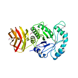
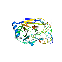
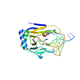
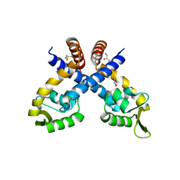
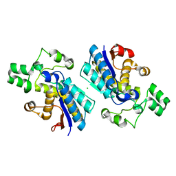
6J7L
 
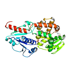 | | Crystal structure of Pseudomonas aeruginosa Earp in complex with TDP | | 分子名称: | 2-(N-MORPHOLINO)-ETHANESULFONIC ACID, Pseudomonas aeruginosa Earp, THYMIDINE-5'-DIPHOSPHATE | | 著者 | He, C, Li, F. | | 登録日 | 2019-01-18 | | 公開日 | 2019-05-15 | | 最終更新日 | 2023-11-22 | | 実験手法 | X-RAY DIFFRACTION (1.851 Å) | | 主引用文献 | Complex Structure ofPseudomonas aeruginosaArginine Rhamnosyltransferase EarP with Its Acceptor Elongation Factor P.
J.Bacteriol., 201, 2019
|
|
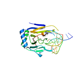
6J7M
 | |
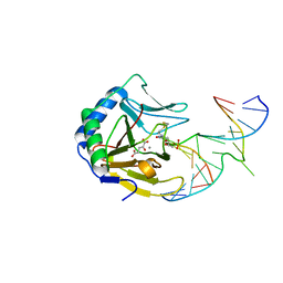
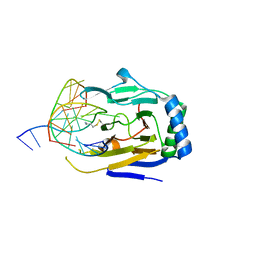
6JJ7
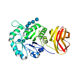
 | | Crystal structure of OsHXK6-Glc complex | | 分子名称: | Rice hexokinase 6, beta-D-glucopyranose | | 著者 | He, C, Wei, P, Chen, J, Wang, H, Wan, Y, Zhou, J, Zhu, Y, Huang, W, Yin, L. | | 登録日 | 2019-02-25 | | 公開日 | 2019-07-03 | | 最終更新日 | 2023-11-22 | | 実験手法 | X-RAY DIFFRACTION (2.9 Å) | | 主引用文献 | Crystal structure of OsHXK6-Glc complex
To Be Published
|
|
8J3Y
 | |
8J3X
 | |
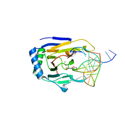
6JJ4
 | | Crystal structure of OsHXK6-apo form | | 分子名称: | Hexokinase-6 | | 著者 | He, C, Wei, P, Chen, J, Wang, H, Wan, Y, Zhou, J, Zhu, Y, Huang, W, Yin, L. | | 登録日 | 2019-02-25 | | 公開日 | 2019-07-03 | | 最終更新日 | 2023-11-22 | | 実験手法 | X-RAY DIFFRACTION (2.6 Å) | | 主引用文献 | Crystal structure of OsHXK6-apo
To Be Published
|
|
6JJ9
 | | Crystal structure of OsHXK6-Glc-ATP-Mg2+ complex | | 分子名称: | ADENOSINE-5'-DIPHOSPHATE, Hexokinase-6, MAGNESIUM ION, ... | | 著者 | He, C, Wei, P, Chen, J, Wang, H, Wan, Y, Zhou, J, Zhu, Y, Huang, W, Yin, L. | | 登録日 | 2019-02-25 | | 公開日 | 2019-07-03 | | 最終更新日 | 2023-11-22 | | 実験手法 | X-RAY DIFFRACTION (3 Å) | | 主引用文献 | Crystal structure of OsHXK6-Glc-ATP-Mg2+ complex
To Be Published
|
|
6JJ8
 | | Crystal structure of OsHXK6-ATP-Mg2+ complex | | 分子名称: | ADENOSINE-5'-DIPHOSPHATE, MAGNESIUM ION, PHOSPHATE ION, ... | | 著者 | He, C, Wei, P, Chen, J, Wang, H, Wan, Y, Zhou, J, Zhu, Y, Huang, W, Yin, L. | | 登録日 | 2019-02-25 | | 公開日 | 2019-07-03 | | 最終更新日 | 2023-11-22 | | 実験手法 | X-RAY DIFFRACTION (2.8 Å) | | 主引用文献 | Crystal structure of OsHXK6-ATP-Mg2+ complex
To Be Published
|
|
7D8M
 | | Crystal structure of DyP | | 分子名称: | Dye-decolorizing peroxidase, OXYGEN MOLECULE, PROTOPORPHYRIN IX CONTAINING FE | | 著者 | He, C, Jia, R, Wang, T, Li, L.Q. | | 登録日 | 2020-10-08 | | 公開日 | 2021-08-18 | | 最終更新日 | 2023-11-29 | | 実験手法 | X-RAY DIFFRACTION (2 Å) | | 主引用文献 | Revealing two important tryptophan residues with completely different roles in a dye-decolorizing peroxidase from Irpex lacteus F17.
Biotechnol Biofuels, 14, 2021
|
|
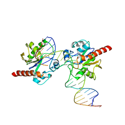
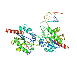
3BTZ
 | | Crystal structure of human ABH2 cross-linked to dsDNA | | 分子名称: | Alpha-ketoglutarate-dependent dioxygenase alkB homolog 2, DNA (5'-D(*AP*GP*GP*TP*GP*AP*(2YR)P*AP*AP*TP*GP*CP*G)-3'), DNA (5'-D(*DTP*DCP*DGP*DCP*DAP*DTP*DTP*DAP*DTP*DCP*DAP*DCP*DC)-3') | | 著者 | Yang, C.-G, Yi, C, He, C. | | 登録日 | 2007-12-31 | | 公開日 | 2008-04-22 | | 最終更新日 | 2021-10-20 | | 実験手法 | X-RAY DIFFRACTION (3 Å) | | 主引用文献 | Crystal structures of DNA/RNA repair enzymes AlkB and ABH2 bound to dsDNA.
Nature, 452, 2008
|
|
3BUC
 | | X-ray structure of human ABH2 bound to dsDNA with Mn(II) and 2KG | | 分子名称: | 2-OXOGLUTARIC ACID, Alpha-ketoglutarate-dependent dioxygenase alkB homolog 2, DNA (5'-D(*DCP*DTP*DGP*DTP*DAP*DTP*(MA7)P*DAP*DCP*DTP*DGP*DCP*DG)-3'), ... | | 著者 | Yang, C.-G, Yi, C, He, C. | | 登録日 | 2008-01-02 | | 公開日 | 2008-04-22 | | 最終更新日 | 2021-10-20 | | 実験手法 | X-RAY DIFFRACTION (2.59 Å) | | 主引用文献 | Crystal structures of DNA/RNA repair enzymes AlkB and ABH2 bound to dsDNA.
Nature, 452, 2008
|
|
3BKZ
 | | X-ray structure of E coli AlkB crosslinked to dsDNA in the active site | | 分子名称: | 2-OXOGLUTARIC ACID, Alpha-ketoglutarate-dependent dioxygenase alkB, DNA (5'-D(*DAP*DAP*DCP*DGP*DAP*DTP*DAP*DTP*DTP*DAP*DCP*DCP*DT)-3'), ... | | 著者 | Yi, C, Yang, C.-G, He, C. | | 登録日 | 2007-12-09 | | 公開日 | 2008-04-22 | | 最終更新日 | 2021-10-20 | | 実験手法 | X-RAY DIFFRACTION (1.65 Å) | | 主引用文献 | Crystal structures of DNA/RNA repair enzymes AlkB and ABH2 bound to dsDNA
Nature, 452, 2008
|
|
3BTX
 | | X-ray structure of human ABH2 bound to dsDNA through active site cross-linking | | 分子名称: | Alpha-ketoglutarate-dependent dioxygenase alkB homolog 2, DNA (5'-D(*CP*TP*GP*TP*AP*TP*(2YR)P*AP*TP*TP*GP*CP*G)-3'), DNA (5'-D(*DTP*DCP*DGP*DCP*DAP*DAP*DTP*DAP*DAP*DTP*DAP*DCP*DA)-3'), ... | | 著者 | Yang, C.-G, Yi, C, He, C. | | 登録日 | 2007-12-31 | | 公開日 | 2008-04-22 | | 最終更新日 | 2021-10-20 | | 実験手法 | X-RAY DIFFRACTION (2 Å) | | 主引用文献 | Crystal structures of DNA/RNA repair enzymes AlkB and ABH2 bound to dsDNA.
Nature, 452, 2008
|
|
3BU0
 | | crystal structure of human ABH2 cross-linked to dsDNA with cofactors | | 分子名称: | 2-OXOGLUTARIC ACID, Alpha-ketoglutarate-dependent dioxygenase alkB homolog 2, DNA (5'-D(*CP*TP*GP*TP*AP*TP*(2YR)P*AP*TP*TP*GP*CP*G)-3'), ... | | 著者 | Yang, C.-G, Yi, C, He, C. | | 登録日 | 2007-12-31 | | 公開日 | 2008-04-22 | | 最終更新日 | 2021-10-20 | | 実験手法 | X-RAY DIFFRACTION (2.5 Å) | | 主引用文献 | Crystal structures of DNA/RNA repair enzymes AlkB and ABH2 bound to dsDNA.
Nature, 452, 2008
|
|
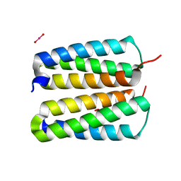
3KPT
 | | Crystal structure of BcpA, the major pilin subunit of Bacillus cereus | | 分子名称: | CALCIUM ION, Collagen adhesion protein | | 著者 | Poor, C.B, Budzik, J.M, Schneewind, O, He, C. | | 登録日 | 2009-11-16 | | 公開日 | 2009-11-24 | | 最終更新日 | 2021-10-13 | | 実験手法 | X-RAY DIFFRACTION (2.102 Å) | | 主引用文献 | Intramolecular amide bonds stabilize pili on the surface of bacilli.
Proc.Natl.Acad.Sci.USA, 106, 2009
|
|
8HOJ
 | | Crystal structure of UGT71AP2 in complex with UDP | | 分子名称: | UGT71AP2, URIDINE-5'-DIPHOSPHATE | | 著者 | Wang, Z.L, He, C, Li, F, Qiao, X, Ye, M. | | 登録日 | 2022-12-10 | | 公開日 | 2023-12-13 | | 実験手法 | X-RAY DIFFRACTION (2.9 Å) | | 主引用文献 | crystal structure of UGT71AP2 in complex with UDP
To Be Published
|
|
8HOK
 | | crystal structure of UGT71AP2 | | 分子名称: | 2-(N-MORPHOLINO)-ETHANESULFONIC ACID, GLYCEROL, UGT71AP2 | | 著者 | Wang, Z.L, He, C, Li, F, Qiao, X, Ye, M. | | 登録日 | 2022-12-10 | | 公開日 | 2023-12-13 | | 実験手法 | X-RAY DIFFRACTION (2.15 Å) | | 主引用文献 | crystal structure of UGT71AP2
To Be Published
|
|
4WQN
 | | Crystal structure of N6-methyladenosine RNA reader YTHDF2 | | 分子名称: | 1,2-ETHANEDIOL, GLYCEROL, YTH domain-containing family protein 2 | | 著者 | Zhu, T, Roundtree, I.A, Wang, P, Wang, X, Wang, L, Sun, C, Tian, Y, Li, J, He, C, Xu, Y. | | 登録日 | 2014-10-22 | | 公開日 | 2014-11-19 | | 最終更新日 | 2023-11-08 | | 実験手法 | X-RAY DIFFRACTION (2.121 Å) | | 主引用文献 | Crystal structure of the YTH domain of YTHDF2 reveals mechanism for recognition of N6-methyladenosine.
Cell Res., 24, 2014
|
|
4GXO
 | | Crystal structure of Staphylococcus aureus protein SarZ mutant C13E | | 分子名称: | GLYCEROL, MarR family regulatory protein | | 著者 | Sun, F, Ding, Y, Ji, Q, Liang, Z, Deng, X, Wong, C.C, Yi, C, Zhang, L, Xie, S, Alvarez, S, Hicks, L.M, Luo, C, Jiang, H, Lan, L, He, C. | | 登録日 | 2012-09-04 | | 公開日 | 2012-09-26 | | 最終更新日 | 2024-02-28 | | 実験手法 | X-RAY DIFFRACTION (2.05 Å) | | 主引用文献 | Protein cysteine phosphorylation of SarA/MgrA family transcriptional regulators mediates bacterial virulence and antibiotic resistance.
Proc.Natl.Acad.Sci.USA, 109, 2012
|
|
3BTY
 | | Crystal structure of human ABH2 bound to dsDNA containing 1meA through cross-linking away from active site | | 分子名称: | Alpha-ketoglutarate-dependent dioxygenase alkB homolog 2, DNA (5'-D(*DCP*DTP*DGP*DTP*DAP*DTP*(MA7)P*DAP*DCP*DTP*DGP*DCP*DG)-3'), DNA (5'-D(*DTP*DCP*DGP*DCP*DAP*DGP*DTP*DTP*DAP*DTP*DAP*DCP*DA)-3'), ... | | 著者 | Yang, C.-G, Yi, C, He, C. | | 登録日 | 2007-12-31 | | 公開日 | 2008-04-22 | | 最終更新日 | 2021-10-20 | | 実験手法 | X-RAY DIFFRACTION (2.35 Å) | | 主引用文献 | Crystal structures of DNA/RNA repair enzymes AlkB and ABH2 bound to dsDNA.
Nature, 452, 2008
|
|
4FZP
 | |
3UO7
 | |
4GPI
 | |
3UOB
 | |
3H8R
 | | Structure determination of DNA methylation lesions N1-meA and N3-meC in duplex DNA using a cross-linked host-guest system | | 分子名称: | 2-AMINO-2-HYDROXYMETHYL-PROPANE-1,3-DIOL, 5'-D(*CP*TP*GP*TP*AP*TP*(2YR)P*AP*TP*(6MA)P*GP*CP*G)-3', 5'-D(*TP*CP*GP*CP*TP*AP*TP*AP*AP*TP*AP*CP*A)-3', ... | | 著者 | Lu, L, Yi, C, Jian, X, Zheng, G, He, C. | | 登録日 | 2009-04-29 | | 公開日 | 2010-03-31 | | 最終更新日 | 2021-10-13 | | 実験手法 | X-RAY DIFFRACTION (1.77 Å) | | 主引用文献 | Structure determination of DNA methylation lesions N1-meA and N3-meC in duplex DNA using a cross-linked protein-DNA system.
Nucleic Acids Res., 38, 2010
|
|


