9GKY
 
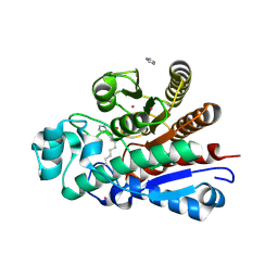 | | Crystal Structure of Histone deacetylase (HdaH) from Vibrio cholerae in complex with decanoic acid | | 分子名称: | DECANOIC ACID, Histone deacetylase, IMIDAZOLE, ... | | 著者 | Graf, L.G, Schulze, S, Palm, G.J, Lammers, M. | | 登録日 | 2024-08-26 | | 公開日 | 2024-11-06 | | 実験手法 | X-RAY DIFFRACTION (1.13 Å) | | 主引用文献 | Distribution and diversity of classical deacylases in bacteria
Nature Communications, 2024
|
|
9GKV
 
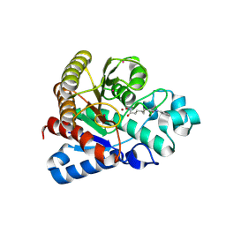 | | Crystal Structure of Deacetylase (HdaH) from Vibrio cholerae in complex with SAHA | | 分子名称: | ACETATE ION, Histone deacetylase, OCTANEDIOIC ACID HYDROXYAMIDE PHENYLAMIDE, ... | | 著者 | Graf, L.G, Schulze, S, Lammers, M, Palm, G.J. | | 登録日 | 2024-08-26 | | 公開日 | 2024-11-06 | | 実験手法 | X-RAY DIFFRACTION (1.9 Å) | | 主引用文献 | Distribution and diversity of classical deacylases in bacteria
Nature Communications, 2024
|
|
9GKX
 
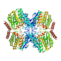 | | Crystal Structure of Rhizorhabdus wittichii Dimethoate hydrolase (DmhA) in complex with SAHA | | 分子名称: | Dimethoate hydrolase, OCTANEDIOIC ACID HYDROXYAMIDE PHENYLAMIDE, OCTANOIC ACID (CAPRYLIC ACID), ... | | 著者 | Graf, L.G, Lammers, M, Schulze, S, Palm, G.J. | | 登録日 | 2024-08-26 | | 公開日 | 2024-11-06 | | 実験手法 | X-RAY DIFFRACTION (1.75 Å) | | 主引用文献 | Distribution and diversity of classical deacylases in bacteria
Nature Communications, 2024
|
|
9GKW
 
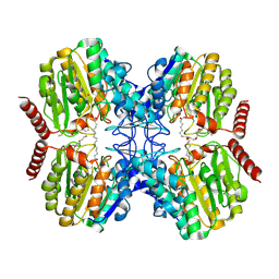 | | Crystal Structure of Dimethoate hydrolase (DmhA) of Rhizorhabdus wittichii in complex with octanoic acid | | 分子名称: | Dimethoate hydrolase, OCTANOIC ACID (CAPRYLIC ACID), PENTAETHYLENE GLYCOL, ... | | 著者 | Graf, L.G, Schulze, S, Palm, G.J, Lammers, M. | | 登録日 | 2024-08-26 | | 公開日 | 2024-11-06 | | 実験手法 | X-RAY DIFFRACTION (2.1 Å) | | 主引用文献 | Distribution and diversity of classical deacylases in bacteria
Nature Communications, 2024
|
|
9GKZ
 
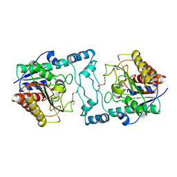 | | Crystal Structure of Acetylpolyamine amidohydrolase (ApaH) from Pseudomonas sp. M30-35 | | 分子名称: | ACETATE ION, Acetylpolyamine amidohydrolase, CHLORIDE ION, ... | | 著者 | Graf, L.G, Schulze, S, Palm, G.J, Lammers, M. | | 登録日 | 2024-08-26 | | 公開日 | 2024-11-06 | | 実験手法 | X-RAY DIFFRACTION (2.72 Å) | | 主引用文献 | Distribution and diversity of classical deacylases in bacteria
Nature Communications, 2024
|
|
9GL0
 
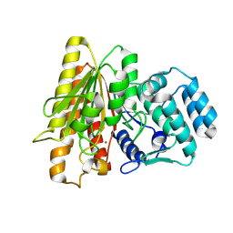 | | Crystal Structure of Acetylpolyamine aminohydrolase (ApaH) from Legionella pneumophila | | 分子名称: | Acetylpolyamine aminohydrolase, POTASSIUM ION, ZINC ION | | 著者 | Graf, L.G, Schulze, S, Palm, G.J, Lammers, M. | | 登録日 | 2024-08-26 | | 公開日 | 2024-11-06 | | 実験手法 | X-RAY DIFFRACTION (2.7 Å) | | 主引用文献 | Distribution and diversity of classical deacylases in bacteria
Nature Communications, 2024
|
|
9GKU
 
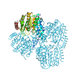 | | Crystal Structure of Propanil hydrolase (PrpH) from Sphingomonas sp. Y57 | | 分子名称: | ACETATE ION, POTASSIUM ION, Propanil hydrolase, ... | | 著者 | Graf, L.G, Lammers, L, Palm, G.J, Schulze, S. | | 登録日 | 2024-08-26 | | 公開日 | 2024-11-06 | | 実験手法 | X-RAY DIFFRACTION (1.48 Å) | | 主引用文献 | Distribution and diversity of classical deacylases in bacteria
Nature Communications, 2024
|
|
9GL1
 
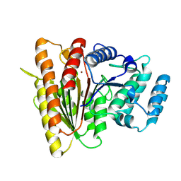 | | Crystal Structure of Acetylpolyamine aminohydrolase (ApaH) from Legionella cherrii | | 分子名称: | Acetylpolyamine aminohydrolase, POTASSIUM ION, ZINC ION | | 著者 | Graf, L.G, Schulze, S, Palm, G.J, Lammers, M. | | 登録日 | 2024-08-26 | | 公開日 | 2024-11-06 | | 実験手法 | X-RAY DIFFRACTION (2.4 Å) | | 主引用文献 | Distribution and diversity of classical deacylases in bacteria
Nature Communications, 2024
|
|
2F91
 
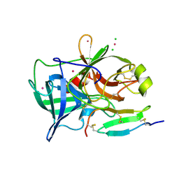 | | 1.2A resolution structure of a crayfish trypsin complexed with a peptide inhibitor, SGTI | | 分子名称: | CADMIUM ION, CHLORIDE ION, Serine protease inhibitor I/II, ... | | 著者 | Fodor, K, Harmat, V, Hetenyi, C, Kardos, J, Antal, J, Perczel, A, Patthy, A, Katona, G, Graf, L. | | 登録日 | 2005-12-05 | | 公開日 | 2006-04-18 | | 最終更新日 | 2023-08-30 | | 実験手法 | X-RAY DIFFRACTION (1.2 Å) | | 主引用文献 | Enzyme:Substrate Hydrogen Bond Shortening during the Acylation Phase of Serine Protease Catalysis.
Biochemistry, 45, 2006
|
|
1KIO
 
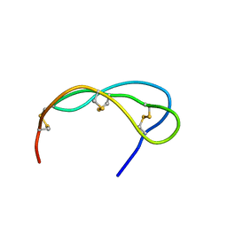 | | SOLUTION STRUCTURE OF THE SMALL SERINE PROTEASE INHIBITOR SGCI[L30R, K31M] | | 分子名称: | SERINE PROTEASE INHIBITOR I | | 著者 | Gaspari, Z, Patthy, A, Graf, L, Perczel, A. | | 登録日 | 2001-12-03 | | 公開日 | 2001-12-12 | | 最終更新日 | 2024-10-09 | | 実験手法 | SOLUTION NMR | | 主引用文献 | Comparative structure analysis of proteinase inhibitors from the desert locust, Schistocerca gregaria.
Eur.J.Biochem., 269, 2002
|
|
1KDQ
 
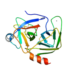 | | Crystal Structure Analysis of the Mutant S189D Rat Chymotrypsin | | 分子名称: | CALCIUM ION, CHYMOTRYPSIN B, B CHAIN, ... | | 著者 | Szabo, E, Bocskei, Z, Naray-Szabo, G, Graf, L, Venekei, I. | | 登録日 | 2001-11-13 | | 公開日 | 2003-06-10 | | 最終更新日 | 2024-10-30 | | 実験手法 | X-RAY DIFFRACTION (2.55 Å) | | 主引用文献 | Three Dimensional Structures of S189D Chymotrypsin and D189S Trypsin Mutants: The Effect of Polarity at Site 189 on a Protease-specific Stabilization of
the Substrate-binding Site
J.Mol.Biol., 331, 2003
|
|
1KGM
 
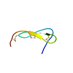 | | SOLUTION STRUCTURE OF THE SMALL SERINE PROTEASE INHIBITOR SGCI | | 分子名称: | SERINE PROTEASE INHIBITOR I | | 著者 | Gaspari, Z, Patthy, A, Graf, L, Perczel, A. | | 登録日 | 2001-11-28 | | 公開日 | 2001-12-12 | | 最終更新日 | 2024-11-06 | | 実験手法 | SOLUTION NMR | | 主引用文献 | Comparative structure analysis of proteinase inhibitors from the desert locust, Schistocerca gregaria.
Eur.J.Biochem., 269, 2002
|
|
2QY0
 
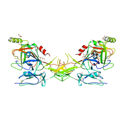 | | Active dimeric structure of the catalytic domain of C1r reveals enzyme-product like contacts | | 分子名称: | Complement C1r subcomponent, GLYCEROL | | 著者 | Kardos, J, Harmat, V, Pallo, A, Barabas, O, Szilagyi, K, Graf, L, Naray-Szabo, G, Goto, Y, Zavodszky, P, Gal, P. | | 登録日 | 2007-08-13 | | 公開日 | 2008-02-05 | | 最終更新日 | 2023-08-30 | | 実験手法 | X-RAY DIFFRACTION (2.6 Å) | | 主引用文献 | Revisiting the mechanism of the autoactivation of the complement protease C1r in the C1 complex: Structure of the active catalytic region of C1r.
Mol.Immunol., 45, 2008
|
|
1KJ0
 
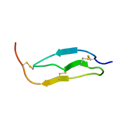 | | SOLUTION STRUCTURE OF THE SMALL SERINE PROTEASE INHIBITOR SGTI | | 分子名称: | SERINE PROTEASE INHIBITOR I | | 著者 | Gaspari, Z, Patthy, A, Graf, L, Perczel, A. | | 登録日 | 2001-12-04 | | 公開日 | 2001-12-12 | | 最終更新日 | 2024-10-30 | | 実験手法 | SOLUTION NMR | | 主引用文献 | Comparative structure analysis of proteinase inhibitors from the desert locust, Schistocerca gregaria.
Eur.J.Biochem., 269, 2002
|
|
8S7Z
 
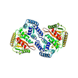 | |
4BNR
 
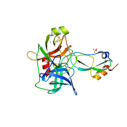 | | Extremely stable complex of crayfish trypsin with bovine trypsin inhibitor | | 分子名称: | CALCIUM ION, HEPATOPANCREAS TRYPSIN, PANCREATIC TRYPSIN INHIBITOR, ... | | 著者 | Molnar, T, Voros, J, Szeder, B, Takats, K, Kardos, J, Katona, G, Graf, L. | | 登録日 | 2013-05-17 | | 公開日 | 2013-09-04 | | 最終更新日 | 2023-12-20 | | 実験手法 | X-RAY DIFFRACTION (2 Å) | | 主引用文献 | Comparison of Complexes Formed by a Crustacean and a Vertebrate Trypsin with Bovine Pancreatic Trypsin Inhibitor - the Key to Achieving Extreme Stability?
FEBS J., 280, 2013
|
|
1H4W
 
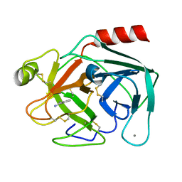 | | Structure of human trypsin IV (brain trypsin) | | 分子名称: | BENZAMIDINE, CALCIUM ION, TRYPSIN IVA | | 著者 | Katona, G, Berglund, G.I, Hajdu, J, Graf, L, Szilagyi, L. | | 登録日 | 2001-05-15 | | 公開日 | 2002-02-11 | | 最終更新日 | 2023-12-13 | | 実験手法 | X-RAY DIFFRACTION (1.7 Å) | | 主引用文献 | Crystal structure reveals basis for the inhibitor resistance of human brain trypsin.
J. Mol. Biol., 315, 2002
|
|
2XTT
 
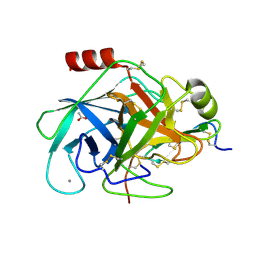 | | Bovine trypsin in complex with evolutionary enhanced Schistocerca gregaria protease inhibitor 1 (SGPI-1-P02) | | 分子名称: | ACETATE ION, CALCIUM ION, CATIONIC TRYPSIN, ... | | 著者 | Wahlgren, W.Y, Pal, G, Kardos, J, Porrogi, P, Szenthe, B, Patthy, A, Graf, L, Katona, G. | | 登録日 | 2010-10-12 | | 公開日 | 2010-11-10 | | 最終更新日 | 2024-11-06 | | 実験手法 | X-RAY DIFFRACTION (0.93 Å) | | 主引用文献 | The catalytic aspartate is protonated in the Michaelis complex formed between trypsin and an in vitro evolved substrate-like inhibitor: a refined mechanism of serine protease action.
J.Biol.Chem., 286, 2011
|
|
2JET
 
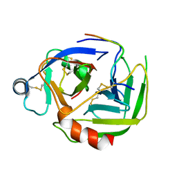 | | Crystal structure of a trypsin-like mutant (S189D , A226G) chymotrypsin. | | 分子名称: | CHYMOTRYPSINOGEN B CHAIN A, CHYMOTRYPSINOGEN B CHAIN B, CHYMOTRYPSINOGEN B CHAIN C | | 著者 | Jelinek, B, Katona, G, Fodor, K, Venekei, I, Graf, L. | | 登録日 | 2007-01-22 | | 公開日 | 2007-09-18 | | 最終更新日 | 2024-11-06 | | 実験手法 | X-RAY DIFFRACTION (2.2 Å) | | 主引用文献 | The Crystal Structure of a Trypsin-Like Mutant Chymotrypsin: The Role of Position 226 in the Activity and Specificity of S189D Chymotrypsin.
Protein J., 27, 2008
|
|
1AMH
 
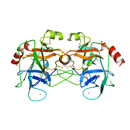 | | UNCOMPLEXED RAT TRYPSIN MUTANT WITH ASP 189 REPLACED WITH SER (D189S) | | 分子名称: | ANIONIC TRYPSIN, CALCIUM ION | | 著者 | Szabo, E, Bocskei, Z.S, Naray-Szabo, G, Graf, L. | | 登録日 | 1997-06-17 | | 公開日 | 1997-12-24 | | 最終更新日 | 2024-10-23 | | 実験手法 | X-RAY DIFFRACTION (2.5 Å) | | 主引用文献 | The three-dimensional structure of Asp189Ser trypsin provides evidence for an inherent structural plasticity of the protease.
Eur.J.Biochem., 263, 1999
|
|
9GN7
 
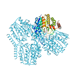 | | Crystal Structure of Deacetylase (HdaH) from Klebsiella pneumoniae subsp. ozaenae in Complex with the inhibitor TSA | | 分子名称: | Deacetylase, GLYCEROL, PHOSPHATE ION, ... | | 著者 | Qin, C, Graf, L.G, Schulze, S, Palm, G.J, Lammers, M. | | 登録日 | 2024-08-30 | | 公開日 | 2024-11-06 | | 実験手法 | X-RAY DIFFRACTION (2.18 Å) | | 主引用文献 | Distribution and diversity of classical deacylases in bacteria
Nature Communications, 2024
|
|
9GN6
 
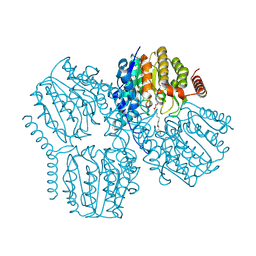 | | Crystal Structure of Deacetylase (HdaH) from Klebsiella pneumoniae subsp. ozaenae in complex with the inhibitor SAHA | | 分子名称: | Deacetylase, OCTANEDIOIC ACID HYDROXYAMIDE PHENYLAMIDE, POTASSIUM ION, ... | | 著者 | Qin, C, Graf, L.G, Schulze, S, Palm, G.J, Lammers, M. | | 登録日 | 2024-08-30 | | 公開日 | 2024-11-06 | | 実験手法 | X-RAY DIFFRACTION (1.95 Å) | | 主引用文献 | Distribution and diversity of classical deacylases in bacteria
Nature Communications, 2024
|
|
9GN1
 
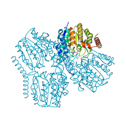 | | Crystal structure of inactive Deacetylase (HdaH) H144A from Klebsiella pneumoniae subsp. ozaenae | | 分子名称: | ACETATE ION, Deacetylase, IMIDAZOLE, ... | | 著者 | Qin, Q, Graf, L.G, Schulze, S, Palm, G.J, Lammers, M. | | 登録日 | 2024-08-30 | | 公開日 | 2024-11-06 | | 実験手法 | X-RAY DIFFRACTION (2.35 Å) | | 主引用文献 | Distribution and diversity of classical deacylases in bacteria
Nature Communications, 2024
|
|
9GLB
 
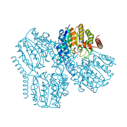 | | Crystal Structure of Deacetylase (HdaH) from Klebsiella pneumoniae subsp. ozaenae | | 分子名称: | ACETATE ION, Deacetylase, GLYCEROL, ... | | 著者 | Qin, C, Graf, L.G, Schulze, S, Palm, G.J, Lammers, M. | | 登録日 | 2024-08-27 | | 公開日 | 2024-11-06 | | 実験手法 | X-RAY DIFFRACTION (2.1 Å) | | 主引用文献 | Distribution and diversity of classical deacylases in bacteria
Nature Communications, 2024
|
|
