2OO9
 
 | |
2OOB
 | |
2OOA
 | |
2QHO
 | |
4GWR
 | |
4IOT
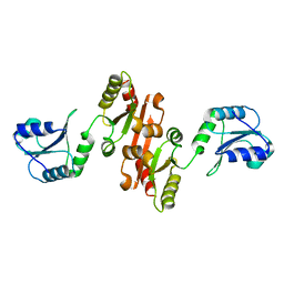
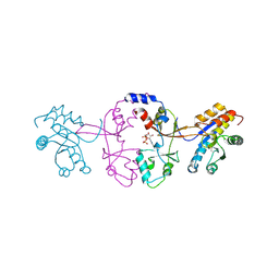
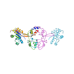
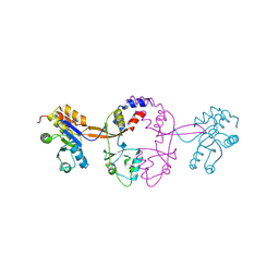
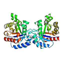 | | High-resolution Structure of Triosephosphate isomerase from E. coli | | 分子名称: | SULFATE ION, Triosephosphate isomerase | | 著者 | Vinaik, R, Kozlov, G, Gehring, K, Montreal-Kingston Bacterial Structural Genomics Initiative (BSGI) | | 登録日 | 2013-01-08 | | 公開日 | 2013-01-23 | | 最終更新日 | 2023-09-20 | | 実験手法 | X-RAY DIFFRACTION (1.85 Å) | | 主引用文献 | Triosephosphate isomerase is a common crystallization contaminant of soluble His-tagged proteins produced in Escherichia coli.
Acta Crystallogr.,Sect.F, 69, 2013
|
|
4I6X
 | |
4JU5
 | |
4JRD
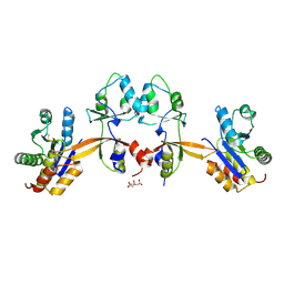
 | | Crystal structure of the parallel double-stranded helix of poly(A) RNA | | 分子名称: | AMMONIUM ION, RNA (5'-R(*AP*AP*AP*AP*AP*AP*AP*AP*AP*AP*A)-3') | | 著者 | Safaee, N, Noronha, A.M, Kozlov, G, Rodionov, D, Wilds, C.J, Sheldrick, G.M, Gehring, K. | | 登録日 | 2013-03-21 | | 公開日 | 2013-06-05 | | 最終更新日 | 2024-02-28 | | 実験手法 | X-RAY DIFFRACTION (1 Å) | | 主引用文献 | Structure of the parallel duplex of poly(A) RNA: evaluation of a 50 year-old prediction.
Angew.Chem.Int.Ed.Engl., 52, 2013
|
|
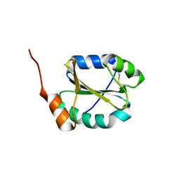
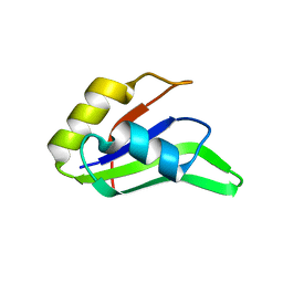
1P9K
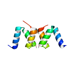
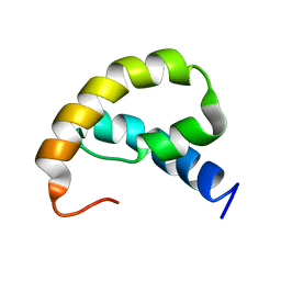
 | | THE SOLUTION STRUCTURE OF YBCJ FROM E. COLI REVEALS A RECENTLY DISCOVERED ALFAL MOTIF INVOLVED IN RNA-BINDING | | 分子名称: | orf, hypothetical protein | | 著者 | Volpon, L, Lievre, C, Osborne, M.J, Gandhi, S, Iannuzzi, P, Larocque, R, Matte, A, Cygler, M, Gehring, K, Ekiel, I, Montreal-Kingston Bacterial Structural Genomics Initiative (BSGI) | | 登録日 | 2003-05-12 | | 公開日 | 2003-11-25 | | 最終更新日 | 2024-05-01 | | 実験手法 | SOLUTION NMR | | 主引用文献 | The solution structure of YbcJ from Escherichia coli reveals a recently discovered alphaL motif involved in RNA binding.
J.Bacteriol., 185, 2003
|
|
1NMR
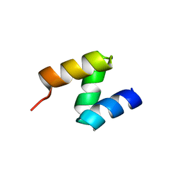
 | | Solution Structure of C-terminal Domain from Trypanosoma cruzi Poly(A)-Binding Protein | | 分子名称: | poly(A)-binding protein | | 著者 | Siddiqui, N, Kozlov, G, D'Orso, I, Trempe, J.F, Frasch, A.C.C, Gehring, K. | | 登録日 | 2003-01-10 | | 公開日 | 2003-09-09 | | 最終更新日 | 2024-05-22 | | 実験手法 | SOLUTION NMR | | 主引用文献 | Solution Structure of the C-terminal Domain from poly(A)-binding protein in Trypanosoma cruzi: A vegetal PABC domain
Protein Sci., 12, 2003
|
|
5VSZ
 | |
5VSX
 | |
1RRZ
 | |
5VMD
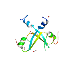 | | Crystal structure of UBR-box from UBR6 in a domain-swapping conformation | | 分子名称: | 1,2-ETHANEDIOL, F-box only protein 11, ZINC ION | | 著者 | Munoz-Escobar, J, Kozlov, G, Gehring, K. | | 登録日 | 2017-04-27 | | 公開日 | 2017-07-12 | | 最終更新日 | 2024-03-13 | | 実験手法 | X-RAY DIFFRACTION (2.202 Å) | | 主引用文献 | Crystal structure of the UBR-box from UBR6/FBXO11 reveals domain swapping mediated by zinc binding.
Protein Sci., 26, 2017
|
|
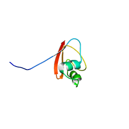
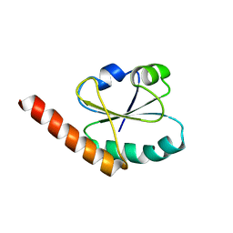
1R6H
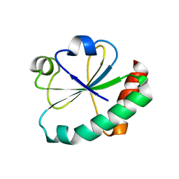
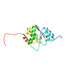 | | Solution Structure of human PRL-3 | | 分子名称: | protein tyrosine phosphatase type IVA, member 3 isoform 1 | | 著者 | Kozlov, G, Gehring, K, Ekiel, I. | | 登録日 | 2003-10-15 | | 公開日 | 2004-01-13 | | 最終更新日 | 2024-05-22 | | 実験手法 | SOLUTION NMR | | 主引用文献 | Structural Insights into Molecular Function of the Metastasis-associated Phosphatase PRL-3.
J.Biol.Chem., 279, 2004
|
|
1RWU
 | |
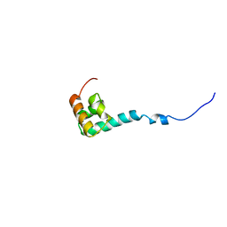
1JH4
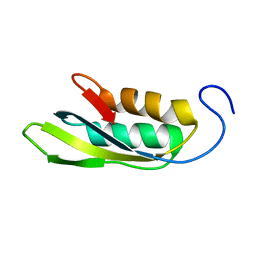
 | | Solution structure of the C-terminal PABC domain of human poly(A)-binding protein in complex with the peptide from Paip1 | | 分子名称: | polyadenylate-binding protein 1, polyadenylate-binding protein-interacting protein-1 | | 著者 | Kozlov, G, Siddiqui, N, Coillet-Matillon, S, Ekiel, I, Gehring, K. | | 登録日 | 2001-06-27 | | 公開日 | 2003-06-24 | | 最終更新日 | 2024-05-22 | | 実験手法 | SOLUTION NMR | | 主引用文献 | Structural basis of ligand recognition by PABC, a highly specific peptide-binding domain found in poly(A)-binding protein and a HECT ubiquitin ligase
EMBO J., 23, 2004
|
|
5K25
 | |
5K22
 | |
5K23
 | |
1JGN
 | | Solution structure of the C-terminal PABC domain of human poly(A)-binding protein in complex with the peptide from Paip2 | | 分子名称: | polyadenylate-binding protein 1, polyadenylate-binding protein-interacting protein 2 | | 著者 | Kozlov, G, Siddiqui, N, Coillet-Matillon, S, Ekiel, I, Gehring, K. | | 登録日 | 2001-06-26 | | 公開日 | 2003-06-24 | | 最終更新日 | 2024-05-22 | | 実験手法 | SOLUTION NMR | | 主引用文献 | Structural basis of ligand recognition by PABC, a highly specific peptide-binding domain found in poly(A)-binding protein and a HECT ubiquitin ligase
EMBO J., 23, 2004
|
|
5K24
 | |
4EF0
 | |
4F26
 | |


