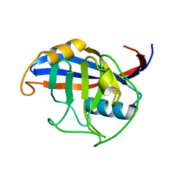
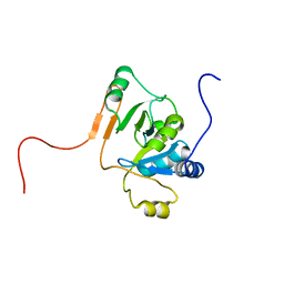
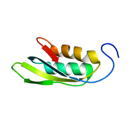
5VRQ
 
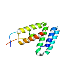
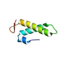
 | | Crystal structure of Legionella pneumophila effector AnkC | | 分子名称: | Ankyrin repeat-containing protein | | 著者 | Kozlov, G, Wong, K, Wang, W, Skubak, P, Munoz-Escobar, J, Liu, Y, Pannu, N.S, Gehring, K, Montreal-Kingston Bacterial Structural Genomics Initiative (BSGI) | | 登録日 | 2017-05-11 | | 公開日 | 2017-11-29 | | 最終更新日 | 2024-03-13 | | 実験手法 | X-RAY DIFFRACTION (3.205 Å) | | 主引用文献 | Ankyrin repeats as a dimerization module.
Biochem. Biophys. Res. Commun., 495, 2018
|
|
5V8Z
 | |
5V90
 | |
1R6H
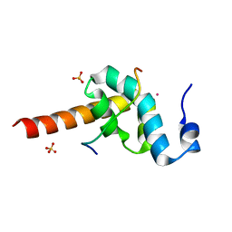
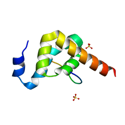
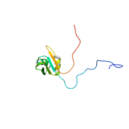
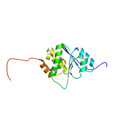 | | Solution Structure of human PRL-3 | | 分子名称: | protein tyrosine phosphatase type IVA, member 3 isoform 1 | | 著者 | Kozlov, G, Gehring, K, Ekiel, I. | | 登録日 | 2003-10-15 | | 公開日 | 2004-01-13 | | 最終更新日 | 2024-05-22 | | 実験手法 | SOLUTION NMR | | 主引用文献 | Structural Insights into Molecular Function of the Metastasis-associated Phosphatase PRL-3.
J.Biol.Chem., 279, 2004
|
|
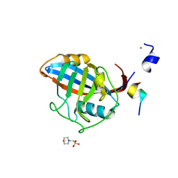
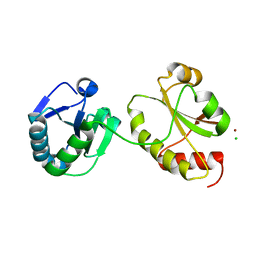
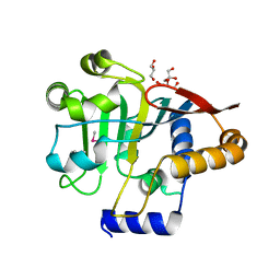
5TDC
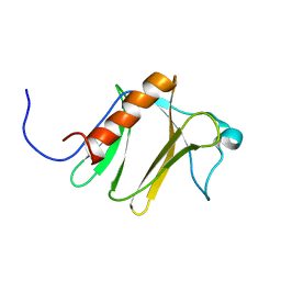
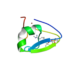 | | Crystal structure of the human UBR-box domain from UBR1 in complex with monomethylated arginine peptide. | | 分子名称: | E3 ubiquitin-protein ligase UBR1, NMM-ILE-PHE-SER peptide, SULFATE ION, ... | | 著者 | Kozlov, G, Munoz-Escobar, J, Matta-Camacho, E, Gehring, K. | | 登録日 | 2016-09-19 | | 公開日 | 2017-03-22 | | 最終更新日 | 2023-10-04 | | 実験手法 | X-RAY DIFFRACTION (1.607 Å) | | 主引用文献 | Bound Waters Mediate Binding of Diverse Substrates to a Ubiquitin Ligase.
Structure, 25, 2017
|
|
3ICH
 | |
3ICI
 | |
3KTR
 | |
3KTP
 | |
1JCU
 | |
3KUJ
 | |
3KUS
 | |
3KUI
 | |
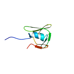
1JE3
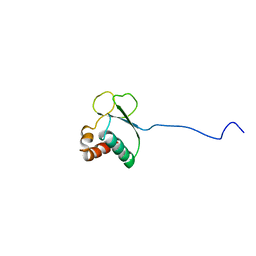
 | | Solution Structure of EC005 from Escherichia coli | | 分子名称: | HYPOTHETICAL 8.6 KDA PROTEIN IN AMYA-FLIE INTERGENIC REGION | | 著者 | Yee, A, Gutierrez, P, Kozlov, G, Denisov, A, Gehring, K, Arrowsmith, C. | | 登録日 | 2001-06-15 | | 公開日 | 2002-03-06 | | 最終更新日 | 2024-05-22 | | 実験手法 | SOLUTION NMR | | 主引用文献 | An NMR approach to structural proteomics.
Proc.Natl.Acad.Sci.USA, 99, 2002
|
|
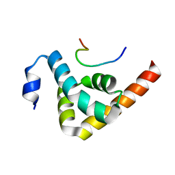
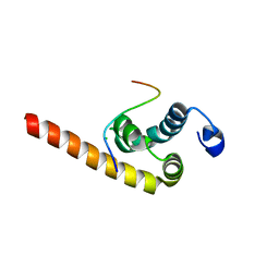
1L1P
 | | Solution Structure of the PPIase Domain from E. coli Trigger Factor | | 分子名称: | trigger factor | | 著者 | Kozlov, G, Trempe, J.-F, Perreault, A, Wong, M, Denisov, A, Ghandi, S, Gehring, K, Ekiel, I, Montreal-Kingston Bacterial Structural Genomics Initiative (BSGI) | | 登録日 | 2002-02-19 | | 公開日 | 2003-06-24 | | 最終更新日 | 2024-05-22 | | 実験手法 | SOLUTION NMR | | 主引用文献 | Solution Structure of the Closed Form of a Peptidyl-Prolyl Isomerase Reveals the Mechanism of Protein Folding
To be Published
|
|
3IDV
 | |
3KUT
 | |
1SG7
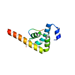
 | | NMR solution structure of the putative cation transport regulator ChaB | | 分子名称: | Putative Cation transport regulator chaB | | 著者 | Osborne, M.J, Siddiqui, N, Cygler, M, Gehring, K, Montreal-Kingston Bacterial Structural Genomics Initiative (BSGI) | | 登録日 | 2004-02-23 | | 公開日 | 2005-03-08 | | 最終更新日 | 2024-05-22 | | 実験手法 | SOLUTION NMR | | 主引用文献 | The solution structure of ChaB, a putative membrane ion antiporter regulator from Escherichia coli
BMC STRUCT.BIOL., 4, 2004
|
|
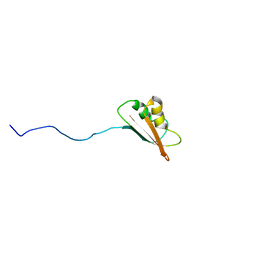
1U6F
 | | NMR solution structure of TcUBP1, a single RBD-unit from Trypanosoma cruzi | | 分子名称: | RNA-binding protein UBP1 | | 著者 | Volpon, L, D'orso, I, Frasch, A, Gehring, K. | | 登録日 | 2004-07-29 | | 公開日 | 2005-01-25 | | 最終更新日 | 2024-05-22 | | 実験手法 | SOLUTION NMR | | 主引用文献 | NMR Structural Study of TcUBP1, a Single RRM Domain Protein from Trypanosoma cruzi: Contribution of a beta Hairpin to RNA Binding
Biochemistry, 44, 2005
|
|
1TQZ
 | |
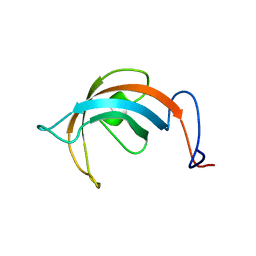
1TVM
 | | NMR structure of enzyme GatB of the galactitol-specific phosphoenolpyruvate-dependent phosphotransferase system | | 分子名称: | PTS system, galactitol-specific IIB component | | 著者 | Volpon, L, Young, C.R, Lim, N.S, Iannuzzi, P, Cygler, M, Gehring, K, Montreal-Kingston Bacterial Structural Genomics Initiative (BSGI) | | 登録日 | 2004-06-29 | | 公開日 | 2005-09-06 | | 最終更新日 | 2024-05-22 | | 実験手法 | SOLUTION NMR | | 主引用文献 | NMR structure of the enzyme GatB of the galactitol-specific phosphoenolpyruvate-dependent phosphotransferase system and its interaction with GatA.
Protein Sci., 15, 2006
|
|
1RWU
 | |
1RYJ
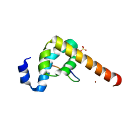
 | | Solution NMR Structure of Protein Mth1743 from Methanobacterium thermoautotrophicum. Ontario Centre for Structural Proteomics target MTH1743_1_70; Northeast Structural Genomics Consortium Target TT526. | | 分子名称: | unknown | | 著者 | Yee, A, Chang, X, Pineda-Lucena, A, Wu, B, Semesi, A, Le, B, Ramelot, T, Lee, G.M, Bhattacharyya, S, Gutierrez, P, Denisov, A, Lee, C.H, Cort, J.R, Kozlov, G, Liao, J, Finak, G, Chen, L, Wishart, D, Lee, W, McIntosh, L.P, Gehring, K, Kennedy, M.A, Edwards, A.M, Arrowsmith, C.H, Northeast Structural Genomics Consortium (NESG) | | 登録日 | 2003-12-22 | | 公開日 | 2004-02-24 | | 最終更新日 | 2024-05-22 | | 実験手法 | SOLUTION NMR | | 主引用文献 | AN NMR APPROACH TO STRUCTURAL PROTEOMICS
Proc.Natl.Acad.Sci.USA, 99, 2002
|
|
5V44
 | |
1SG5
 | |


