1Y7X
 
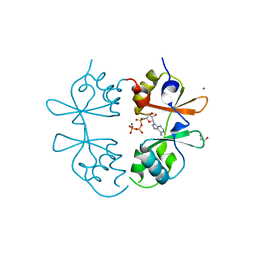
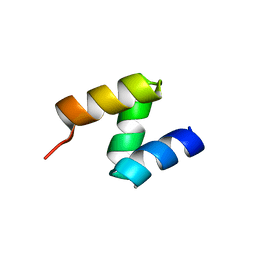
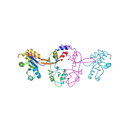
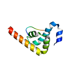
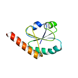
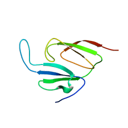 | | Solution structure of a two-repeat fragment of major vault protein | | 分子名称: | Major vault protein | | 著者 | Kozlov, G, Vavelyuk, O, Minailiuc, O, Banville, D, Gehring, K, Ekiel, I. | | 登録日 | 2004-12-10 | | 公開日 | 2005-12-20 | | 最終更新日 | 2024-05-22 | | 実験手法 | SOLUTION NMR | | 主引用文献 | Solution structure of a two-repeat fragment of major vault protein.
J.Mol.Biol., 356, 2006
|
|
1ZY3
 | |
4JRD
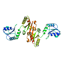
 | | Crystal structure of the parallel double-stranded helix of poly(A) RNA | | 分子名称: | AMMONIUM ION, RNA (5'-R(*AP*AP*AP*AP*AP*AP*AP*AP*AP*AP*A)-3') | | 著者 | Safaee, N, Noronha, A.M, Kozlov, G, Rodionov, D, Wilds, C.J, Sheldrick, G.M, Gehring, K. | | 登録日 | 2013-03-21 | | 公開日 | 2013-06-05 | | 最終更新日 | 2024-02-28 | | 実験手法 | X-RAY DIFFRACTION (1 Å) | | 主引用文献 | Structure of the parallel duplex of poly(A) RNA: evaluation of a 50 year-old prediction.
Angew.Chem.Int.Ed.Engl., 52, 2013
|
|
3EC3
 | |
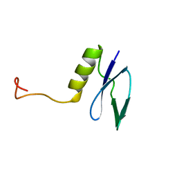
1G9L
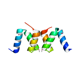
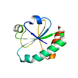
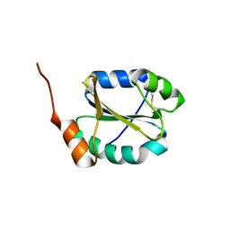
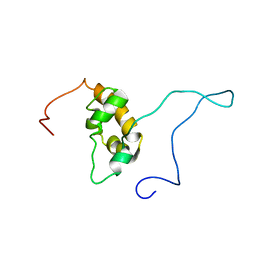 | | SOLUTION STRUCTURE OF THE PABC DOMAIN OF HUMAN POLY(A) BINDING PROTEIN | | 分子名称: | POLYADENYLATE-BINDING PROTEIN 1 | | 著者 | Kozlov, G, Trempe, J.-F, Khaleghpour, K, Kahvejian, A, Ekiel, I, Gehring, K. | | 登録日 | 2000-11-24 | | 公開日 | 2001-03-14 | | 最終更新日 | 2024-05-22 | | 実験手法 | SOLUTION NMR | | 主引用文献 | Structure and function of the C-terminal PABC domain of human poly(A)-binding protein.
Proc.Natl.Acad.Sci.USA, 98, 2001
|
|
7MSU
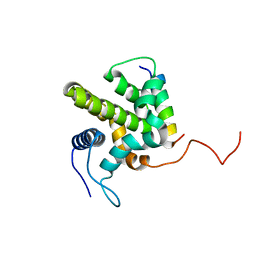
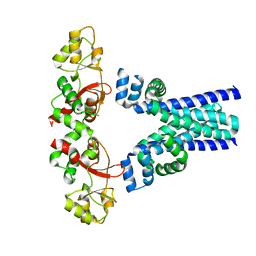
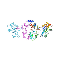
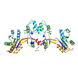
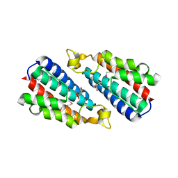
 | | Crystal structure of an archaeal CNNM, MtCorB, CBS-pair domain in complex with Mg2+-ATP | | 分子名称: | 1,2-ETHANEDIOL, ADENOSINE-5'-TRIPHOSPHATE, Hemolysin, ... | | 著者 | Chen, Y.S, Gehring, K. | | 登録日 | 2021-05-12 | | 公開日 | 2021-06-16 | | 最終更新日 | 2023-10-18 | | 実験手法 | X-RAY DIFFRACTION (2.07 Å) | | 主引用文献 | Crystal structure of an archaeal CorB magnesium transporter.
Nat Commun, 12, 2021
|
|
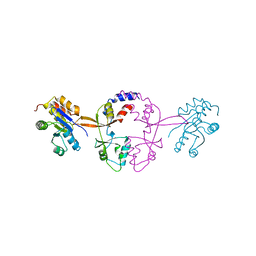
7M1U
 | | Crystal structure of an archaeal CNNM, MtCorB, R235L mutant with C-terminal deletion | | 分子名称: | Hemolysin, contains CBS domains | | 著者 | Chen, Y.S, Kozlov, G, Gehring, K. | | 登録日 | 2021-03-15 | | 公開日 | 2021-06-16 | | 最終更新日 | 2023-10-18 | | 実験手法 | X-RAY DIFFRACTION (3.8 Å) | | 主引用文献 | Crystal structure of an archaeal CorB magnesium transporter.
Nat Commun, 12, 2021
|
|
3GZH
 | |
2OO9
 | |
2OOA
 | |
1GH9
 | |
1JH4
 | | Solution structure of the C-terminal PABC domain of human poly(A)-binding protein in complex with the peptide from Paip1 | | 分子名称: | polyadenylate-binding protein 1, polyadenylate-binding protein-interacting protein-1 | | 著者 | Kozlov, G, Siddiqui, N, Coillet-Matillon, S, Ekiel, I, Gehring, K. | | 登録日 | 2001-06-27 | | 公開日 | 2003-06-24 | | 最終更新日 | 2024-05-22 | | 実験手法 | SOLUTION NMR | | 主引用文献 | Structural basis of ligand recognition by PABC, a highly specific peptide-binding domain found in poly(A)-binding protein and a HECT ubiquitin ligase
EMBO J., 23, 2004
|
|
1JGN
 | | Solution structure of the C-terminal PABC domain of human poly(A)-binding protein in complex with the peptide from Paip2 | | 分子名称: | polyadenylate-binding protein 1, polyadenylate-binding protein-interacting protein 2 | | 著者 | Kozlov, G, Siddiqui, N, Coillet-Matillon, S, Ekiel, I, Gehring, K. | | 登録日 | 2001-06-26 | | 公開日 | 2003-06-24 | | 最終更新日 | 2024-05-22 | | 実験手法 | SOLUTION NMR | | 主引用文献 | Structural basis of ligand recognition by PABC, a highly specific peptide-binding domain found in poly(A)-binding protein and a HECT ubiquitin ligase
EMBO J., 23, 2004
|
|
5K25
 | |
5K22
 | |
5K23
 | |
4GWR
 | |
4IOT
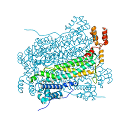
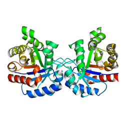 | | High-resolution Structure of Triosephosphate isomerase from E. coli | | 分子名称: | SULFATE ION, Triosephosphate isomerase | | 著者 | Vinaik, R, Kozlov, G, Gehring, K, Montreal-Kingston Bacterial Structural Genomics Initiative (BSGI) | | 登録日 | 2013-01-08 | | 公開日 | 2013-01-23 | | 最終更新日 | 2023-09-20 | | 実験手法 | X-RAY DIFFRACTION (1.85 Å) | | 主引用文献 | Triosephosphate isomerase is a common crystallization contaminant of soluble His-tagged proteins produced in Escherichia coli.
Acta Crystallogr.,Sect.F, 69, 2013
|
|
5K24
 | |
3PKN
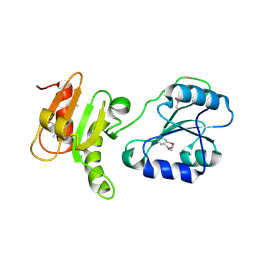
 | | Crystal structure of MLLE domain of poly(A) binding protein in complex with PAM2 motif of La-related protein 4 (LARP4) | | 分子名称: | IODIDE ION, La-related protein 4, Polyadenylate-binding protein 1, ... | | 著者 | Xie, J, Kozlov, G, Gehring, K. | | 登録日 | 2010-11-11 | | 公開日 | 2011-01-12 | | 最終更新日 | 2023-09-06 | | 実験手法 | X-RAY DIFFRACTION (1.8 Å) | | 主引用文献 | La-Related Protein 4 Binds Poly(A), Interacts with the Poly(A)-Binding Protein MLLE Domain via a Variant PAM2w Motif, and Can Promote mRNA Stability.
Mol.Cell.Biol., 31, 2011
|
|
4JU5
 | |
4I6X
 | |
3NY2
 | | Structure of the ubr-box of UBR2 ubiquitin ligase | | 分子名称: | E3 ubiquitin-protein ligase UBR2, ZINC ION | | 著者 | Matta-Camacho, E, Kozlov, G, Li, F, Gehring, K. | | 登録日 | 2010-07-14 | | 公開日 | 2010-08-11 | | 最終更新日 | 2024-02-21 | | 実験手法 | X-RAY DIFFRACTION (2.61 Å) | | 主引用文献 | Structural basis of substrate recognition and specificity in the N-end rule pathway.
Nat.Struct.Mol.Biol., 17, 2010
|
|
3O10
 | |
4EF0
 | |


