6PZQ
 
 | |
8QQK
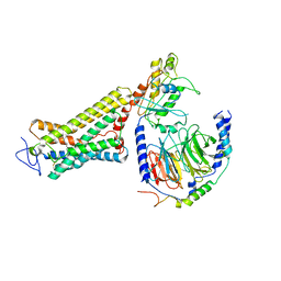
 | | Cryo-EM structure of E. coli cytochrome bo3 quinol oxidase assembled in peptidiscs | | 分子名称: | (2S)-3-(hexadecanoyloxy)-2-[(9Z)-octadec-9-enoyloxy]propyl 2-(trimethylammonio)ethyl phosphate, 1,2-Distearoyl-sn-glycerophosphoethanolamine, COPPER (II) ION, ... | | 著者 | Gao, Y, Zhang, Y, Hakke, S, Peters, P.J, Ravelli, R.B.G. | | 登録日 | 2023-10-05 | | 公開日 | 2024-04-24 | | 最終更新日 | 2024-05-01 | | 実験手法 | ELECTRON MICROSCOPY (2.8 Å) | | 主引用文献 | Cryo-EM structure of cytochrome bo 3 quinol oxidase assembled in peptidiscs reveals an "open" conformation for potential ubiquinone-8 release.
Biochim Biophys Acta Bioenerg, 1865, 2024
|
|
5L9X
 | |
6OYA
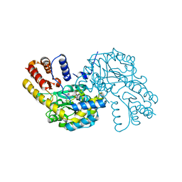
 | | Structure of the Rhodopsin-Transducin-Nanobody Complex | | 分子名称: | Camelid antibody VHH fragment, Gt-alpha/Gi1-alpha chimera, Guanine nucleotide-binding protein G(I)/G(S)/G(T) subunit beta-1, ... | | 著者 | Gao, Y, Hu, H, Ramachandran, S, Erickson, J.W, Cerione, R.A, Skiniotis, G. | | 登録日 | 2019-05-14 | | 公開日 | 2019-07-24 | | 最終更新日 | 2019-12-04 | | 実験手法 | ELECTRON MICROSCOPY (3.3 Å) | | 主引用文献 | Structures of the Rhodopsin-Transducin Complex: Insights into G-Protein Activation.
Mol.Cell, 75, 2019
|
|
6OY9
 | | Structure of the Rhodopsin-Transducin Complex | | 分子名称: | Gt-alpha/Gi1-alpha chimera, Guanine nucleotide-binding protein G(I)/G(S)/G(T) subunit beta-1, Guanine nucleotide-binding protein G(T) subunit gamma-T1, ... | | 著者 | Gao, Y, Hu, H, Ramachandran, S, Erickson, J.W, Cerione, R.A, Skiniotis, G. | | 登録日 | 2019-05-14 | | 公開日 | 2019-07-24 | | 最終更新日 | 2019-12-04 | | 実験手法 | ELECTRON MICROSCOPY (3.9 Å) | | 主引用文献 | Structures of the Rhodopsin-Transducin Complex: Insights into G-Protein Activation.
Mol.Cell, 75, 2019
|
|
1CL4
 | |
8HR7
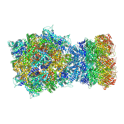 | | Structure of RdrA-RdrB complex | | 分子名称: | Adenosine deaminase, Archaeal ATPase | | 著者 | Gao, Y. | | 登録日 | 2022-12-15 | | 公開日 | 2023-02-01 | | 最終更新日 | 2023-08-16 | | 実験手法 | ELECTRON MICROSCOPY (3.96 Å) | | 主引用文献 | Molecular basis of RADAR anti-phage supramolecular assemblies.
Cell, 186, 2023
|
|
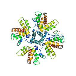
8HR8
 | | Structure of heptameric RdrA ring | | 分子名称: | ADENOSINE-5'-TRIPHOSPHATE, Archaeal ATPase | | 著者 | Gao, Y. | | 登録日 | 2022-12-15 | | 公開日 | 2023-02-01 | | 最終更新日 | 2023-08-16 | | 実験手法 | ELECTRON MICROSCOPY (3.3 Å) | | 主引用文献 | Molecular basis of RADAR anti-phage supramolecular assemblies.
Cell, 186, 2023
|
|
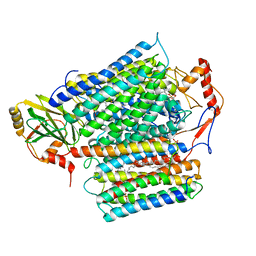
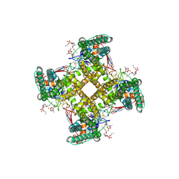
8HRC
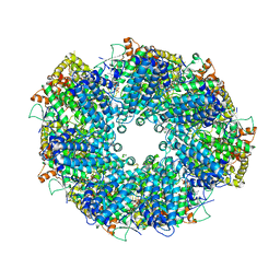
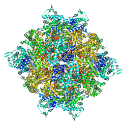 | | Structure of dodecameric RdrB cage | | 分子名称: | Adenosine deaminase | | 著者 | Gao, Y. | | 登録日 | 2022-12-15 | | 公開日 | 2023-02-01 | | 最終更新日 | 2023-08-16 | | 実験手法 | ELECTRON MICROSCOPY (2.58 Å) | | 主引用文献 | Molecular basis of RADAR anti-phage supramolecular assemblies.
Cell, 186, 2023
|
|
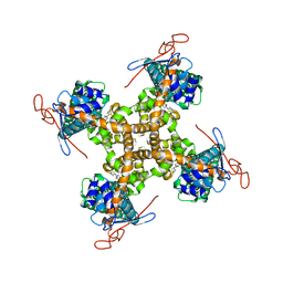
8HRB
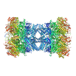 | | Structure of tetradecameric RdrA ring in RNA-loading state | | 分子名称: | ADENOSINE-5'-TRIPHOSPHATE, Archaeal ATPase, RNA (5'-R(*GP*UP*CP*CP*AP*GP*CP*GP*UP*CP*AP*UP*CP*GP*CP*UP*GP*GP*AP*C)-3') | | 著者 | Gao, Y. | | 登録日 | 2022-12-15 | | 公開日 | 2023-02-01 | | 最終更新日 | 2023-08-16 | | 実験手法 | ELECTRON MICROSCOPY (3.78 Å) | | 主引用文献 | Molecular basis of RADAR anti-phage supramolecular assemblies.
Cell, 186, 2023
|
|
8HR9
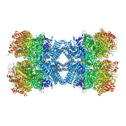 | | Structure of tetradecameric RdrA ring | | 分子名称: | ADENOSINE-5'-TRIPHOSPHATE, Archaeal ATPase | | 著者 | Gao, Y. | | 登録日 | 2022-12-15 | | 公開日 | 2023-02-01 | | 最終更新日 | 2023-08-16 | | 実験手法 | ELECTRON MICROSCOPY (3.03 Å) | | 主引用文献 | Molecular basis of RADAR anti-phage supramolecular assemblies.
Cell, 186, 2023
|
|
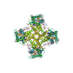
8HRA
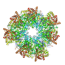 | | Structure of heptameric RdrA ring in RNA-loading state | | 分子名称: | ADENOSINE-5'-TRIPHOSPHATE, Archaeal ATPase, RNA (5'-R(P*GP*UP*CP*CP*AP*GP*CP*GP*UP*CP*AP*UP*CP*GP*CP*UP*GP*GP*AP*C)-3') | | 著者 | Gao, Y. | | 登録日 | 2022-12-15 | | 公開日 | 2023-02-01 | | 最終更新日 | 2023-08-16 | | 実験手法 | ELECTRON MICROSCOPY (3.76 Å) | | 主引用文献 | Molecular basis of RADAR anti-phage supramolecular assemblies.
Cell, 186, 2023
|
|
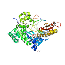
2DU9
 | | crystal structure of the transcriptional factor from C.glutamicum | | 分子名称: | (4S)-2-METHYL-2,4-PENTANEDIOL, Predicted transcriptional regulators | | 著者 | Gao, Y, Yao, M, Tanaka, I. | | 登録日 | 2006-07-20 | | 公開日 | 2007-07-24 | | 最終更新日 | 2011-07-13 | | 実験手法 | X-RAY DIFFRACTION (2.28 Å) | | 主引用文献 | The structures of transcription factor CGL2947 from Corynebacterium glutamicum in two crystal forms: A novel homodimer assembling and the implication for effector-binding mode
Protein Sci., 16, 2007
|
|
8FTI
 | |
2MDJ
 | |
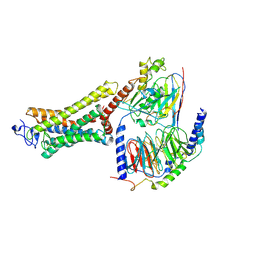
8E2L
 | | Structure of Lates calcarifer Twinkle helicase with ATP and DNA | | 分子名称: | ADENOSINE-5'-TRIPHOSPHATE, DNA (5'-D(P*TP*TP*TP*TP*TP*TP*TP*TP*TP*TP*TP*T)-3'), MAGNESIUM ION, ... | | 著者 | Gao, Y, Li, Z. | | 登録日 | 2022-08-15 | | 公開日 | 2022-11-02 | | 最終更新日 | 2024-06-12 | | 実験手法 | ELECTRON MICROSCOPY (3.51 Å) | | 主引用文献 | Structural and dynamic basis of DNA capture and translocation by mitochondrial Twinkle helicase.
Nucleic Acids Res., 50, 2022
|
|
7M3G
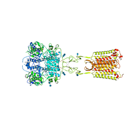 | | Asymmetric Activation of the Calcium Sensing Receptor Homodimer | | 分子名称: | 2-[4-[(3S)-3-[[(1R)-1-naphthalen-1-ylethyl]amino]pyrrolidin-1-yl]phenyl]ethanoic acid, 2-acetamido-2-deoxy-beta-D-glucopyranose, 2-acetamido-2-deoxy-beta-D-glucopyranose-(1-4)-2-acetamido-2-deoxy-beta-D-glucopyranose, ... | | 著者 | Gao, Y, Robertson, M.J, Zhang, C, Meyerowitz, J.G, Panova, O, Skiniotis, G. | | 登録日 | 2021-03-18 | | 公開日 | 2021-06-30 | | 最終更新日 | 2021-07-21 | | 実験手法 | ELECTRON MICROSCOPY (2.5 Å) | | 主引用文献 | Asymmetric activation of the calcium-sensing receptor homodimer.
Nature, 595, 2021
|
|
7M3J
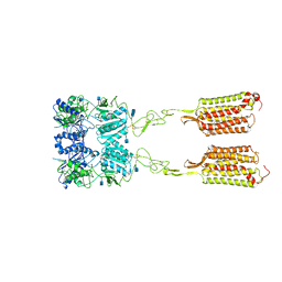 | | Asymmetric Activation of the Calcium Sensing Receptor Homodimer | | 分子名称: | 2-acetamido-2-deoxy-beta-D-glucopyranose, 2-acetamido-2-deoxy-beta-D-glucopyranose-(1-4)-2-acetamido-2-deoxy-beta-D-glucopyranose, 2-chloro-6-[(2R)-2-hydroxy-3-{[2-methyl-1-(naphthalen-2-yl)propan-2-yl]amino}propoxy]benzonitrile, ... | | 著者 | Gao, Y, Robertson, M.J, Zhang, C, Meyerowitz, J.G, Panova, O, Skiniotis, G. | | 登録日 | 2021-03-18 | | 公開日 | 2021-06-30 | | 最終更新日 | 2021-07-21 | | 実験手法 | ELECTRON MICROSCOPY (4.1 Å) | | 主引用文献 | Asymmetric activation of the calcium-sensing receptor homodimer.
Nature, 595, 2021
|
|
7M3E
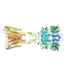 | | Asymmetric Activation of the Calcium Sensing Receptor Homodimer | | 分子名称: | 2-acetamido-2-deoxy-beta-D-glucopyranose, 2-acetamido-2-deoxy-beta-D-glucopyranose-(1-4)-2-acetamido-2-deoxy-beta-D-glucopyranose, 2-chloro-6-[(2R)-2-hydroxy-3-{[2-methyl-1-(naphthalen-2-yl)propan-2-yl]amino}propoxy]benzonitrile, ... | | 著者 | Gao, Y, Robertson, M.J, Zhang, C, Meyerowitz, J.G, Panova, O, Skiniotis, G. | | 登録日 | 2021-03-18 | | 公開日 | 2021-06-30 | | 最終更新日 | 2021-07-21 | | 実験手法 | ELECTRON MICROSCOPY (3.2 Å) | | 主引用文献 | Asymmetric activation of the calcium-sensing receptor homodimer.
Nature, 595, 2021
|
|
7M3F
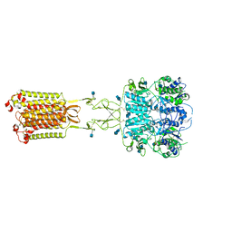 | | Asymmetric Activation of the Calcium Sensing Receptor Homodimer | | 分子名称: | 2-acetamido-2-deoxy-beta-D-glucopyranose, 2-acetamido-2-deoxy-beta-D-glucopyranose-(1-4)-2-acetamido-2-deoxy-beta-D-glucopyranose, CALCIUM ION, ... | | 著者 | Gao, Y, Robertson, M.J, Zhang, C, Meyerowitz, J.G, Panova, O, Skiniotis, G. | | 登録日 | 2021-03-18 | | 公開日 | 2021-06-30 | | 最終更新日 | 2021-07-21 | | 実験手法 | ELECTRON MICROSCOPY (2.8 Å) | | 主引用文献 | Asymmetric activation of the calcium-sensing receptor homodimer.
Nature, 595, 2021
|
|
8WKJ
 | |
5IRX
 | | Structure of TRPV1 in complex with DkTx and RTX, determined in lipid nanodisc | | 分子名称: | (2S)-2-(acetyloxy)-3-{[(R)-(2-aminoethoxy)(hydroxy)phosphoryl]oxy}propyl pentanoate, (2S)-3-{[(S)-(2-aminoethoxy)(hydroxy)phosphoryl]oxy}-2-(hexanoyloxy)propyl hexanoate, (4R,7S)-4-hydroxy-N,N,N-trimethyl-4,9-dioxo-7-[(pentanoyloxy)methyl]-3,5,8-trioxa-4lambda~5~-phosphatetradecan-1-aminium, ... | | 著者 | Gao, Y, Cao, E, Julius, D, Cheng, Y. | | 登録日 | 2016-03-14 | | 公開日 | 2016-05-25 | | 最終更新日 | 2019-12-18 | | 実験手法 | ELECTRON MICROSCOPY (2.95 Å) | | 主引用文献 | TRPV1 structures in nanodiscs reveal mechanisms of ligand and lipid action.
Nature, 534, 2016
|
|
5IS0
 | | Structure of TRPV1 in complex with capsazepine, determined in lipid nanodisc | | 分子名称: | Transient receptor potential cation channel subfamily V member 1, capsazepine | | 著者 | Gao, Y, Cao, E, Julius, D, Cheng, Y. | | 登録日 | 2016-03-15 | | 公開日 | 2016-05-25 | | 最終更新日 | 2024-03-06 | | 実験手法 | ELECTRON MICROSCOPY (3.43 Å) | | 主引用文献 | TRPV1 structures in nanodiscs reveal mechanisms of ligand and lipid action.
Nature, 534, 2016
|
|
5IRZ
 | | Structure of TRPV1 determined in lipid nanodisc | | 分子名称: | (2S)-1-{[(R)-hydroxy{[(1R,2R,3S,4S,5S,6S)-2,3,4,5,6-pentahydroxycyclohexyl]oxy}phosphoryl]oxy}-3-(pentanoyloxy)propan-2-yl decanoate, (2S)-3-{[(S)-(2-aminoethoxy)(hydroxy)phosphoryl]oxy}-2-(hexanoyloxy)propyl hexanoate, (4R,7S)-4-hydroxy-N,N,N-trimethyl-4,9-dioxo-7-[(pentanoyloxy)methyl]-3,5,8-trioxa-4lambda~5~-phosphatetradecan-1-aminium, ... | | 著者 | Gao, Y, Cao, E, Julius, D, Cheng, Y. | | 登録日 | 2016-03-15 | | 公開日 | 2016-05-25 | | 最終更新日 | 2024-03-06 | | 実験手法 | ELECTRON MICROSCOPY (3.28 Å) | | 主引用文献 | TRPV1 structures in nanodiscs reveal mechanisms of ligand and lipid action.
Nature, 534, 2016
|
|
3UBU
 | | Crystal structure of agkisacucetin, a GpIb-binding snaclec (snake C-type lectin) that inhibits platelet | | 分子名称: | Agglucetin subunit alpha-1, Agglucetin subunit beta-2, GLYCEROL, ... | | 著者 | Gao, Y, Ge, H, Chen, H, Li, H, Liu, Y, Niu, L, Teng, M. | | 登録日 | 2011-10-25 | | 公開日 | 2012-04-11 | | 実験手法 | X-RAY DIFFRACTION (1.91 Å) | | 主引用文献 | Crystal structure of agkisacucetin, a Gpib-binding snake C-type lectin that inhibits platelet adhesion and aggregation.
Proteins, 2012
|
|


