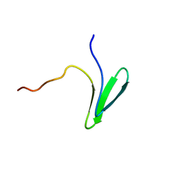
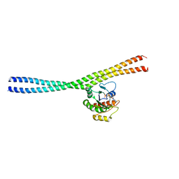
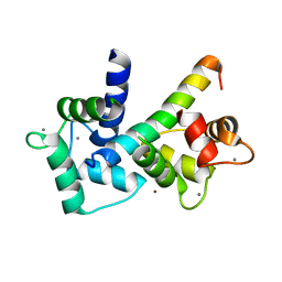
1HQB
 
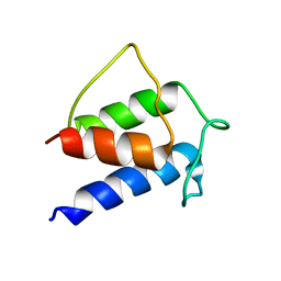 | | TERTIARY STRUCTURE OF APO-D-ALANYL CARRIER PROTEIN | | 分子名称: | APO-D-ALANYL CARRIER PROTEIN | | 著者 | Volkman, B.F, Zhang, Q, Debabov, D.V, Rivera, E, Kresheck, G, Neuhaus, F.C. | | 登録日 | 2000-12-14 | | 公開日 | 2001-08-01 | | 最終更新日 | 2024-05-22 | | 実験手法 | SOLUTION NMR | | 主引用文献 | Biosynthesis of D-alanyl-lipoteichoic acid: the tertiary structure of apo-D-alanyl carrier protein.
Biochemistry, 40, 2001
|
|
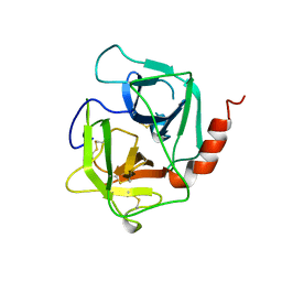
5WKA
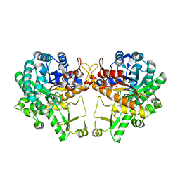 | | Crystal structure of a GH1 beta-glucosidase retrieved from microbial metagenome of Poraque Amazon lake | | 分子名称: | Beta-glucosidase, DI(HYDROXYETHYL)ETHER, GLYCEROL | | 著者 | Morais, M.A.B, Toyama, D, Ramos, F.C, Zanphorlin, L.M, Tonoli, C.C.C, Miranda, F.P, Ruller, R, Henrique-Silva, F, Murakami, M.T. | | 登録日 | 2017-07-24 | | 公開日 | 2018-03-07 | | 最終更新日 | 2023-10-04 | | 実験手法 | X-RAY DIFFRACTION (2.75 Å) | | 主引用文献 | A novel beta-glucosidase isolated from the microbial metagenome of Lake Poraque (Amazon, Brazil).
Biochim. Biophys. Acta, 1866, 2018
|
|
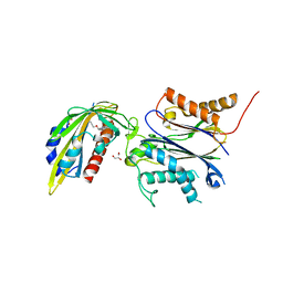
5I7Z
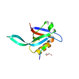 | | Crystal structure of a Par-6 PDZ-Crumbs 3 C-terminal peptide complex | | 分子名称: | Crb-3, DI(HYDROXYETHYL)ETHER, LD29223p | | 著者 | Whitney, D.S, Peterson, F.C, Prehoda, K.E, Volkman, B.F. | | 登録日 | 2016-02-18 | | 公開日 | 2016-03-16 | | 最終更新日 | 2024-03-06 | | 実験手法 | X-RAY DIFFRACTION (1.801 Å) | | 主引用文献 | Binding of Crumbs to the Par-6 CRIB-PDZ Module Is Regulated by Cdc42.
Biochemistry, 55, 2016
|
|
5JNE
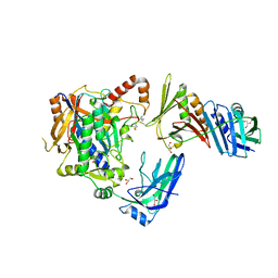 | | E2-SUMO-Siz1 E3-SUMO-PCNA complex | | 分子名称: | E3 SUMO-protein ligase SIZ1,Ubiquitin-like protein SMT3, GLYCEROL, Proliferating cell nuclear antigen, ... | | 著者 | Lima, C.D, Streich Jr, F.C. | | 登録日 | 2016-04-29 | | 公開日 | 2016-08-10 | | 最終更新日 | 2024-10-09 | | 実験手法 | X-RAY DIFFRACTION (2.85 Å) | | 主引用文献 | Capturing a substrate in an activated RING E3/E2-SUMO complex.
Nature, 536, 2016
|
|
7LP4
 | | Structure of Nedd4L WW3 domain | | 分子名称: | E3 ubiquitin-protein ligase NEDD4-like | | 著者 | Alam, S.L, Alian, A, Thompson, T, Rheinemann, L, Volkman, B.F, Peterson, F.C, Sundquist, W.I. | | 登録日 | 2021-02-11 | | 公開日 | 2021-07-28 | | 最終更新日 | 2024-05-15 | | 実験手法 | SOLUTION NMR | | 主引用文献 | Interactions between AMOT PPxY motifs and NEDD4L WW domains function in HIV-1 release.
J.Biol.Chem., 297, 2021
|
|
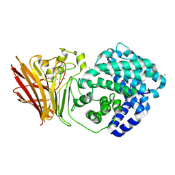
1AE5
 | | HUMAN HEPARIN BINDING PROTEIN | | 分子名称: | 2-acetamido-2-deoxy-beta-D-glucopyranose, HEPARIN BINDING PROTEIN | | 著者 | Iversen, L.F, Kastrup, J.S, Bjorn, S.E, Rasmussen, P.B, Wiberg, F.C, Flodgaard, H.J, Larsen, I.K. | | 登録日 | 1997-03-05 | | 公開日 | 1998-03-11 | | 最終更新日 | 2023-08-02 | | 実験手法 | X-RAY DIFFRACTION (2.3 Å) | | 主引用文献 | Structure of HBP, a multifunctional protein with a serine proteinase fold.
Nat.Struct.Biol., 4, 1997
|
|
8EY0
 | | Structure of an orthogonal PYR1*:HAB1* chemical-induced dimerization module in complex with mandipropamid | | 分子名称: | (2S)-2-(4-chlorophenyl)-N-{2-[3-methoxy-4-(prop-2-yn-1-yloxy)phenyl]ethyl}-2-(prop-2-yn-1-yloxy)ethanamide, Abscisic acid receptor PYR1, GLYCEROL, ... | | 著者 | Park, S.-Y, Volkman, B.F, Cutler, S.R, Peterson, F.C. | | 登録日 | 2022-10-26 | | 公開日 | 2023-11-08 | | 最終更新日 | 2024-01-10 | | 実験手法 | X-RAY DIFFRACTION (2.4 Å) | | 主引用文献 | An orthogonalized PYR1-based CID module with reprogrammable ligand-binding specificity.
Nat.Chem.Biol., 20, 2024
|
|
4Z8Y
 | |
4ZDW
 | |
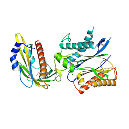
4NDL
 | | Computational design and experimental verification of a symmetric homodimer | | 分子名称: | ENH-c2b, computational designed homodimer | | 著者 | Mou, Y, Huang, P.S, Hsu, F.C, Huang, S.J, Mayo, S.L. | | 登録日 | 2013-10-26 | | 公開日 | 2014-11-05 | | 最終更新日 | 2024-02-28 | | 実験手法 | X-RAY DIFFRACTION (3.5 Å) | | 主引用文献 | Computational design and experimental verification of a symmetric protein homodimer.
Proc.Natl.Acad.Sci.USA, 112, 2015
|
|
8T3N
 | | Solution NMR structure of synthetic peptide AMPCry10Aa_5 rational designed from Cry10Aa bacterial protein | | 分子名称: | Pesticidal crystal protein Cry10Aa peptide | | 著者 | Barra, J.B, Freitas, C.D.P, Rios, T.B, Maximiano, M.R, Fernandes, F.C, Amorim, G.C, Porto, W.F, Grossi-de-Sa, M.F, Franco, O.F, Liao, L.M. | | 登録日 | 2023-06-07 | | 公開日 | 2024-06-12 | | 最終更新日 | 2024-07-24 | | 実験手法 | SOLUTION NMR | | 主引用文献 | Anti-Staphy Peptides Rationally Designed from Cry10Aa Bacterial Protein.
Acs Omega, 9, 2024
|
|
8T3H
 | | Solution NMR structure alpha-helix 3 of Cry10Aa protein | | 分子名称: | Pesticidal crystal protein Cry10Aa peptide | | 著者 | Barra, J.B, Freitas, C.D.P, Rios, T.B, Maximiano, M.R, Fernandes, F.C, Amorim, G.C, Porto, W.F, Grossi-de-Sa, M.F, Franco, O.F, Liao, L.M. | | 登録日 | 2023-06-07 | | 公開日 | 2024-06-12 | | 最終更新日 | 2024-10-30 | | 実験手法 | SOLUTION NMR | | 主引用文献 | Alpha-helix 3 of Cry10Aa protein
To Be Published
|
|
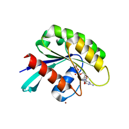
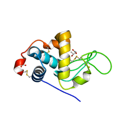
2BE6
 | | 2.0 A crystal structure of the CaV1.2 IQ domain-Ca/CaM complex | | 分子名称: | CALCIUM ION, Calmodulin 2, NICKEL (II) ION, ... | | 著者 | Van Petegem, F, Chatelain, F.C, Minor Jr, D.L. | | 登録日 | 2005-10-23 | | 公開日 | 2005-11-15 | | 最終更新日 | 2024-05-22 | | 実験手法 | X-RAY DIFFRACTION (2 Å) | | 主引用文献 | Insights into voltage-gated calcium channel regulation from the structure of the Ca(V)1.2 IQ domain-Ca(2+)/calmodulin complex
Nat.Struct.Mol.Biol., 12, 2005
|
|
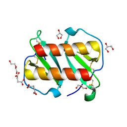
4WWC
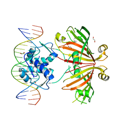 | | Crystal structure of full length YvoA in complex with palindromic operator DNA | | 分子名称: | 1,2-ETHANEDIOL, DNA (5'-D(P*CP*AP*GP*TP*GP*GP*TP*CP*TP*AP*GP*AP*CP*CP*AP*CP*TP*GP*G)-3'), HTH-type transcriptional repressor YvoA | | 著者 | Grau, F.C, Fillenberg, S.B, Muller, Y.A. | | 登録日 | 2014-11-10 | | 公開日 | 2015-01-14 | | 最終更新日 | 2024-01-10 | | 実験手法 | X-RAY DIFFRACTION (2.903 Å) | | 主引用文献 | Structural insight into operator dre-sites recognition and effector binding in the GntR/HutC transcription regulator NagR.
Nucleic Acids Res., 43, 2015
|
|
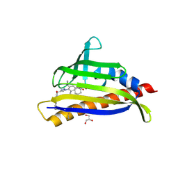
4WVO
 | | An engineered PYR1 mandipropamid receptor in complex with mandipropamid and HAB1 | | 分子名称: | (2S)-2-(4-chlorophenyl)-N-{2-[3-methoxy-4-(prop-2-yn-1-yloxy)phenyl]ethyl}-2-(prop-2-yn-1-yloxy)ethanamide, Abscisic acid receptor PYR1, MAGNESIUM ION, ... | | 著者 | Peterson, F.C, Volkman, B.F, Cutler, S.R. | | 登録日 | 2014-11-06 | | 公開日 | 2015-02-11 | | 最終更新日 | 2023-10-18 | | 実験手法 | X-RAY DIFFRACTION (2.251 Å) | | 主引用文献 | Agrochemical control of plant water use using engineered abscisic acid receptors.
Nature, 520, 2015
|
|
5KTF
 | | Structure of the C-terminal transmembrane domain of scavenger receptor BI (SR-BI) | | 分子名称: | Scavenger receptor class B member 1 | | 著者 | Chadwick, A.C, Peterson, F.C, Volkman, B.F, Sahoo, D. | | 登録日 | 2016-07-11 | | 公開日 | 2017-03-08 | | 最終更新日 | 2024-05-15 | | 実験手法 | SOLUTION NMR | | 主引用文献 | NMR Structure of the C-Terminal Transmembrane Domain of the HDL Receptor, SR-BI, and a Functionally Relevant Leucine Zipper Motif.
Structure, 25, 2017
|
|
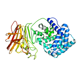
3CB7
 | | The crystallographic structure of the digestive lysozyme 2 from Musca domestica at 1.9 Ang. | | 分子名称: | ACETIC ACID, GLYCEROL, ISOPROPYL ALCOHOL, ... | | 著者 | Cancado, F.C, Valerio, A.A, Marana, S.R, Barbosa, J.A.R.G. | | 登録日 | 2008-02-21 | | 公開日 | 2009-02-24 | | 最終更新日 | 2024-10-16 | | 実験手法 | X-RAY DIFFRACTION (1.9 Å) | | 主引用文献 | The crystallographic structure of the digestive lysozyme 2 from Musca domestica at 1.9 Ang.
To be Published
|
|
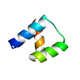
1DV5
 | | TERTIARY STRUCTURE OF APO-D-ALANYL CARRIER PROTEIN | | 分子名称: | APO-D-ALANYL CARRIER PROTEIN | | 著者 | Volkman, B.F, Zhang, Q, Debabov, D.V, Rivera, E, Kresheck, G.C, Neuhaus, F.C. | | 登録日 | 2000-01-19 | | 公開日 | 2001-08-01 | | 最終更新日 | 2024-04-10 | | 実験手法 | SOLUTION NMR | | 主引用文献 | Biosynthesis of D-alanyl-lipoteichoic acid: the tertiary structure of apo-D-alanyl carrier protein.
Biochemistry, 40, 2001
|
|
8KHV
 | |
8KHW
 | |
7T1E
 | | Structure of monomeric and dimeric human CCL20 | | 分子名称: | C-C motif chemokine 20, DI(HYDROXYETHYL)ETHER, GLYCEROL, ... | | 著者 | Peterson, F.C, Riutta, S.J, Volkman, B.F. | | 登録日 | 2021-12-01 | | 公開日 | 2022-12-14 | | 最終更新日 | 2024-10-16 | | 実験手法 | X-RAY DIFFRACTION (1.459 Å) | | 主引用文献 | The Chemokine, CCL20, and Its Receptor, CCR6, in the Pathogenesis and Treatment of Psoriasis and Psoriatic Arthritis
J Psoriasis Psoriatic Arthritis, 8, 2023
|
|
7MLC
 | | PYL10 bound to the ABA pan-antagonist 4a | | 分子名称: | 1-{2-[3,5-dicyclopropyl-4-(4-{[(quinoxaline-2-carbonyl)amino]methyl}-1H-1,2,3-triazol-1-yl)phenyl]acetamido}cyclohexane-1-carboxylic acid, Abscisic acid receptor PYL10, GLYCEROL | | 著者 | Peterson, F.C, Vaidya, A.S, Volkman, B.F, Cutler, S.R. | | 登録日 | 2021-04-28 | | 公開日 | 2021-09-29 | | 最終更新日 | 2023-10-18 | | 実験手法 | X-RAY DIFFRACTION (1.77 Å) | | 主引用文献 | Click-to-lead design of a picomolar ABA receptor antagonist with potent activity in vivo.
Proc.Natl.Acad.Sci.USA, 118, 2021
|
|
7MLD
 | | PYL10 bound to the ABA pan-antagonist antabactin | | 分子名称: | Abscisic acid receptor PYL10, CITRIC ACID, antabactin | | 著者 | Peterson, F.C, Vaidya, A.S, Volkman, B.F, Cutler, S.R. | | 登録日 | 2021-04-28 | | 公開日 | 2021-09-29 | | 最終更新日 | 2023-10-18 | | 実験手法 | X-RAY DIFFRACTION (1.8 Å) | | 主引用文献 | Click-to-lead design of a picomolar ABA receptor antagonist with potent activity in vivo.
Proc.Natl.Acad.Sci.USA, 118, 2021
|
|
7MWN
 | | An engineered PYL2-based WIN 55,212-2 synthetic cannabinoid sensor with a stabilized HAB1 variant | | 分子名称: | ACETATE ION, Abscisic acid receptor PYL2, CHLORIDE ION, ... | | 著者 | Peterson, F.C, Beltran, J, Bedewitz, M, Steiner, P.J, Cutler, S.R, Whitehead, T.A. | | 登録日 | 2021-05-17 | | 公開日 | 2022-06-01 | | 最終更新日 | 2023-10-25 | | 実験手法 | X-RAY DIFFRACTION (1.902 Å) | | 主引用文献 | Rapid biosensor development using plant hormone receptors as reprogrammable scaffolds.
Nat.Biotechnol., 40, 2022
|
|
2ADC
 | | Solution structure of Polypyrimidine Tract Binding protein RBD34 complexed with CUCUCU RNA | | 分子名称: | 5'-R(*CP*UP*CP*UP*CP*U)-3', Polypyrimidine tract-binding protein 1 | | 著者 | Oberstrass, F.C, Auweter, S.D, Erat, M, Hargous, Y, Henning, A, Wenter, P, Reymond, L, Pitsch, S, Black, D.L, Allain, F.H.T. | | 登録日 | 2005-07-20 | | 公開日 | 2005-10-04 | | 最終更新日 | 2024-05-29 | | 実験手法 | SOLUTION NMR | | 主引用文献 | Structure of PTB bound to RNA: specific binding and implications for splicing regulation
Science, 309, 2005
|
|


