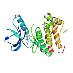
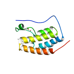
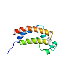
6E8V
 
 | |
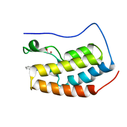
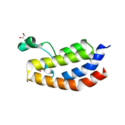
6E9H
 | |
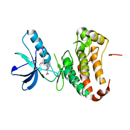
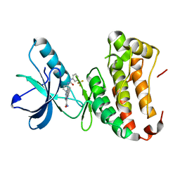
2AEM
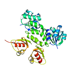
 | | Crystal Structures of the MthK RCK Domain | | 分子名称: | Calcium-gated potassium channel mthK | | 著者 | Dong, J, Shi, N, Berke, I, Chen, L, Jiang, Y. | | 登録日 | 2005-07-22 | | 公開日 | 2005-10-25 | | 最終更新日 | 2023-08-23 | | 実験手法 | X-RAY DIFFRACTION (2.8 Å) | | 主引用文献 | Structures of the MthK RCK Domain and the Effect of Ca2+ on Gating Ring Stability
J.Biol.Chem., 280, 2005
|
|
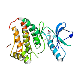
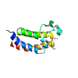
2AEJ
 | | Crystal Structures of the MthK RCK Domain in no Ca2+ bound form | | 分子名称: | Calcium-gated potassium channel mthK | | 著者 | Dong, J, Shi, N, Berke, I, Chen, L, Jiang, Y. | | 登録日 | 2005-07-22 | | 公開日 | 2005-10-25 | | 最終更新日 | 2023-08-23 | | 実験手法 | X-RAY DIFFRACTION (2.1 Å) | | 主引用文献 | Structures of the MthK RCK Domain and the Effect of Ca2+ on Gating Ring Stability
J.Biol.Chem., 280, 2005
|
|
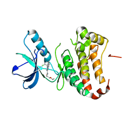
2AEF
 | | Crystal Structures of the MthK RCK Domain in Ca2+ bound form | | 分子名称: | CALCIUM ION, Calcium-gated potassium channel mthK | | 著者 | Dong, J, Shi, N, Berke, I, Chen, L, Jiang, Y. | | 登録日 | 2005-07-22 | | 公開日 | 2005-10-25 | | 最終更新日 | 2023-08-23 | | 実験手法 | X-RAY DIFFRACTION (1.7 Å) | | 主引用文献 | Structures of the MthK RCK Domain and the Effect of Ca2+ on Gating Ring Stability
J.Biol.Chem., 280, 2005
|
|
4GF6
 | | crystal structure of GFP with cuprum bound at the Incorporated metal Chelating Amino Acid PYZ151 | | 分子名称: | CALCIUM ION, COPPER (II) ION, green fluorescent protein | | 著者 | Dong, J, Liu, X, Li, J, Wang, J, Gong, W. | | 登録日 | 2012-08-03 | | 公開日 | 2012-08-29 | | 最終更新日 | 2023-11-15 | | 実験手法 | X-RAY DIFFRACTION (1.1 Å) | | 主引用文献 | Genetic incorporation of a metal-chelating amino Acid as a probe for protein electron transfer.
Angew.Chem.Int.Ed.Engl., 51, 2012
|
|
4GES
 | | crystal structure of GFP-TYR151PYZ with an unnatural amino acid incorporation | | 分子名称: | Green fluorescent protein | | 著者 | Dong, J, Liu, X, Li, J, Wang, J, Gong, W. | | 登録日 | 2012-08-02 | | 公開日 | 2012-08-29 | | 最終更新日 | 2023-11-15 | | 実験手法 | X-RAY DIFFRACTION (1.23 Å) | | 主引用文献 | Genetic incorporation of a metal-chelating amino Acid as a probe for protein electron transfer.
Angew.Chem.Int.Ed.Engl., 51, 2012
|
|
5OBO
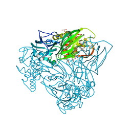 | | Crystal structure of nitrite bound D97N mutant of three-domain heme-Cu nitrite reductase from Ralstonia pickettii | | 分子名称: | COPPER (II) ION, GLYCEROL, HEME C, ... | | 著者 | Dong, J, Sasaki, D, Eady, R, Antonyuk, S.V, Hasnain, S.S. | | 登録日 | 2017-06-28 | | 公開日 | 2018-08-01 | | 最終更新日 | 2024-01-17 | | 実験手法 | X-RAY DIFFRACTION (1.89 Å) | | 主引用文献 | Activation of redox tyrosine switch is required for ligand binding at the catalytic site in heme-cu nitrite reductases
To be published
|
|
5OCB
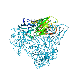 | | Crystal structure of nitric oxide bound D97N mutant of three-domain heme-Cu nitrite reductase from Ralstonia pickettii | | 分子名称: | COPPER (II) ION, HEME C, NITRIC OXIDE, ... | | 著者 | Dong, J, Sasaki, D, Eady, R, Antonyuk, S.V, Hasnain, S.S. | | 登録日 | 2017-06-30 | | 公開日 | 2018-06-27 | | 最終更新日 | 2024-01-17 | | 実験手法 | X-RAY DIFFRACTION (1.78 Å) | | 主引用文献 | Activation of redox tyrosine switch is required for ligand binding at the catalytic site in heme-cu nitrite reductases
To be published
|
|
5OCF
 | | Crystal structure of nitric oxide bound to three-domain heme-Cu nitrite reductase from Ralstonia pickettii | | 分子名称: | COPPER (II) ION, HEME C, NITRIC OXIDE, ... | | 著者 | Dong, J, Sasaki, D, Eady, R, Antonyuk, S.V, Hasnain, S.S. | | 登録日 | 2017-06-30 | | 公開日 | 2018-06-27 | | 最終更新日 | 2024-01-17 | | 実験手法 | X-RAY DIFFRACTION (1.8 Å) | | 主引用文献 | Activation of redox tyrosine switch is required for ligand binding at the catalytic site in heme-cu nitrite reductases
To be published
|
|
5ZM6
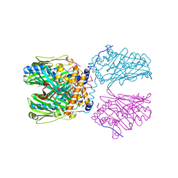 | | Crystal structure of ORP1-ORD in complex with PI(4,5)P2 | | 分子名称: | ACETATE ION, Oxysterol-binding protein-related protein 1, [(2~{S})-1-octadecanoyloxy-3-[oxidanyl-[(1~{R},2~{R},3~{S},4~{S},5~{S},6~{S})-2,3,6-tris(oxidanyl)-4,5-diphosphonooxy-cyclohexyl]oxy-phosphoryl]oxy-propan-2-yl] icosa-5,8,11,14-tetraenoate | | 著者 | Dong, J, Wang, J, Luo, Z, Wu, J.W. | | 登録日 | 2018-04-01 | | 公開日 | 2019-02-27 | | 最終更新日 | 2023-11-22 | | 実験手法 | X-RAY DIFFRACTION (2.7 Å) | | 主引用文献 | Allosteric enhancement of ORP1-mediated cholesterol transport by PI(4,5)P2/PI(3,4)P2.
Nat Commun, 10, 2019
|
|
5ZM7
 | | Crystal structure of ORP1-ORD in complex with cholesterol at 3.4 A resolution | | 分子名称: | CHOLESTEROL, Oxysterol-binding protein-related protein 1 | | 著者 | Dong, J, Wang, J, Wu, J.W. | | 登録日 | 2018-04-01 | | 公開日 | 2019-02-27 | | 最終更新日 | 2023-11-22 | | 実験手法 | X-RAY DIFFRACTION (3.401 Å) | | 主引用文献 | Allosteric enhancement of ORP1-mediated cholesterol transport by PI(4,5)P2/PI(3,4)P2.
Nat Commun, 10, 2019
|
|
5ZM5
 | | Crystal structure of human ORP1-ORD in complex with cholesterol at 2.6 A resolution | | 分子名称: | CHOLESTEROL, Oxysterol-binding protein-related protein 1 | | 著者 | Dong, J, Wang, J, Wu, J.W. | | 登録日 | 2018-04-01 | | 公開日 | 2019-02-27 | | 最終更新日 | 2023-11-22 | | 実験手法 | X-RAY DIFFRACTION (2.6 Å) | | 主引用文献 | Allosteric enhancement of ORP1-mediated cholesterol transport by PI(4,5)P2/PI(3,4)P2.
Nat Commun, 10, 2019
|
|
4G2F
 | | Human EphA3 kinase domain in complex with compound 7 | | 分子名称: | 1-amino-5-(5-hydroxy-2-methylphenyl)-7,8,9,10-tetrahydropyrimido[4,5-c]isoquinolin-6(5H)-one, EPH receptor A3 | | 著者 | Dong, J, Caflisch, A. | | 登録日 | 2012-07-12 | | 公開日 | 2012-10-24 | | 最終更新日 | 2023-11-08 | | 実験手法 | X-RAY DIFFRACTION (1.699 Å) | | 主引用文献 | Discovery of a novel chemotype of tyrosine kinase inhibitors by fragment-based docking and molecular dynamics.
ACS MED.CHEM.LETT., 3, 2012
|
|
4GK3
 | | Human EphA3 Kinase domain in complex with ligand 87 | | 分子名称: | 8-butyl-1-methyl-7-(2-methylphenyl)-1H-imidazo[2,1-f]purine-2,4(3H,8H)-dione, EPH receptor A3 | | 著者 | Dong, J, Caflisch, A. | | 登録日 | 2012-08-10 | | 公開日 | 2013-01-23 | | 最終更新日 | 2023-11-08 | | 実験手法 | X-RAY DIFFRACTION (1.898 Å) | | 主引用文献 | Optimization of Inhibitors of the Tyrosine Kinase EphB4. 2. Cellular Potency Improvement and Binding Mode Validation by X-ray Crystallography.
J.Med.Chem., 56, 2013
|
|
4GK2
 | | Human EphA3 Kinase domain in complex with ligand 66 | | 分子名称: | 7-(5-hydroxy-2-methylphenyl)-8-(2-methoxyphenyl)-1-methyl-1H-imidazo[2,1-f]purine-2,4(3H,8H)-dione, EPH receptor A3 | | 著者 | Dong, J, Caflisch, A. | | 登録日 | 2012-08-10 | | 公開日 | 2013-01-23 | | 最終更新日 | 2023-11-08 | | 実験手法 | X-RAY DIFFRACTION (2.195 Å) | | 主引用文献 | Optimization of Inhibitors of the Tyrosine Kinase EphB4. 2. Cellular Potency Improvement and Binding Mode Validation by X-ray Crystallography.
J.Med.Chem., 56, 2013
|
|
4GK4
 | | Human EphA3 Kinase domain in complex with ligand 90 | | 分子名称: | 8-butyl-1-methyl-7-(5-methyl-1H-indazol-4-yl)-1H-imidazo[2,1-f]purine-2,4(3H,8H)-dione, EPH receptor A3 | | 著者 | Dong, J, Caflisch, A. | | 登録日 | 2012-08-10 | | 公開日 | 2013-01-23 | | 最終更新日 | 2023-11-08 | | 実験手法 | X-RAY DIFFRACTION (2.1 Å) | | 主引用文献 | Optimization of Inhibitors of the Tyrosine Kinase EphB4. 2. Cellular Potency Improvement and Binding Mode Validation by X-ray Crystallography.
J.Med.Chem., 56, 2013
|
|
4P5Q
 | | Human EphA3 Kinase domain in complex with quinoxaline derivatives | | 分子名称: | 2-amino-1-(2-chlorophenyl)-N-(3-ethoxypropyl)-1H-pyrrolo[2,3-b]quinoxaline-3-carboxamide, Ephrin type-A receptor 3 | | 著者 | Dong, J, Caflisch, A. | | 登録日 | 2014-03-19 | | 公開日 | 2014-08-13 | | 最終更新日 | 2023-12-20 | | 実験手法 | X-RAY DIFFRACTION (1.35 Å) | | 主引用文献 | Pyrrolo[3,2-b]quinoxaline Derivatives as Types I1/2 and II Eph Tyrosine Kinase Inhibitors: Structure-Based Design, Synthesis, and in Vivo Validation.
J.Med.Chem., 57, 2014
|
|
4P4C
 | | Human EphA3 Kinase domain in complex with quinoxaline derivatives | | 分子名称: | 2-amino-1-(3-methoxyphenyl)-1H-pyrrolo[2,3-b]quinoxaline-3-carboxamide, EPH receptor A3 | | 著者 | Dong, J, Caflisch, A. | | 登録日 | 2014-03-12 | | 公開日 | 2014-08-13 | | 最終更新日 | 2023-09-27 | | 実験手法 | X-RAY DIFFRACTION (1.599 Å) | | 主引用文献 | Pyrrolo[3,2-b]quinoxaline Derivatives as Types I1/2 and II Eph Tyrosine Kinase Inhibitors: Structure-Based Design, Synthesis, and in Vivo Validation.
J.Med.Chem., 57, 2014
|
|
4PCE
 | | Crystal Structure of the first bromodomain of human BRD4 in complex with compound B13 | | 分子名称: | 1,2-ETHANEDIOL, 1-benzyl-2-ethyl-1,5,6,7-tetrahydro-4H-indol-4-one, Bromodomain-containing protein 4 | | 著者 | Dong, J, Caflisch, A. | | 登録日 | 2014-04-15 | | 公開日 | 2014-05-07 | | 最終更新日 | 2023-12-27 | | 実験手法 | X-RAY DIFFRACTION (1.293 Å) | | 主引用文献 | Discovery of BRD4 bromodomain inhibitors by fragment-based high-throughput docking.
Bioorg.Med.Chem.Lett., 24, 2014
|
|
4PCI
 | | Crystal Structure of the first bromodomain of BRD4 in complex with B16 | | 分子名称: | (4S)-1-methyl-4-phenyl-1,3,4,5-tetrahydro-2H-1,5-benzodiazepin-2-one, 1,2-ETHANEDIOL, Bromodomain-containing protein 4 | | 著者 | Dong, J, Caflisch, A. | | 登録日 | 2014-04-15 | | 公開日 | 2014-05-14 | | 最終更新日 | 2023-09-27 | | 実験手法 | X-RAY DIFFRACTION (1.25 Å) | | 主引用文献 | Discovery of BRD4 bromodomain inhibitors by fragment-based high-throughput docking.
Bioorg.Med.Chem.Lett., 24, 2014
|
|
4P5Z
 | | Human EphA3 Kinase domain in complex with quinoxaline derivatives | | 分子名称: | 2-amino-1-[4-({[3-(trifluoromethyl)phenyl]carbamoyl}amino)phenyl]-1H-pyrrolo[2,3-b]quinoxaline-3-carboxamide, Ephrin type-A receptor 3 | | 著者 | Dong, J, Caflisch, A. | | 登録日 | 2014-03-20 | | 公開日 | 2014-08-13 | | 最終更新日 | 2023-12-20 | | 実験手法 | X-RAY DIFFRACTION (2.002 Å) | | 主引用文献 | Pyrrolo[3,2-b]quinoxaline Derivatives as Types I1/2 and II Eph Tyrosine Kinase Inhibitors: Structure-Based Design, Synthesis, and in Vivo Validation.
J.Med.Chem., 57, 2014
|
|
5ENG
 | |
5EP7
 | |
5EIC
 | |


