6JPF
 
 | |
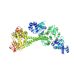
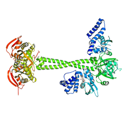
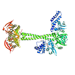
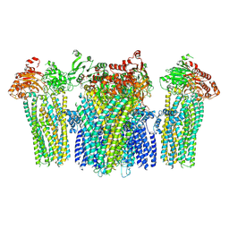
6JB3
 | | Structure of SUR1 subunit bound with repaglinide | | 分子名称: | ATP-binding cassette sub-family C member 8 isoform X2, Digitonin, Repaglinide | | 著者 | Chen, L, Ding, D, Wang, M, Wu, J.-X, Kang, Y. | | 登録日 | 2019-01-25 | | 公開日 | 2019-05-22 | | 最終更新日 | 2024-05-29 | | 実験手法 | ELECTRON MICROSCOPY (3.53 Å) | | 主引用文献 | The Structural Basis for the Binding of Repaglinide to the Pancreatic KATPChannel.
Cell Rep, 27, 2019
|
|
6JT2
 | | Structure of human soluble guanylate cyclase in the NO activated state | | 分子名称: | Guanylate cyclase soluble subunit alpha-1, Guanylate cyclase soluble subunit beta-1, MAGNESIUM ION, ... | | 著者 | Chen, L, Kang, Y, Liu, R, Wu, J.-X. | | 登録日 | 2019-04-08 | | 公開日 | 2019-09-04 | | 最終更新日 | 2024-03-27 | | 実験手法 | ELECTRON MICROSCOPY (3.8 Å) | | 主引用文献 | Structural insights into the mechanism of human soluble guanylate cyclase.
Nature, 574, 2019
|
|
6JT0
 | | Structure of human soluble guanylate cyclase in the unliganded state | | 分子名称: | Guanylate cyclase soluble subunit alpha-1, Guanylate cyclase soluble subunit beta-1, PROTOPORPHYRIN IX CONTAINING FE | | 著者 | Chen, L, Kang, Y, Liu, R, Wu, J.-X. | | 登録日 | 2019-04-08 | | 公開日 | 2019-08-28 | | 最終更新日 | 2024-03-27 | | 実験手法 | ELECTRON MICROSCOPY (4 Å) | | 主引用文献 | Structural insights into the mechanism of human soluble guanylate cyclase.
Nature, 574, 2019
|
|
6JT1
 | | Structure of human soluble guanylate cyclase in the heme oxidised state | | 分子名称: | Guanylate cyclase soluble subunit alpha-1, Guanylate cyclase soluble subunit beta-1, PROTOPORPHYRIN IX CONTAINING FE | | 著者 | Chen, L, Kang, Y, Liu, R, Wu, J.-X. | | 登録日 | 2019-04-08 | | 公開日 | 2019-08-28 | | 最終更新日 | 2024-03-27 | | 実験手法 | ELECTRON MICROSCOPY (3.9 Å) | | 主引用文献 | Structural insights into the mechanism of human soluble guanylate cyclase.
Nature, 574, 2019
|
|
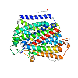
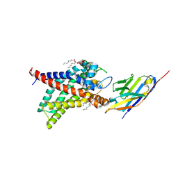
6L72
 | | Sirtuin 2 demyristoylation native final product | | 分子名称: | NAD-dependent protein deacetylase sirtuin-2, ZINC ION, [(2S,3R,4R,5R)-5-[[[[(2R,3S,4R,5R)-5-(6-aminopurin-9-yl)-3,4-bis(oxidanyl)oxolan-2-yl]methoxy-oxidanyl-phosphoryl]oxy-oxidanyl-phosphoryl]oxymethyl]-2,4-bis(oxidanyl)oxolan-3-yl] tetradecanoate | | 著者 | Chen, L.F. | | 登録日 | 2019-10-30 | | 公開日 | 2021-03-31 | | 最終更新日 | 2023-11-22 | | 実験手法 | X-RAY DIFFRACTION (2.501 Å) | | 主引用文献 | Sirtuin 2 protein with H3K18 myristoylated peptide
To Be Published
|
|
6L71
 | |
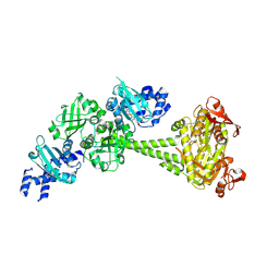
7WIG
 | | Cryo-EM structure of the L-054,264-bound human SSTR2-Gi1 complex | | 分子名称: | Guanine nucleotide-binding protein G(I)/G(S)/G(O) subunit gamma-2, Guanine nucleotide-binding protein G(I)/G(S)/G(T) subunit beta-1, Guanine nucleotide-binding protein G(i) subunit alpha-1, ... | | 著者 | Chen, L, Wang, W, Dong, Y, Shen, D, Guo, J, Qin, J, Zhang, H, Shen, Q, Zhang, Y, Mao, C. | | 登録日 | 2022-01-03 | | 公開日 | 2022-06-01 | | 最終更新日 | 2022-08-17 | | 実験手法 | ELECTRON MICROSCOPY (2.7 Å) | | 主引用文献 | Structures of the endogenous peptide- and selective non-peptide agonist-bound SSTR2 signaling complexes.
Cell Res., 32, 2022
|
|
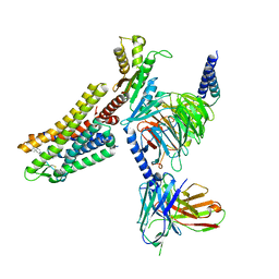
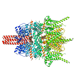
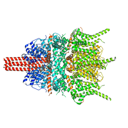
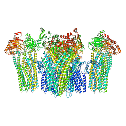
7VSI
 | | Structure of human SGLT2-MAP17 complex bound with empagliflozin | | 分子名称: | (2S,3R,4R,5S,6R)-2-[4-chloranyl-3-[[4-[(3S)-oxolan-3-yl]oxyphenyl]methyl]phenyl]-6-(hydroxymethyl)oxane-3,4,5-triol, PALMITIC ACID, PDZK1-interacting protein 1, ... | | 著者 | Chen, L, Niu, Y, Liu, R. | | 登録日 | 2021-10-26 | | 公開日 | 2021-12-15 | | 最終更新日 | 2022-02-16 | | 実験手法 | ELECTRON MICROSCOPY (2.95 Å) | | 主引用文献 | Structural basis of inhibition of the human SGLT2-MAP17 glucose transporter.
Nature, 601, 2022
|
|
5ZBG
 | |
5YX9
 | |
8HBE
 | |
8HBH
 | |
8HBF
 | |
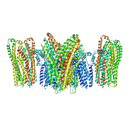
8HBW
 | | Structure of human UCP1 in the ATP-bound state | | 分子名称: | 1,2-DIACYL-SN-GLYCERO-3-PHOSPHOCHOLINE, ADENOSINE-5'-TRIPHOSPHATE, CARDIOLIPIN, ... | | 著者 | Chen, L, Kang, Y. | | 登録日 | 2022-10-31 | | 公開日 | 2023-06-21 | | 最終更新日 | 2023-08-30 | | 実験手法 | ELECTRON MICROSCOPY (2.57 Å) | | 主引用文献 | Structural basis for the binding of DNP and purine nucleotides onto UCP1.
Nature, 620, 2023
|
|
8HBV
 | | Structure of human UCP1 in the nucleotide-free state | | 分子名称: | 1,2-DIACYL-SN-GLYCERO-3-PHOSPHOCHOLINE, CARDIOLIPIN, Mitochondrial brown fat uncoupling protein 1, ... | | 著者 | Chen, L, Kang, Y. | | 登録日 | 2022-10-31 | | 公開日 | 2023-06-21 | | 最終更新日 | 2023-08-30 | | 実験手法 | ELECTRON MICROSCOPY (2.51 Å) | | 主引用文献 | Structural basis for the binding of DNP and purine nucleotides onto UCP1.
Nature, 620, 2023
|
|
5YKF
 | |
5YKG
 | |
5YKE
 | |
8J1N
 | | Structure of human UCP1 in the DNP-bound state | | 分子名称: | 1,2-DIACYL-SN-GLYCERO-3-PHOSPHOCHOLINE, 2,4-DINITROPHENOL, CARDIOLIPIN, ... | | 著者 | Chen, L, Kang, Y. | | 登録日 | 2023-04-13 | | 公開日 | 2023-06-21 | | 最終更新日 | 2023-08-30 | | 実験手法 | ELECTRON MICROSCOPY (2.51 Å) | | 主引用文献 | Structural basis for the binding of DNP and purine nucleotides onto UCP1.
Nature, 620, 2023
|
|
5YWC
 | |
5YW9
 | |
5YWB
 | |
5YW8
 | |
5YWA
 | |


