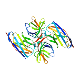
8I6C
 
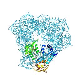
 | | Crystal structure of Mycobacterium tuberculosis Uracil-DNA glycosylase in complex with 6-Formyl-uracil, Form III | | 分子名称: | 6-[bis(oxidanyl)methyl]-5~{H}-pyrimidine-2,4-dione, Uracil-DNA glycosylase | | 著者 | Raj, P, Paul, A, Gopal, B. | | 登録日 | 2023-01-27 | | 公開日 | 2023-07-12 | | 最終更新日 | 2024-05-08 | | 実験手法 | X-RAY DIFFRACTION (2.28 Å) | | 主引用文献 | Crystal structures of non-uracil ring fragments in complex with Mycobacterium tuberculosis uracil DNA glycosylase (MtUng) as a starting point for novel inhibitor design: A case study with the barbituric acid fragment.
Eur.J.Med.Chem., 258, 2023
|
|
7OCJ
 | | Crystal structure of E.coli LexA in complex with nanobody NbSOS2(Nb14509) | | 分子名称: | 1,2-ETHANEDIOL, LexA repressor, NbSOS2 (14509) | | 著者 | Maso, L, Vascon, F, Chinellato, M, Pardon, E, Steyaert, J, Angelini, A, Tondi, D, Cendron, L. | | 登録日 | 2021-04-27 | | 公開日 | 2022-10-26 | | 最終更新日 | 2024-01-31 | | 実験手法 | X-RAY DIFFRACTION (2.7 Å) | | 主引用文献 | Nanobodies targeting LexA autocleavage disclose a novel suppression strategy of SOS-response pathway.
Structure, 30, 2022
|
|
8DBW
 | |
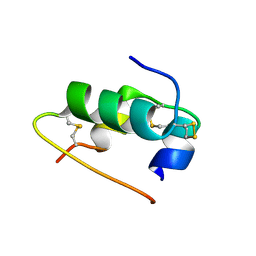
7Q2U
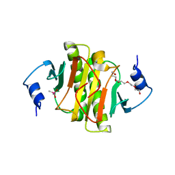 | | The crystal structure of the HINT1 Q62A mutant. | | 分子名称: | CACODYLATE ION, HEXAETHYLENE GLYCOL, Histidine triad nucleotide-binding protein 1, ... | | 著者 | Dolot, R.M, Strom, A.M, Wagner, C.R. | | 登録日 | 2021-10-26 | | 公開日 | 2021-11-03 | | 最終更新日 | 2024-02-07 | | 実験手法 | X-RAY DIFFRACTION (2.27 Å) | | 主引用文献 | Dynamic Long-Range Interactions Influence Substrate Binding and Catalysis by Human Histidine Triad Nucleotide-Binding Proteins (HINTs), Key Regulators of Multiple Cellular Processes and Activators of Antiviral ProTides.
Biochemistry, 61, 2022
|
|
5LUI
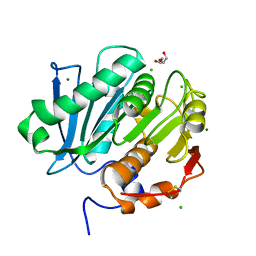 | | Structure of cutinase 1 from Thermobifida cellulosilytica | | 分子名称: | CHLORIDE ION, Cutinase 1, DI(HYDROXYETHYL)ETHER, ... | | 著者 | Hromic, A, Lyskowski, A, Gruber, K. | | 登録日 | 2016-09-08 | | 公開日 | 2017-07-12 | | 最終更新日 | 2024-01-17 | | 実験手法 | X-RAY DIFFRACTION (1.5 Å) | | 主引用文献 | Small cause, large effect: Structural characterization of cutinases from Thermobifida cellulosilytica.
Biotechnol. Bioeng., 114, 2017
|
|
6VUT
 | | Crystal structure of Eis from Mycobacterium tuberculosis in complex with inhibitor SGT392 | | 分子名称: | 2-[(4-amino-5,6,7,8-tetrahydro[1]benzothieno[2,3-d]pyrimidin-2-yl)sulfanyl]-N-[2-(morpholin-4-yl)ethyl]acetamide, DI(HYDROXYETHYL)ETHER, GLYCEROL, ... | | 著者 | Punetha, A, Hou, C, Ngo, H.X, Garneau-Tsodikova, S, Tsodikov, O.V. | | 登録日 | 2020-02-16 | | 公開日 | 2020-06-03 | | 最終更新日 | 2023-10-11 | | 実験手法 | X-RAY DIFFRACTION (2.73 Å) | | 主引用文献 | Structure-Guided Optimization of Inhibitors of Acetyltransferase Eis fromMycobacterium tuberculosis.
Acs Chem.Biol., 15, 2020
|
|
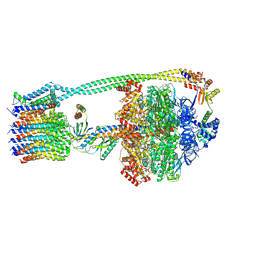
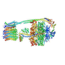
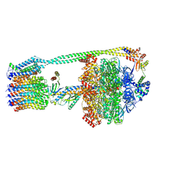
6ZNL
 | | Cryo-EM structure of the dynactin complex | | 分子名称: | ADENOSINE-5'-DIPHOSPHATE, ADENOSINE-5'-TRIPHOSPHATE, ARP1 actin related protein 1 homolog A, ... | | 著者 | Lau, C.K, Lacey, S.E, Carter, A.P. | | 登録日 | 2020-07-06 | | 公開日 | 2020-07-29 | | 最終更新日 | 2024-05-01 | | 実験手法 | ELECTRON MICROSCOPY (3.8 Å) | | 主引用文献 | Cryo-EM reveals the complex architecture of dynactin's shoulder region and pointed end.
Embo J., 40, 2021
|
|
6VVD
 | |
6NOP
 | | Structure of Cyanothece McdA(D38A)-ATP complex | | 分子名称: | ADENOSINE-5'-TRIPHOSPHATE, Cobyrinic acid ac-diamide synthase, MAGNESIUM ION | | 著者 | Schumacher, M.A. | | 登録日 | 2019-01-16 | | 公開日 | 2019-04-24 | | 最終更新日 | 2023-10-11 | | 実験手法 | X-RAY DIFFRACTION (1.7 Å) | | 主引用文献 | Structures of maintenance of carboxysome distribution Walker-box McdA and McdB adaptor homologs.
Nucleic Acids Res., 47, 2019
|
|
8I64
 | | Crystal structure of Mycobacterium tuberculosis Uracil-DNA glycosylase in complex with Barbituric acid, Form II | | 分子名称: | 1,2-ETHANEDIOL, BARBITURIC ACID, Uracil-DNA glycosylase | | 著者 | Raj, P, Paul, A, Gopal, B. | | 登録日 | 2023-01-27 | | 公開日 | 2023-07-12 | | 最終更新日 | 2024-05-08 | | 実験手法 | X-RAY DIFFRACTION (2.26 Å) | | 主引用文献 | Crystal structures of non-uracil ring fragments in complex with Mycobacterium tuberculosis uracil DNA glycosylase (MtUng) as a starting point for novel inhibitor design: A case study with the barbituric acid fragment.
Eur.J.Med.Chem., 258, 2023
|
|
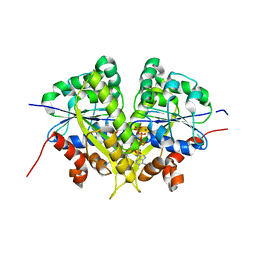
8I68
 | | Crystal structure of Mycobacterium tuberculosis Uracil-DNA glycosylase in complex with Uric acid, Form III | | 分子名称: | 1,2-ETHANEDIOL, URIC ACID, Uracil-DNA glycosylase | | 著者 | Raj, P, Paul, A, Gopal, B. | | 登録日 | 2023-01-27 | | 公開日 | 2023-07-12 | | 最終更新日 | 2024-05-08 | | 実験手法 | X-RAY DIFFRACTION (1.88 Å) | | 主引用文献 | Crystal structures of non-uracil ring fragments in complex with Mycobacterium tuberculosis uracil DNA glycosylase (MtUng) as a starting point for novel inhibitor design: A case study with the barbituric acid fragment.
Eur.J.Med.Chem., 258, 2023
|
|
6QNR
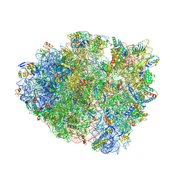 | | 70S ribosome elongation complex (EC) with experimentally assigned potassium ions | | 分子名称: | 16S ribosomal RNA, 23S ribosomal RNA, 30S ribosomal protein S10, ... | | 著者 | Rozov, A, Khusainov, I, Yusupov, M, Yusupova, G. | | 登録日 | 2019-02-11 | | 公開日 | 2019-06-19 | | 最終更新日 | 2024-04-24 | | 実験手法 | X-RAY DIFFRACTION (3.1 Å) | | 主引用文献 | Importance of potassium ions for ribosome structure and function revealed by long-wavelength X-ray diffraction.
Nat Commun, 10, 2019
|
|
8DBU
 | |
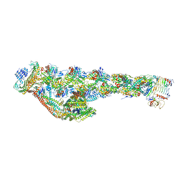
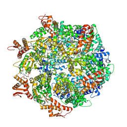
6QBX
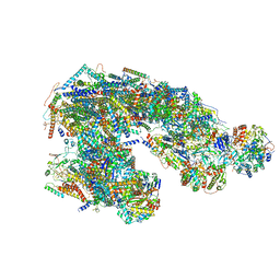 | | Ovine respiratory supercomplex I+III2 closed class. | | 分子名称: | 1,2-DIACYL-SN-GLYCERO-3-PHOSPHOCHOLINE, 1,2-DIACYL-SN-GLYCERO-3-PHOSPHOETHANOLAMINE, Acyl carrier protein, ... | | 著者 | Letts, J.A, Sazanov, L.A. | | 登録日 | 2018-12-24 | | 公開日 | 2019-08-21 | | 最終更新日 | 2019-12-18 | | 実験手法 | ELECTRON MICROSCOPY (4.2 Å) | | 主引用文献 | Structures of Respiratory Supercomplex I+III2Reveal Functional and Conformational Crosstalk.
Mol.Cell, 75, 2019
|
|
7PGT
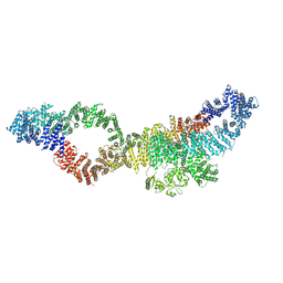
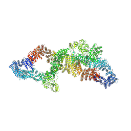 | | The structure of human neurofibromin isoform 2 in opened conformation. | | 分子名称: | Neurofibromin, ZINC ION | | 著者 | Naschberger, A, Baradaran, R, Carroni, M, Rupp, B. | | 登録日 | 2021-08-15 | | 公開日 | 2021-11-17 | | 最終更新日 | 2024-07-17 | | 実験手法 | ELECTRON MICROSCOPY (4.8 Å) | | 主引用文献 | The structure of neurofibromin isoform 2 reveals different functional states.
Nature, 599, 2021
|
|
7PGS
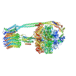
 | | Consensus structure of human Neurofibromin isoform 2 | | 分子名称: | Neurofibromin, ZINC ION | | 著者 | Naschberger, A, Baradaran, R, Carroni, M, Rupp, B. | | 登録日 | 2021-08-15 | | 公開日 | 2021-11-17 | | 最終更新日 | 2024-07-17 | | 実験手法 | ELECTRON MICROSCOPY (3.4 Å) | | 主引用文献 | The structure of neurofibromin isoform 2 reveals different functional states.
Nature, 599, 2021
|
|
8DBQ
 | |
7PGR
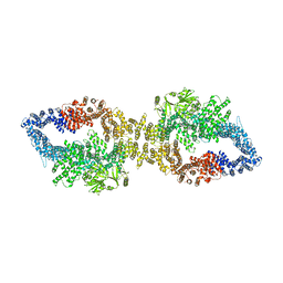 | | The structure of human neurofibromin isoform 2 in closed conformation | | 分子名称: | Neurofibromin, ZINC ION | | 著者 | Naschberger, A, Baradaran, R, Carroni, M, Rupp, B. | | 登録日 | 2021-08-15 | | 公開日 | 2021-11-17 | | 最終更新日 | 2024-07-17 | | 実験手法 | ELECTRON MICROSCOPY (4 Å) | | 主引用文献 | The structure of neurofibromin isoform 2 reveals different functional states.
Nature, 599, 2021
|
|
7PGU
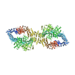 | | Autoinhibited structure of human neurofibromin isoform 2 stabilized by Zinc. | | 分子名称: | (1S)-2-{[(2-AMINOETHOXY)(HYDROXY)PHOSPHORYL]OXY}-1-[(PALMITOYLOXY)METHYL]ETHYL STEARATE, Neurofibromin, ZINC ION | | 著者 | Naschberger, A, Baradaran, R, Carroni, M, Rupp, B. | | 登録日 | 2021-08-15 | | 公開日 | 2021-11-17 | | 最終更新日 | 2024-07-17 | | 実験手法 | ELECTRON MICROSCOPY (3.3 Å) | | 主引用文献 | The structure of neurofibromin isoform 2 reveals different functional states.
Nature, 599, 2021
|
|
6W24
 | | ClpA Engaged2 State bound to RepA-GFP (Focused Classification) | | 分子名称: | ADENOSINE-5'-DIPHOSPHATE, ADENOSINE-5'-TRIPHOSPHATE, ATP-dependent Clp protease ATP-binding subunit ClpA, ... | | 著者 | Lopez, K.L, Rizo, A.N, Tse, E, Lin, J, Scull, N.W, Thwin, A.C, Lucius, A.L, Shorter, J, Southworth, D.R. | | 登録日 | 2020-03-04 | | 公開日 | 2020-05-06 | | 最終更新日 | 2024-03-06 | | 実験手法 | ELECTRON MICROSCOPY (3.4 Å) | | 主引用文献 | Conformational plasticity of the ClpAP AAA+ protease couples protein unfolding and proteolysis.
Nat.Struct.Mol.Biol., 27, 2020
|
|
8DBV
 | |
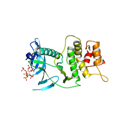
6PTA
 | | Crystal structure of the ARF family small GTPase ARF1 from Candida albicans in complex with GDP | | 分子名称: | ADP-ribosylation factor, GUANOSINE-5'-DIPHOSPHATE | | 著者 | Stogios, P.J, Evdokimova, E, Di Leo, R, Savchenko, A, Joachimiak, A, Satchell, K.J.F, Center for Structural Genomics of Infectious Diseases (CSGID) | | 登録日 | 2019-07-15 | | 公開日 | 2019-07-24 | | 最終更新日 | 2023-10-11 | | 実験手法 | X-RAY DIFFRACTION (2.5 Å) | | 主引用文献 | Crystal structure of the ARF family small GTPase ARF1 from Candida albicans in complex with GDP
To Be Published
|
|
6YJX
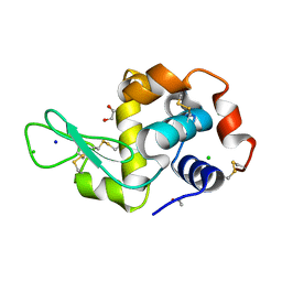 | | Structure of Hen egg-white lysozyme crystallized with PAS polypeptide | | 分子名称: | 1,2-ETHANEDIOL, ACETATE ION, CHLORIDE ION, ... | | 著者 | Schiefner, A, Skerra, A. | | 登録日 | 2020-04-05 | | 公開日 | 2020-07-22 | | 最終更新日 | 2024-01-24 | | 実験手法 | X-RAY DIFFRACTION (1.2 Å) | | 主引用文献 | Proline/alanine-rich sequence (PAS) polypeptides as an alternative to PEG precipitants for protein crystallization.
Acta Crystallogr.,Sect.F, 76, 2020
|
|
5LIS
 | |
6Y84
 | | SARS-CoV-2 main protease with unliganded active site (2019-nCoV, coronavirus disease 2019, COVID-19) | | 分子名称: | 3C-like proteinase nsp5, DIMETHYL SULFOXIDE | | 著者 | Owen, C.D, Lukacik, P, Strain-Damerell, C.M, Douangamath, A, Powell, A.J, Fearon, D, Brandao-Neto, J, Crawshaw, A.D, Aragao, D, Williams, M, Flaig, R, Hall, D.R, McAuley, K.E, Mazzorana, M, Stuart, D.I, von Delft, F, Walsh, M.A. | | 登録日 | 2020-03-03 | | 公開日 | 2020-03-11 | | 最終更新日 | 2024-02-07 | | 実験手法 | X-RAY DIFFRACTION (1.39 Å) | | 主引用文献 | COVID-19 main protease with unliganded active site
To Be Published
|
|


