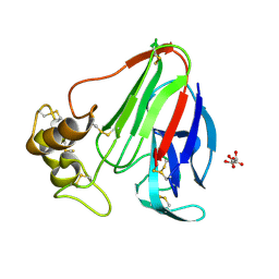
6C1H
 
 | | High-Resolution Cryo-EM Structures of Actin-bound Myosin States Reveal the Mechanism of Myosin Force Sensing | | 分子名称: | ADENOSINE-5'-DIPHOSPHATE, Actin, alpha skeletal muscle, ... | | 著者 | Mentes, A, Huehn, A, Liu, X, Zwolak, A, Dominguez, R, Shuman, H, Ostap, E.M, Sindelar, C.V. | | 登録日 | 2018-01-04 | | 公開日 | 2018-01-31 | | 最終更新日 | 2024-03-13 | | 実験手法 | ELECTRON MICROSCOPY (3.9 Å) | | 主引用文献 | High-resolution cryo-EM structures of actin-bound myosin states reveal the mechanism of myosin force sensing.
Proc. Natl. Acad. Sci. U.S.A., 115, 2018
|
|
4YPZ
 | |
7QQY
 | |
8DH7
 | |
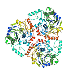
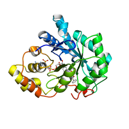
6C4A
 | | Crystal structure of 3-nitropropionate modified isocitrate lyase from Mycobacterium tuberculosis with pyruvate | | 分子名称: | 3-NITROPROPANOIC ACID, 4-(2-HYDROXYETHYL)-1-PIPERAZINE ETHANESULFONIC ACID, DI(HYDROXYETHYL)ETHER, ... | | 著者 | Kreitler, D.F, Ray, S, Murkin, A.S, Gulick, A.M. | | 登録日 | 2018-01-11 | | 公開日 | 2018-06-06 | | 最終更新日 | 2023-11-15 | | 実験手法 | X-RAY DIFFRACTION (1.8 Å) | | 主引用文献 | The Nitro Group as a Masked Electrophile in Covalent Enzyme Inhibition.
ACS Chem. Biol., 13, 2018
|
|
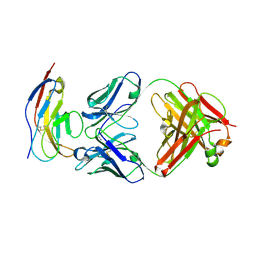
8DGL
 | | Crystal Structure of the RdfS Excisionase | | 分子名称: | CHLORIDE ION, GLYCEROL, Recombination Directionality Factor RdfS | | 著者 | Verdonk, C.J, Ramsay, J.P, Marshall, A.C, Bond, C.S. | | 登録日 | 2022-06-24 | | 公開日 | 2022-10-05 | | 最終更新日 | 2024-04-03 | | 実験手法 | X-RAY DIFFRACTION (2.45 Å) | | 主引用文献 | Crystallographic and X-ray scattering study of RdfS, a recombination directionality factor from an integrative and conjugative element.
Acta Crystallogr D Struct Biol, 78, 2022
|
|
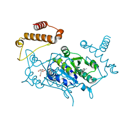
8QN9
 | | OPR3 variant - R283E | | 分子名称: | (4S)-2-METHYL-2,4-PENTANEDIOL, 12-oxophytodienoate reductase 3, FLAVIN MONONUCLEOTIDE, ... | | 著者 | Bijelic, A, Macheroux, P, Kerschbaumer, B. | | 登録日 | 2023-09-26 | | 公開日 | 2024-08-14 | | 最終更新日 | 2024-08-21 | | 実験手法 | X-RAY DIFFRACTION (1.6 Å) | | 主引用文献 | Analysis of homodimer formation in 12-oxophytodienoate reductase 3 in solutio and crystallo challenges the physiological role of the dimer.
Sci Rep, 14, 2024
|
|
6HPR
 | |
6HPX
 | | Crystal structure of ENL (MLLT1) in complex with compound 19 | | 分子名称: | 1,2-ETHANEDIOL, Protein ENL, ~{N}-[(3-chlorophenyl)methyl]-1-(2-pyrrolidin-1-ylethyl)benzimidazole-5-carboxamide | | 著者 | Heidenreich, D, Chaikuad, A, Arrowsmith, C.H, Edwards, A.M, Bountra, C, Knapp, S, Structural Genomics Consortium (SGC) | | 登録日 | 2018-09-22 | | 公開日 | 2018-11-28 | | 最終更新日 | 2024-01-24 | | 実験手法 | X-RAY DIFFRACTION (2.3 Å) | | 主引用文献 | Structure-Based Approach toward Identification of Inhibitory Fragments for Eleven-Nineteen-Leukemia Protein (ENL).
J.Med.Chem., 61, 2018
|
|
6BIA
 | | Crystal structure of Ps i-CgsB | | 分子名称: | 1,2-ETHANEDIOL, CALCIUM ION, CITRIC ACID, ... | | 著者 | Hettle, A.G, Boraston, A.B. | | 登録日 | 2017-11-01 | | 公開日 | 2018-03-14 | | 最終更新日 | 2023-10-04 | | 実験手法 | X-RAY DIFFRACTION (2.8 Å) | | 主引用文献 | The Molecular Basis of Polysaccharide Sulfatase Activity and a Nomenclature for Catalytic Subsites in this Class of Enzyme.
Structure, 26, 2018
|
|
6HQ2
 | |
4YPY
 | |
4RNU
 | | G303 Circular Permutation of Old Yellow Enzyme | | 分子名称: | FLAVIN MONONUCLEOTIDE, NADPH dehydrogenase 1, PHOSPHATE ION | | 著者 | Horton, J.R, Daugherty, A.B, Cheng, X, Lutz, S. | | 登録日 | 2014-10-26 | | 公開日 | 2015-01-14 | | 最終更新日 | 2023-09-20 | | 実験手法 | X-RAY DIFFRACTION (2.677 Å) | | 主引用文献 | STRUCTURAL AND FUNCTIONAL CONSEQUENCES OF CIRCULAR PERMUTATION ON THE ACTIVE SITE OF OLD YELLOW ENZYME.
ACS Catal, 5, 2015
|
|
4YS1
 | | Human Aldose Reductase complexed with a ligand with an IDD structure (2) at 1.07 A. | | 分子名称: | 3-({[2-(carboxymethoxy)-4-fluorobenzoyl]amino}methyl)benzoic acid, Aldose reductase, NADP NICOTINAMIDE-ADENINE-DINUCLEOTIDE PHOSPHATE | | 著者 | Rechlin, C, Heine, A, Klebe, G. | | 登録日 | 2015-03-16 | | 公開日 | 2016-03-23 | | 最終更新日 | 2024-01-10 | | 実験手法 | X-RAY DIFFRACTION (1.07 Å) | | 主引用文献 | Price for Opening the Transient Specificity Pocket in Human Aldose Reductase upon Ligand Binding: Structural, Thermodynamic, Kinetic, and Computational Analysis.
ACS Chem. Biol., 12, 2017
|
|
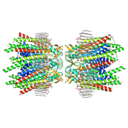
6BIT
 | | SIRPalpha antibody complex | | 分子名称: | KWAR23 Fab heavy chain, KWAR23 Fab light chain, Tyrosine-protein phosphatase non-receptor type substrate 1 | | 著者 | Ring, N.G, Herndler-Brandstetter, D, Weiskopf, K, Shan, L, Volkmer, J.P, George, B.M, Lietzenmayer, M, McKenna, K.M, Naik, T.J, McCarty, A, Zheng, Y, Ring, A.M, Flavell, R.A, Weissman, I.L. | | 登録日 | 2017-11-03 | | 公開日 | 2017-12-06 | | 最終更新日 | 2019-11-20 | | 実験手法 | X-RAY DIFFRACTION (2.191 Å) | | 主引用文献 | Anti-SIRP alpha antibody immunotherapy enhances neutrophil and macrophage antitumor activity.
Proc. Natl. Acad. Sci. U.S.A., 114, 2017
|
|
4YQ5
 | |
7JN0
 | | Sheep Connexin-46 at 2.5 angstroms resolution, Lipid Class 2 | | 分子名称: | 1,2-DIMYRISTOYL-RAC-GLYCERO-3-PHOSPHOCHOLINE, Gap junction alpha-3 protein | | 著者 | Flores, J.A, Haddad, B.G, Dolan, K.A, Myers, J.A, Yoshioka, C.C, Copperman, J, Zuckerman, D.M, Reichow, S.L. | | 登録日 | 2020-08-03 | | 公開日 | 2020-09-09 | | 実験手法 | ELECTRON MICROSCOPY (2.5 Å) | | 主引用文献 | Connexin-46/50 in a dynamic lipid environment resolved by CryoEM at 1.9 angstrom.
Nat Commun, 11, 2020
|
|
5KW3
 | | T. danielli thaumatin at 278K, Data set 1 | | 分子名称: | L(+)-TARTARIC ACID, Thaumatin-1 | | 著者 | Russi, S, Gonzalez, A, Kenner, L.R, Keedy, D.A, Fraser, J.S, van den Bedem, H. | | 登録日 | 2016-07-15 | | 公開日 | 2016-08-10 | | 最終更新日 | 2023-10-04 | | 実験手法 | X-RAY DIFFRACTION (1.55 Å) | | 主引用文献 | Conformational variation of proteins at room temperature is not dominated by radiation damage.
J Synchrotron Radiat, 24, 2017
|
|
4YU0
 | | Crystal structure of a tetramer of GluA2 TR mutant ligand binding domains bound with glutamate at 1.26 Angstrom resolution | | 分子名称: | DI(HYDROXYETHYL)ETHER, GLUTAMIC ACID, Glutamate receptor 2,Glutamate receptor 2, ... | | 著者 | Chebli, M, Salazar, H, Baranovic, J, Carbone, A.L, Ghisi, V, Faelber, K, Lau, A.Y, Daumke, O, Plested, A.J.R. | | 登録日 | 2015-03-18 | | 公開日 | 2016-01-13 | | 最終更新日 | 2024-01-10 | | 実験手法 | X-RAY DIFFRACTION (1.26 Å) | | 主引用文献 | Crystal structure of the tetrameric GluA2 ligand-binding domain in complex with glutamate at 1.26 Angstroms resolution
To Be Published
|
|
6R5W
 | | Crystal structure of the receptor binding protein (gp15) of Listeria phage PSA | | 分子名称: | ACETATE ION, CADMIUM ION, Gp15 protein, ... | | 著者 | Dunne, M, Ernst, P, Pluckthun, A, Loessner, M.J, Kilcher, S. | | 登録日 | 2019-03-25 | | 公開日 | 2019-10-30 | | 最終更新日 | 2024-05-15 | | 実験手法 | X-RAY DIFFRACTION (1.7 Å) | | 主引用文献 | Reprogramming Bacteriophage Host Range through Structure-Guided Design of Chimeric Receptor Binding Proteins.
Cell Rep, 29, 2019
|
|
6HS9
 | |
6ZWO
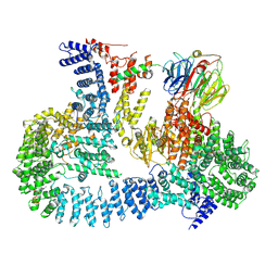 | | cryo-EM structure of human mTOR complex 2, focused on one half | | 分子名称: | ACETYL GROUP, INOSITOL HEXAKISPHOSPHATE, PHOSPHOTHIOPHOSPHORIC ACID-ADENYLATE ESTER, ... | | 著者 | Scaiola, A, Mangia, F, Imseng, S, Boehringer, D, Ban, N, Maier, T. | | 登録日 | 2020-07-28 | | 公開日 | 2020-11-18 | | 実験手法 | ELECTRON MICROSCOPY (3 Å) | | 主引用文献 | The 3.2- angstrom resolution structure of human mTORC2.
Sci Adv, 6, 2020
|
|
5EZ8
 | |
5Y1Q
 | | Crystal structure of Plasmodium falciparum aminopeptidase N in complex with (S)-2-(3-(3-chlorobenzyl)ureido)-N-hydroxy-4-methylpentanamide | | 分子名称: | (2S)-2-[(3-chlorophenyl)methylcarbamoylamino]-4-methyl-N-oxidanyl-pentanamide, M1 family aminopeptidase, MAGNESIUM ION, ... | | 著者 | Marapaka, A.K, Zhang, Y, Addlagatta, A. | | 登録日 | 2017-07-21 | | 公開日 | 2018-08-01 | | 最終更新日 | 2023-11-22 | | 実験手法 | X-RAY DIFFRACTION (2.14 Å) | | 主引用文献 | Development of peptidomimetic hydroxamates as PfA-M1 and PfA-M17 dual inhibitors: Biological evaluation and structural characterization by cocrystallization
Chin.Chem.Lett., 33, 2022
|
|
7JWF
 | | Crystal structure of PdGH110B D344N in complex with alpha-(1,3)-galactobiose | | 分子名称: | 1,2-ETHANEDIOL, 4-(2-HYDROXYETHYL)-1-PIPERAZINE ETHANESULFONIC ACID, ACETATE ION, ... | | 著者 | Hettle, A.G, Boraston, A.B. | | 登録日 | 2020-08-25 | | 公開日 | 2020-11-04 | | 最終更新日 | 2023-10-18 | | 実験手法 | X-RAY DIFFRACTION (2.187 Å) | | 主引用文献 | The structure of a family 110 glycoside hydrolase provides insight into the hydrolysis of alpha-1,3-galactosidic linkages in lambda-carrageenan and blood group antigens.
J.Biol.Chem., 295, 2020
|
|


