4V6E
 
 | | Crystal structure of the E. coli 70S ribosome in an intermediate state of ratcheting | | 分子名称: | 16S rRNA, 23S rRNA, 30S ribosomal protein S10, ... | | 著者 | Zhang, W, Dunkle, J.A, Cate, J.H.D. | | 登録日 | 2009-06-28 | | 公開日 | 2014-07-09 | | 最終更新日 | 2014-12-10 | | 実験手法 | X-RAY DIFFRACTION (3.712 Å) | | 主引用文献 | Structures of the ribosome in intermediate States of ratcheting.
Science, 325, 2009
|
|
4V6D
 | | Crystal structure of the E. coli 70S ribosome in an intermediate state of ratcheting | | 分子名称: | 16S rRNA, 23S rRNA, 30S ribosomal protein S10, ... | | 著者 | Zhang, W, Dunkle, J.A, Cate, J.H.D. | | 登録日 | 2009-06-27 | | 公開日 | 2014-07-09 | | 最終更新日 | 2014-12-10 | | 実験手法 | X-RAY DIFFRACTION (3.814 Å) | | 主引用文献 | Structures of the ribosome in intermediate States of ratcheting.
Science, 325, 2009
|
|
4WW5
 | | Crystal structure of binary complex Bud32-Cgi121 in complex with AMPP | | 分子名称: | ACETATE ION, EKC/KEOPS complex subunit BUD32, EKC/KEOPS complex subunit CGI121, ... | | 著者 | Zhang, W. | | 登録日 | 2014-11-10 | | 公開日 | 2015-03-18 | | 最終更新日 | 2015-04-15 | | 実験手法 | X-RAY DIFFRACTION (1.997 Å) | | 主引用文献 | Crystal structures of the Gon7/Pcc1 and Bud32/Cgi121 complexes provide a model for the complete yeast KEOPS complex.
Nucleic Acids Res., 43, 2015
|
|
4WW9
 | |
4X3X
 | |
6QJP
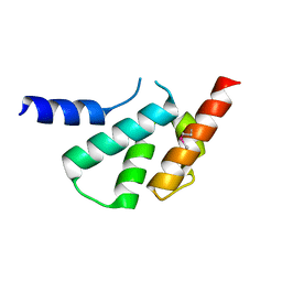
 | | Cryo-EM structure of heparin-induced 2N4R tau jagged filaments | | 分子名称: | Microtubule-associated protein tau | | 著者 | Zhang, W, Falcon, B, Murzin, A.G, Fan, J, Crowther, R.A, Goedert, M, Scheres, S.H.W. | | 登録日 | 2019-01-24 | | 公開日 | 2019-02-20 | | 実験手法 | ELECTRON MICROSCOPY (3.5 Å) | | 主引用文献 | Heparin-induced tau filaments are polymorphic and differ from those in Alzheimer's and Pick's diseases.
Elife, 8, 2019
|
|
6QJH
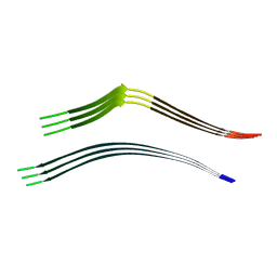
 | | Cryo-EM structure of heparin-induced 2N4R tau snake filaments | | 分子名称: | Microtubule-associated protein tau | | 著者 | Zhang, W, Falcon, B, Murzin, A.G, Fan, J, Crowther, R.A, Goedert, M, Scheres, S.H.W. | | 登録日 | 2019-01-24 | | 公開日 | 2019-02-20 | | 実験手法 | ELECTRON MICROSCOPY (3.3 Å) | | 主引用文献 | Heparin-induced tau filaments are polymorphic and differ from those in Alzheimer's and Pick's diseases.
Elife, 8, 2019
|
|
6QJM
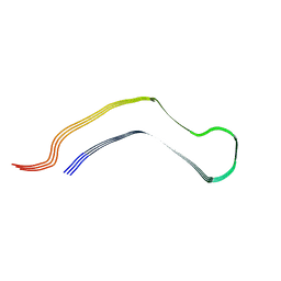
 | | Cryo-EM structure of heparin-induced 2N4R tau twister filaments | | 分子名称: | Microtubule-associated protein tau | | 著者 | Zhang, W, Falcon, B, Murzin, A.G, Fan, J, Crowther, R.A, Goedert, M, Scheres, S.H.W. | | 登録日 | 2019-01-24 | | 公開日 | 2019-02-27 | | 実験手法 | ELECTRON MICROSCOPY (3.3 Å) | | 主引用文献 | Heparin-induced tau filaments are polymorphic and differ from those in Alzheimer's and Pick's diseases.
Elife, 8, 2019
|
|
6QJQ
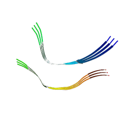
 | | Cryo-EM structure of heparin-induced 2N3R tau filaments | | 分子名称: | Microtubule-associated protein tau | | 著者 | Zhang, W, Falcon, B, Murzin, A.G, Fan, J, Crowther, R.A, Goedert, M, Scheres, S.H.W. | | 登録日 | 2019-01-24 | | 公開日 | 2019-02-20 | | 実験手法 | ELECTRON MICROSCOPY (3.7 Å) | | 主引用文献 | Heparin-induced tau filaments are polymorphic and differ from those in Alzheimer's and Pick's diseases.
Elife, 8, 2019
|
|
6TJO
 | | Cryo-EM structure of TypeI tau filaments extracted from the brains of individuals with Corticobasal degeneration | | 分子名称: | Microtubule-associated protein tau | | 著者 | Zhang, W, Murzin, A.G, Falcon, B, Shi, Y, Goedert, M, Scheres, S.H.W. | | 登録日 | 2019-11-26 | | 公開日 | 2020-02-05 | | 最終更新日 | 2020-04-22 | | 実験手法 | ELECTRON MICROSCOPY (3.2 Å) | | 主引用文献 | Novel tau filament fold in corticobasal degeneration.
Nature, 580, 2020
|
|
6TJX
 | | Cryo-EM structure of TypeII tau filaments extracted from the brains of individuals with Corticobasal degeneration | | 分子名称: | Microtubule-associated protein tau | | 著者 | Zhang, W, Murzin, A.G, Falcon, B, Shi, Y, Goedert, M, Scheres, S.H.W. | | 登録日 | 2019-11-27 | | 公開日 | 2020-02-05 | | 最終更新日 | 2020-04-22 | | 実験手法 | ELECTRON MICROSCOPY (3 Å) | | 主引用文献 | Novel tau filament fold in corticobasal degeneration.
Nature, 580, 2020
|
|
1P58
 | | Complex Organization of Dengue Virus Membrane Proteins as Revealed by 9.5 Angstrom Cryo-EM reconstruction | | 分子名称: | Envelope protein M, Major envelope protein E | | 著者 | Zhang, W, Chipman, P.R, Corver, J, Johnson, P.R, Zhang, Y, Mukhopadhyay, S, Baker, T.S, Strauss, J.H, Rossmann, M.G, Kuhn, R.J. | | 登録日 | 2003-04-25 | | 公開日 | 2003-11-04 | | 最終更新日 | 2024-02-14 | | 実験手法 | ELECTRON MICROSCOPY (9.5 Å) | | 主引用文献 | Visualization of membrane protein domains by cryo-electron microscopy of dengue virus
Nat.Struct.Biol., 10, 2003
|
|
2K9X
 | | Solution structure of Urm1 from Trypanosoma brucei | | 分子名称: | Uncharacterized protein | | 著者 | Zhang, W, Zhang, J, Xu, C, Wang, T, Zhang, X, Tu, X. | | 登録日 | 2008-10-27 | | 公開日 | 2009-03-10 | | 最終更新日 | 2022-03-16 | | 実験手法 | SOLUTION NMR | | 主引用文献 | Solution structure of Urm1 from Trypanosoma brucei
Proteins, 75, 2009
|
|
5KRG
 | | RNA 15mer duplex binding with PZG monomer | | 分子名称: | RNA (5'-R(*(LCC)P*(LCC)P*(LCC)P*(LCG)P*AP*CP*UP*UP*AP*AP*GP*UP*CP*GP*G)-3'), [(2~{R},3~{S},4~{R},5~{R})-5-(2-azanyl-6-oxidanylidene-1~{H}-purin-9-yl)-3,4-bis(oxidanyl)oxolan-2-yl]methoxy-(3-methyl-1~{H}-pyrazol-4-yl)phosphinic acid | | 著者 | Zhang, W, Tam, C.P, Szostak, J.W. | | 登録日 | 2016-07-07 | | 公開日 | 2016-12-07 | | 最終更新日 | 2023-10-04 | | 実験手法 | X-RAY DIFFRACTION (1.6 Å) | | 主引用文献 | Unusual Base-Pairing Interactions in Monomer-Template Complexes.
ACS Cent Sci, 2, 2016
|
|
5L00
 | |
2L42
 | |
3E6U
 | | Crystal structure of Human LanCL1 | | 分子名称: | LanC-like protein 1, ZINC ION | | 著者 | Zhang, W, Zhu, G, Li, X, Rao, Z, Zhang, C. | | 登録日 | 2008-08-16 | | 公開日 | 2009-06-23 | | 最終更新日 | 2024-03-20 | | 実験手法 | X-RAY DIFFRACTION (2.6 Å) | | 主引用文献 | Structure of human lanthionine synthetase C-like protein 1 and its interaction with Eps8 and glutathione
Genes Dev., 23, 2009
|
|
3E73
 | | Crystal Structure of Human LanCL1 complexed with GSH | | 分子名称: | GLUTATHIONE, LanC-like protein 1, ZINC ION | | 著者 | Zhang, W, Zhu, G, Li, X, Rao, Z, Zhang, C. | | 登録日 | 2008-08-17 | | 公開日 | 2009-06-30 | | 最終更新日 | 2023-11-01 | | 実験手法 | X-RAY DIFFRACTION (2.8 Å) | | 主引用文献 | Structure of human lanthionine synthetase C-like protein 1 and its interaction with Eps8 and glutathione
Genes Dev., 23, 2009
|
|
2L7E
 | |
5CB3
 | | Structural Insights into the Mechanism of Escherichia coli Ymdb | | 分子名称: | ADENOSINE-5-DIPHOSPHORIBOSE, O-acetyl-ADP-ribose deacetylase | | 著者 | Zhang, W, Wang, C, Song, Y, Shao, C, Zhang, X, Zang, J. | | 登録日 | 2015-06-30 | | 公開日 | 2015-11-04 | | 最終更新日 | 2023-11-08 | | 実験手法 | X-RAY DIFFRACTION (1.8 Å) | | 主引用文献 | Structural insights into the mechanism of Escherichia coli YmdB: A 2'-O-acetyl-ADP-ribose deacetylase
J.Struct.Biol., 192, 2015
|
|
5CB5
 | | Structural Insights into the Mechanism of Escherichia coli Ymdb | | 分子名称: | ACETATE ION, ADENOSINE-5-DIPHOSPHORIBOSE, O-acetyl-ADP-ribose deacetylase, ... | | 著者 | Zhang, W, Wang, C, Song, Y, Shao, C, Zhang, X, Zang, J. | | 登録日 | 2015-06-30 | | 公開日 | 2015-11-04 | | 最終更新日 | 2023-11-08 | | 実験手法 | X-RAY DIFFRACTION (2.8 Å) | | 主引用文献 | Structural insights into the mechanism of Escherichia coli YmdB: A 2'-O-acetyl-ADP-ribose deacetylase
J.Struct.Biol., 192, 2015
|
|
5CMS
 | | Structural Insights into the Mechanism of Escherichia coli Ymdb | | 分子名称: | ADENOSINE-5-DIPHOSPHORIBOSE, O-acetyl-ADP-ribose deacetylase, SULFATE ION | | 著者 | Zhang, W, Wang, C, Song, Y, Shao, C, Zhang, X, Zang, J. | | 登録日 | 2015-07-17 | | 公開日 | 2015-11-04 | | 最終更新日 | 2023-11-08 | | 実験手法 | X-RAY DIFFRACTION (2.98 Å) | | 主引用文献 | Structural insights into the mechanism of Escherichia coli YmdB: A 2'-O-acetyl-ADP-ribose deacetylase
J.Struct.Biol., 192, 2015
|
|
5GQ0
 | |
5GQT
 | |
1LD4
 | | Placement of the Structural Proteins in Sindbis Virus | | 分子名称: | Coat protein C, GENERAL CONTROL PROTEIN GCN4, Spike glycoprotein E1, ... | | 著者 | Zhang, W, Mukhopadhyay, S, Pletnev, S.V, Baker, T.S, Kuhn, R.J, Rossmann, M.G. | | 登録日 | 2002-04-08 | | 公開日 | 2002-11-04 | | 最終更新日 | 2019-11-06 | | 実験手法 | ELECTRON MICROSCOPY (11.4 Å) | | 主引用文献 | Placement of the Structural Proteins in Sindbis virus
J.VIROL., 76, 2002
|
|


