Loading
PDBj
MenuPDBj@FacebookPDBj@TwitterPDBj@YouTubewwPDB FoundationwwPDB
RCSB PDBPDBeBMRBAdv. SearchSearch help
Search by PDB author
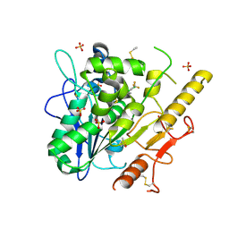
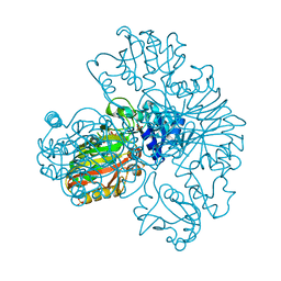
1S7C
DownloadVisualize
BU of 1s7c by Molmil
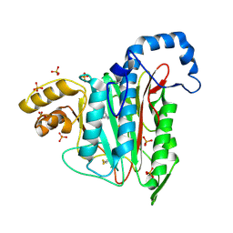
Crystal structure of MES buffer bound form of glyceraldehyde 3-phosphate dehydrogenase from Escherichia coli
Descriptor: 2-(N-MORPHOLINO)-ETHANESULFONIC ACID, Glyceraldehyde 3-phosphate dehydrogenase A, SULFATE ION
Authors:Shin, D.H, Thor, J, Yokota, H, Kim, R, Kim, S.H, Berkeley Structural Genomics Center (BSGC)
Deposit date:2004-01-29
Release date:2004-08-10
Last modified:2024-02-14
Method:X-RAY DIFFRACTION (2.04 Å)
Cite:Crystal structure of MES buffer bound form of glyceraldehyde 3-phosphate dehydrogenase from Escherichia coli
To be Published
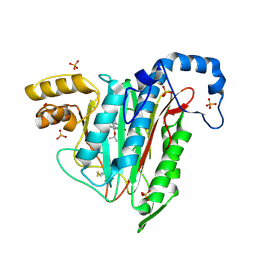
4X8G
DownloadVisualize
BU of 4x8g by Molmil
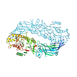
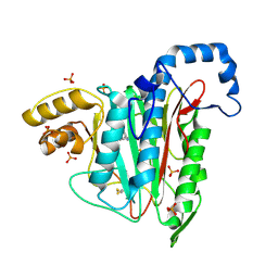
Crystal structure of human peptidylarginine deiminase type4 (PAD4) in complex with GSK199
Descriptor: CALCIUM ION, Protein-arginine deiminase type-4, [(3R)-3-aminopiperidin-1-yl][2-(1-ethyl-1H-pyrrolo[2,3-b]pyridin-2-yl)-7-methoxy-1-methyl-1H-benzimidazol-5-yl]methanone
Authors:Lewis, H.D, Bax, B.D, Chung, C.-W, Polyakova, O, Thorpe, J.
Deposit date:2014-12-10
Release date:2015-01-28
Last modified:2024-02-28
Method:X-RAY DIFFRACTION (3.29 Å)
Cite:Inhibition of PAD4 activity is sufficient to disrupt mouse and human NET formation.
Nat.Chem.Biol., 11, 2015
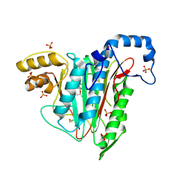
4X8C
DownloadVisualize
BU of 4x8c by Molmil
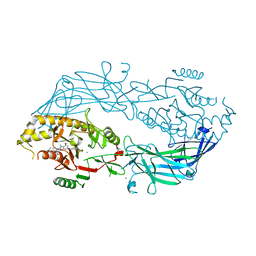
Crystal structure of human peptidylarginine deiminase type4 (PAD4) in complex with GSK147
Descriptor: CALCIUM ION, Protein-arginine deiminase type-4, [(3S,4R)-3-amino-4-hydroxypiperidin-1-yl]{2-[1-(cyclopropylmethyl)-1H-pyrrolo[2,3-b]pyridin-2-yl]-7-methoxy-1-methyl-1H-benzimidazol-5-yl}methanone
Authors:Lewis, H.D, Bax, B.D, Chung, C.-W, Polyakova, O, Thorpe, J.
Deposit date:2014-12-10
Release date:2015-01-28
Last modified:2023-09-27
Method:X-RAY DIFFRACTION (3.1 Å)
Cite:Inhibition of PAD4 activity is sufficient to disrupt mouse and human NET formation.
Nat.Chem.Biol., 11, 2015
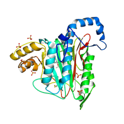
1FPW
DownloadVisualize
BU of 1fpw by Molmil
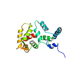
STRUCTURE OF YEAST FREQUENIN
Descriptor: CALCIUM ION, CALCIUM-BINDING PROTEIN NCS-1
Authors:Ames, J.B, Hendricks, K.B, Strahl, T, Huttner, I.G, Thorner, J.
Deposit date:2000-08-31
Release date:2000-10-18
Last modified:2024-05-22
Method:SOLUTION NMR
Cite:Structure and calcium-binding properties of Frq1, a novel calcium sensor in the yeast Saccharomyces cerevisiae.
Biochemistry, 39, 2000
1K7J
DownloadVisualize
BU of 1k7j by Molmil
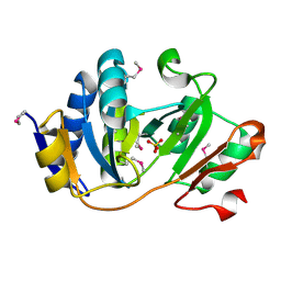
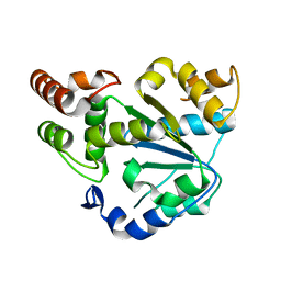
Structural Genomics, protein TF1
Descriptor: Protein yciO, SULFATE ION
Authors:Zhang, R, Dementieva, I, Thorn, J, Donnelly, M, Joachimiak, A, Midwest Center for Structural Genomics (MCSG)
Deposit date:2001-10-19
Release date:2002-08-14
Last modified:2011-07-13
Method:X-RAY DIFFRACTION (1.4 Å)
Cite:Structural Genomics, protein TF1
To be Published
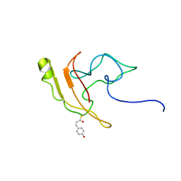
2KX6
DownloadVisualize
BU of 2kx6 by Molmil
Signaling state of Photoactive Yellow Protein
Descriptor: 4'-HYDROXYCINNAMIC ACID, Photoactive yellow protein
Authors:Ramachandran, P.L, Lovett, J.E, Carl, P.J, Cammarata, M, Lee, J.H, Yang, J.O, Ihee, H, Timmel, C.R, van Thor, J.
Deposit date:2010-04-27
Release date:2011-06-15
Last modified:2012-07-18
Method:SOLUTION NMR, SOLUTION SCATTERING
Cite:The short-lived signaling state of the photoactive yellow protein photoreceptor revealed by combined structural probes.
J.Am.Chem.Soc., 133, 2011
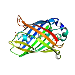
8A83
DownloadVisualize
BU of 8a83 by Molmil
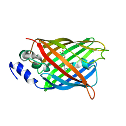
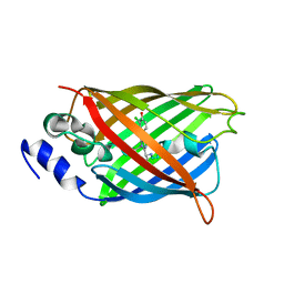
rsEGFP2 with a chlorinated chromophore in the fluorescent ON-state in a crystal dehydrated after illumination
Descriptor: Green fluorescent protein, SULFATE ION
Authors:Fadini, A, van Thor, J, Chang, J.
Deposit date:2022-06-21
Release date:2023-07-19
Last modified:2024-02-07
Method:X-RAY DIFFRACTION (1.81 Å)
Cite:Serial Femtosecond Crystallography Reveals that Photoactivation in a Fluorescent Protein Proceeds via the Hula Twist Mechanism.
J.Am.Chem.Soc., 145, 2023
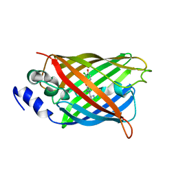
8A6O
DownloadVisualize
BU of 8a6o by Molmil
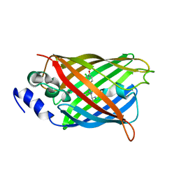
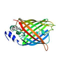
Room temperature rsEGFP2 with a chlorinated chromophore 600 fs after Photoexcitation
Descriptor: Green fluorescent protein
Authors:Fadini, A, van Thor, J.
Deposit date:2022-06-18
Release date:2023-07-19
Last modified:2024-02-07
Method:X-RAY DIFFRACTION (1.63 Å)
Cite:Serial Femtosecond Crystallography Reveals that Photoactivation in a Fluorescent Protein Proceeds via the Hula Twist Mechanism.
J.Am.Chem.Soc., 145, 2023
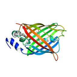
8A6R
DownloadVisualize
BU of 8a6r by Molmil
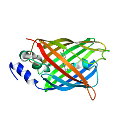
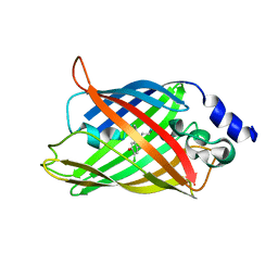
Room temperature rsEGFP2 with a chlorinated chromophore 100 ps after Photoexcitation
Descriptor: Green fluorescent protein
Authors:Fadini, A, van Thor, J.
Deposit date:2022-06-18
Release date:2023-07-19
Last modified:2024-02-07
Method:X-RAY DIFFRACTION (1.63 Å)
Cite:Serial Femtosecond Crystallography Reveals that Photoactivation in a Fluorescent Protein Proceeds via the Hula Twist Mechanism.
J.Am.Chem.Soc., 145, 2023
8A6S
DownloadVisualize
BU of 8a6s by Molmil
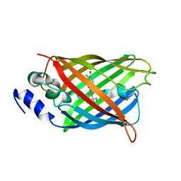
Room temperature rsEGFP2 with a chlorinated chromophore 1 microsecond after Photoexcitation
Descriptor: Green fluorescent protein
Authors:Fadini, A, van Thor, J.
Deposit date:2022-06-19
Release date:2023-07-19
Last modified:2024-02-07
Method:X-RAY DIFFRACTION (1.63 Å)
Cite:Serial Femtosecond Crystallography Reveals that Photoactivation in a Fluorescent Protein Proceeds via the Hula Twist Mechanism.
J.Am.Chem.Soc., 145, 2023
8A6G
DownloadVisualize
BU of 8a6g by Molmil
Room temperature rsEGFP2 with a chlorinated chromophore in the non-fluorescent OFF-state
Descriptor: Green fluorescent protein
Authors:Fadini, A, van Thor, J.
Deposit date:2022-06-17
Release date:2023-07-19
Last modified:2024-02-07
Method:X-RAY DIFFRACTION (1.63 Å)
Cite:Serial Femtosecond Crystallography Reveals that Photoactivation in a Fluorescent Protein Proceeds via the Hula Twist Mechanism.
J.Am.Chem.Soc., 145, 2023
8A6N
DownloadVisualize
BU of 8a6n by Molmil
Room temperature rsEGFP2 with a chlorinated chromophore 300 fs after Photoexcitation
Descriptor: Green fluorescent protein
Authors:Fadini, A, van Thor, J.
Deposit date:2022-06-18
Release date:2023-07-19
Last modified:2024-02-07
Method:X-RAY DIFFRACTION (1.63 Å)
Cite:Serial Femtosecond Crystallography Reveals that Photoactivation in a Fluorescent Protein Proceeds via the Hula Twist Mechanism.
J.Am.Chem.Soc., 145, 2023
8A6P
DownloadVisualize
BU of 8a6p by Molmil
Room temperature rsEGFP2 with a chlorinated chromophore 900 fs after photoexcitation
Descriptor: Green fluorescent protein
Authors:Fadini, A, van Thor, J.
Deposit date:2022-06-18
Release date:2023-07-19
Last modified:2024-02-07
Method:X-RAY DIFFRACTION (1.63 Å)
Cite:Serial Femtosecond Crystallography Reveals that Photoactivation in a Fluorescent Protein Proceeds via the Hula Twist Mechanism.
J.Am.Chem.Soc., 145, 2023
8A6Q
DownloadVisualize
BU of 8a6q by Molmil
Room temperature rsEGFP2 with a chlorinated chromophore 5 ps after photoexcitation
Descriptor: Green fluorescent protein
Authors:Fadini, A, van Thor, J.
Deposit date:2022-06-18
Release date:2023-07-19
Last modified:2024-02-07
Method:X-RAY DIFFRACTION (1.63 Å)
Cite:Serial Femtosecond Crystallography Reveals that Photoactivation in a Fluorescent Protein Proceeds via the Hula Twist Mechanism.
J.Am.Chem.Soc., 145, 2023
8A7V
DownloadVisualize
BU of 8a7v by Molmil
Room temperature rsEGFP2 in its OFF-state obtained with SFX
Descriptor: Green fluorescent protein
Authors:Fadini, A, van Thor, J.
Deposit date:2022-06-21
Release date:2023-07-19
Last modified:2024-02-07
Method:X-RAY DIFFRACTION (1.46 Å)
Cite:Serial Femtosecond Crystallography Reveals that Photoactivation in a Fluorescent Protein Proceeds via the Hula Twist Mechanism.
J.Am.Chem.Soc., 145, 2023
8AM4
DownloadVisualize
BU of 8am4 by Molmil
Cl-rsEGFP2 Long Wavelength Structure
Descriptor: Green fluorescent protein
Authors:Orr, C.M, Fadini, A, van Thor, J.
Deposit date:2022-08-02
Release date:2023-08-02
Last modified:2024-01-31
Method:X-RAY DIFFRACTION (2.02 Å)
Cite:Serial Femtosecond Crystallography Reveals that Photoactivation in a Fluorescent Protein Proceeds via the Hula Twist Mechanism.
J.Am.Chem.Soc., 2023
6T2K
DownloadVisualize
BU of 6t2k by Molmil
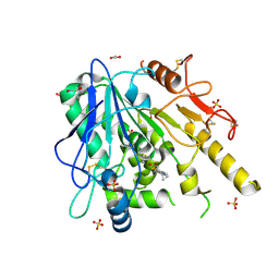
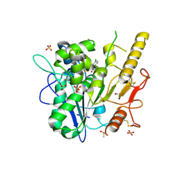
Furano[2,3-d]prymidine amides as Notum inhibitors
Descriptor: 1,2-ETHANEDIOL, 2-(6-chloranyl-7-cyclopropyl-thieno[3,2-d]pyrimidin-4-yl)sulfanylethanoic acid, 2-acetamido-2-deoxy-beta-D-glucopyranose, ...
Authors:Zhao, Y, Jones, E.Y.
Deposit date:2019-10-08
Release date:2020-01-01
Last modified:2024-01-24
Method:X-RAY DIFFRACTION (1.38 Å)
Cite:Scaffold-hopping identifies furano[2,3-d]pyrimidine amides as potent Notum inhibitors.
Bioorg.Med.Chem.Lett., 30, 2020
7PK3
DownloadVisualize
BU of 7pk3 by Molmil
Notum_ARUK3001185
Descriptor: 1-[2,4-bis(chloranyl)-3-(trifluoromethyl)phenyl]-1,2,3-triazole, 2-acetamido-2-deoxy-beta-D-glucopyranose, DIMETHYL SULFOXIDE, ...
Authors:Vecchia, L, Hillier, J, Zhao, Y, Fish, P, Jones, E.Y.
Deposit date:2021-08-25
Release date:2022-09-07
Last modified:2024-02-07
Method:X-RAY DIFFRACTION (1.41 Å)
Cite:Design of a Potent, Selective, and Brain-Penetrant Inhibitor of Wnt-Deactivating Enzyme Notum by Optimization of a Crystallographic Fragment Hit.
J.Med.Chem., 65, 2022
7A16
DownloadVisualize
BU of 7a16 by Molmil
CRYSTAL STRUCTURE OF HUMAN METHIONINE AMINOPEPTIDASE-2 IN COMPLEX WITH AN INHIBITOR GSK2229238A (COMPOUND 43)
Descriptor: 5,6-bis(fluoranyl)-3-(4-piperazin-1-yl-2-propan-2-yloxy-phenyl)-1~{H}-indole-2-carboxamide, MANGANESE (II) ION, Methionine aminopeptidase 2, ...
Authors:Thorpe, J.H.
Deposit date:2020-08-11
Release date:2020-09-23
Last modified:2024-01-31
Method:X-RAY DIFFRACTION (1.9 Å)
Cite:Structure-based optimisation of orally active & reversible MetAP-2 inhibitors maintaining a tight 'molecular budget'.
Bioorg.Med.Chem.Lett., 30, 2020
7A15
DownloadVisualize
BU of 7a15 by Molmil
CRYSTAL STRUCTURE OF HUMAN METHIONINE AMINOPEPTIDASE-2 IN COMPLEX WITH AN INHIBITOR GSK2224863A (COMPOUND 42)
Descriptor: 5-chloranyl-6-fluoranyl-3-(4-piperazin-1-yl-2-propan-2-yloxy-phenyl)-1~{H}-indole-2-carboxamide, MANGANESE (II) ION, Methionine aminopeptidase 2, ...
Authors:Thorpe, J.H.
Deposit date:2020-08-11
Release date:2020-09-23
Last modified:2024-01-31
Method:X-RAY DIFFRACTION (2.15 Å)
Cite:Structure-based optimisation of orally active & reversible MetAP-2 inhibitors maintaining a tight 'molecular budget'.
Bioorg.Med.Chem.Lett., 30, 2020
7A14
DownloadVisualize
BU of 7a14 by Molmil
CRYSTAL STRUCTURE OF HUMAN METHIONINE AMINOPEPTIDASE-2 IN COMPLEX WITH AN INHIBITOR GSK2218325A (COMPOUND 32)
Descriptor: 5-chloranyl-6-fluoranyl-3-(2-propan-2-yloxyphenyl)-1~{H}-indole-2-carboxamide, MANGANESE (II) ION, Methionine aminopeptidase 2, ...
Authors:Thorpe, J.H.
Deposit date:2020-08-11
Release date:2020-09-23
Last modified:2024-01-31
Method:X-RAY DIFFRACTION (2.14 Å)
Cite:Structure-based optimisation of orally active & reversible MetAP-2 inhibitors maintaining a tight 'molecular budget'.
Bioorg.Med.Chem.Lett., 30, 2020
7A12
DownloadVisualize
BU of 7a12 by Molmil
CRYSTAL STRUCTURE OF HUMAN METHIONINE AMINOPEPTIDASE-2 IN COMPLEX WITH AN INHIBITOR GW557358X (COMPOUND 9)
Descriptor: 5-chloranyl-3-phenyl-1~{H}-indole-2-carboxamide, MANGANESE (II) ION, Methionine aminopeptidase 2, ...
Authors:Thorpe, J.H.
Deposit date:2020-08-11
Release date:2020-09-23
Last modified:2024-01-31
Method:X-RAY DIFFRACTION (2 Å)
Cite:Structure-based optimisation of orally active & reversible MetAP-2 inhibitors maintaining a tight 'molecular budget'.
Bioorg.Med.Chem.Lett., 30, 2020
7A13
DownloadVisualize
BU of 7a13 by Molmil
CRYSTAL STRUCTURE OF HUMAN METHIONINE AMINOPEPTIDASE-2 IN COMPLEX WITH AN INHIBITOR GSK1978537A (COMPOUND 27)
Descriptor: 5-chloranyl-3-(3-methoxyphenyl)-1~{H}-indole-2-carboxamide, MANGANESE (II) ION, Methionine aminopeptidase 2, ...
Authors:Thorpe, J.H.
Deposit date:2020-08-11
Release date:2020-09-23
Last modified:2024-01-31
Method:X-RAY DIFFRACTION (2.04 Å)
Cite:Structure-based optimisation of orally active & reversible MetAP-2 inhibitors maintaining a tight 'molecular budget'.
Bioorg.Med.Chem.Lett., 30, 2020
2AD1
DownloadVisualize
BU of 2ad1 by Molmil
Human Sulfotransferase SULT1C2
Descriptor: Sulfotransferase 1C2
Authors:Dong, A, Dombrovski, L, Loppnau, P, Edwards, A.M, Arrowsmith, C.H, Weigelt, J, Sundstrom, M, Bochkarev, A, Plotnikov, A.N, Structural Genomics Consortium (SGC)
Deposit date:2005-07-19
Release date:2005-08-02
Last modified:2023-08-23
Method:X-RAY DIFFRACTION (2 Å)
Cite:Structural and chemical profiling of the human cytosolic sulfotransferases.
Plos Biol., 5, 2007
6ZVL
DownloadVisualize
BU of 6zvl by Molmil
ARUK3000263 complex with Notum
Descriptor: 1,2-ETHANEDIOL, 2-acetamido-2-deoxy-beta-D-glucopyranose, 5-[4-chloranyl-3-(trifluoromethyl)phenyl]-3~{H}-1,3,4-oxadiazol-2-one, ...
Authors:Zhao, Y, Ruza, R.
Deposit date:2020-07-24
Release date:2020-11-11
Last modified:2024-01-31
Method:X-RAY DIFFRACTION (1.3 Å)
Cite:5-Phenyl-1,3,4-oxadiazol-2(3 H )-ones Are Potent Inhibitors of Notum Carboxylesterase Activity Identified by the Optimization of a Crystallographic Fragment Screening Hit.
J.Med.Chem., 63, 2020


1234>

223166

PDB entries from 2024-07-31

PDB statisticsPDBj update infoContact PDBjnumon