Loading
PDBj
MenuPDBj@FacebookPDBj@TwitterPDBj@YouTubewwPDB FoundationwwPDB
RCSB PDBPDBeBMRBAdv. SearchSearch help
Search by PDB author
6TX5
DownloadVisualize
BU of 6tx5 by Molmil
CRYSTAL STRUCTURE OF HUMAN FKBP51 FK1 DOMAIN A19T MUTANT IN COMPLEX WITH 4-METHYLIMIDAZOLE
Descriptor: 4-METHYLIMIDAZOLE, Peptidyl-prolyl cis-trans isomerase FKBP5, SODIUM ION
Authors:Fiegen, D, Draxler, S.W.
Deposit date:2020-01-13
Release date:2020-05-27
Last modified:2024-01-24
Method:X-RAY DIFFRACTION (1.08 Å)
Cite:Hybrid Screening Approach for Very Small Fragments: X-ray and Computational Screening on FKBP51.
J.Med.Chem., 63, 2020
6OPJ
DownloadVisualize
BU of 6opj by Molmil
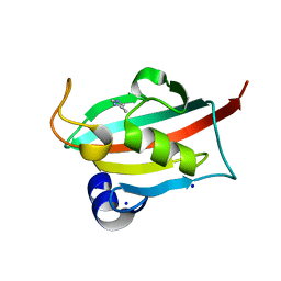
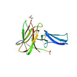
Menin in complex with peptide inhibitor 25
Descriptor: DIMETHYL SULFOXIDE, Menin, Peptide inhibitor 25, ...
Authors:Linhares, B.M, Fortuna, P, Cierpicki, T, Grembecka, J, Berlicki, L.
Deposit date:2019-04-25
Release date:2020-09-02
Last modified:2023-11-15
Method:X-RAY DIFFRACTION (1.5006572 Å)
Cite:Covalent and noncovalent constraints yield a figure eight-like conformation of a peptide inhibiting the menin-MLL interaction.
Eur.J.Med.Chem., 207, 2020
6PHJ
DownloadVisualize
BU of 6phj by Molmil
Crystal structure of native glucagon in space group P213 at 1.99 A resolution
Descriptor: Glucagon
Authors:Mroz, P.A, Gonzalez-Gutierrez, G, DiMarchi, R.D.
Deposit date:2019-06-25
Release date:2020-07-01
Last modified:2023-10-11
Method:X-RAY DIFFRACTION (1.99 Å)
Cite:High resolution X-ray structure of glucagon and selected stereo-inversed analogs in novel crystallographic packing arrangement.
To Be Published
6PHO
DownloadVisualize
BU of 6pho by Molmil
Crystal structure of glucagon analog with selenomethionine substitutions at position 1 and 27 in space group I41 at 1.42 A resolution
Descriptor: Glucagon
Authors:Mroz, P.A, Gonzalez-Gutierrez, G, DiMarchi, R.D.
Deposit date:2019-06-25
Release date:2020-07-01
Method:X-RAY DIFFRACTION (1.42 Å)
Cite:High resolution X-ray structure of glucagon and selected stereo-inversed analogs in novel crystallographic packing arrangement.
To Be Published
6P0J
DownloadVisualize
BU of 6p0j by Molmil
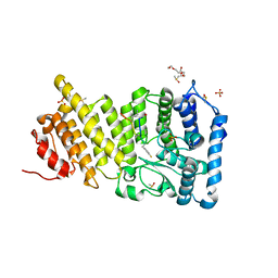
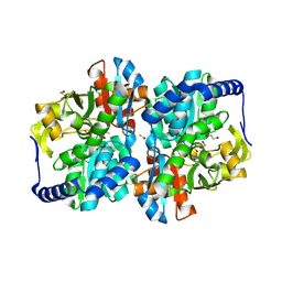
Crystal structure of GDP-bound human RalA
Descriptor: CALCIUM ION, GUANOSINE-5'-DIPHOSPHATE, Ras-related protein Ral-A
Authors:Bum-Erdene, K, Gonzalez-Gutierrez, G, Liu, D, Meroueh, S.O.
Deposit date:2019-05-17
Release date:2020-03-04
Last modified:2023-10-11
Method:X-RAY DIFFRACTION (1.31 Å)
Cite:Small-molecule covalent bond formation at tyrosine creates a binding site and inhibits activation of Ral GTPases.
Proc.Natl.Acad.Sci.USA, 117, 2020
6PHK
DownloadVisualize
BU of 6phk by Molmil
Crystal structure of glucagon analog with mono-stereoinversion at position 21 (D-Asp21) in space group I41 at 1.18 A resolution
Descriptor: Glucagon
Authors:Mroz, P.A, Gonzalez-Gutierrez, G, DiMarchi, R.D.
Deposit date:2019-06-25
Release date:2020-07-01
Last modified:2023-10-11
Method:X-RAY DIFFRACTION (1.18 Å)
Cite:High resolution X-ray structure of glucagon and selected stereo-inversed analogs in novel crystallographic packing arrangement.
To Be Published
6PHP
DownloadVisualize
BU of 6php by Molmil
Crystal structure of glucagon analog with 4-bromo-phenylalanine substitutions at position 6 and 22 in space group I41 at 1.65 A resolution
Descriptor: Glucagon
Authors:Mroz, P.A, Gonzalez-Gutierrez, G, DiMarchi, R.D.
Deposit date:2019-06-25
Release date:2020-07-01
Last modified:2023-11-15
Method:X-RAY DIFFRACTION (1.65 Å)
Cite:High resolution X-ray structure of glucagon and selected stereo-inversed analogs in novel crystallographic packing arrangement.
To Be Published
6PQ7
DownloadVisualize
BU of 6pq7 by Molmil
Structure of the iMango-III fluorescent aptamer at room temperature.
Descriptor: MAGNESIUM ION, POTASSIUM ION, RNA (37-MER), ...
Authors:Trachman III, R.J, Ferre-D'Amare, A.R.
Deposit date:2019-07-08
Release date:2019-07-31
Last modified:2023-10-11
Method:X-RAY DIFFRACTION (3 Å)
Cite:Co-crystal structure of the iMango-III fluorescent RNA aptamer using an X-ray free-electron laser.
Acta Crystallogr.,Sect.F, 75, 2019
6PHN
DownloadVisualize
BU of 6phn by Molmil
Crystal structure of glucagon analog composed of D-amino acids with mono-stereoinversion at position 23 (L-Val23) in space group I41 at 1.33 A resolution
Descriptor: D-glucagon L-Val23, SODIUM ION
Authors:Mroz, P.A, Gonzalez-Gutierrez, G, DiMarchi, R.D.
Deposit date:2019-06-25
Release date:2020-07-01
Last modified:2023-11-15
Method:X-RAY DIFFRACTION (1.33 Å)
Cite:High resolution X-ray structure of glucagon and selected stereo-inversed analogs in novel crystallographic packing arrangement.
To Be Published
6PHL
DownloadVisualize
BU of 6phl by Molmil
Crystal structure of glucagon analog with mono-stereoinversion at position 23 (D-Val23) in space group I41 at 1.44 A resolution
Descriptor: Glucagon
Authors:Mroz, P.A, Gonzalez-Gutierrez, G, DiMarchi, R.D.
Deposit date:2019-06-25
Release date:2020-07-01
Last modified:2023-10-11
Method:X-RAY DIFFRACTION (1.443 Å)
Cite:High resolution X-ray structure of glucagon and selected stereo-inversed analogs in novel crystallographic packing arrangement.
To Be Published
6PHQ
DownloadVisualize
BU of 6phq by Molmil
Crystal structure of glucagon analog fully composed of D-amino acids with 4-bromo-D-phenylalanine substitutions at position 6 and 22 in space group I41 at 1.1 A resolution
Descriptor: D-glucagon D-BrPhe 6,22
Authors:Mroz, P.A, Gonzalez-Gutierrez, G, DiMarchi, R.D.
Deposit date:2019-06-25
Release date:2020-07-01
Last modified:2023-11-15
Method:X-RAY DIFFRACTION (1.32 Å)
Cite:High resolution X-ray structure of glucagon and selected stereo-inversed analogs in novel crystallographic packing arrangement.
To Be Published
6PHI
DownloadVisualize
BU of 6phi by Molmil
Crystal structure of native glucagon in space group I41 at 1.1 A resolution
Descriptor: CHLORIDE ION, Glucagon
Authors:Mroz, P.A, Gonzalez-Gutierrez, G, DiMarchi, R.D.
Deposit date:2019-06-25
Release date:2020-07-01
Last modified:2023-10-11
Method:X-RAY DIFFRACTION (1.1 Å)
Cite:High resolution X-ray structure of glucagon and selected stereo-inversed analogs in novel crystallographic packing arrangement.
To Be Published
6PHM
DownloadVisualize
BU of 6phm by Molmil
Crystal structure of glucagon analog fully composed of D-amino acids in space group I41 at 1.1 A resolution
Descriptor: D-glucagon, SULFATE ION
Authors:Mroz, P.A, Gonzalez-Gutierrez, G, DiMarchi, R.D.
Deposit date:2019-06-25
Release date:2020-07-01
Last modified:2023-11-15
Method:X-RAY DIFFRACTION (1.1 Å)
Cite:High resolution X-ray structure of glucagon and selected stereo-inversed analogs in novel crystallographic packing arrangement.
To Be Published
6P7M
DownloadVisualize
BU of 6p7m by Molmil
Cryo-EM structure of LbCas12a-crRNA: AcrVA4 (1:2 complex)
Descriptor: Cas12a, MAGNESIUM ION, anti-CRISPR VA4, ...
Authors:Knott, G.J, Liu, J.J, Doudna, J.A.
Deposit date:2019-06-06
Release date:2019-08-21
Last modified:2024-03-20
Method:ELECTRON MICROSCOPY (3 Å)
Cite:Structural basis for AcrVA4 inhibition of specific CRISPR-Cas12a.
Elife, 8, 2019
6P7N
DownloadVisualize
BU of 6p7n by Molmil
Cryo-EM structure of LbCas12a-crRNA: AcrVA4 (2:2 complex)
Descriptor: Cas12a, MAGNESIUM ION, anti-CRISPR VA4, ...
Authors:Knott, G.J, Liu, J.J, Doudna, J.A.
Deposit date:2019-06-06
Release date:2019-08-21
Last modified:2023-08-16
Method:ELECTRON MICROSCOPY (4.9 Å)
Cite:Structural basis for AcrVA4 inhibition of specific CRISPR-Cas12a.
Elife, 8, 2019
7QKB
DownloadVisualize
BU of 7qkb by Molmil
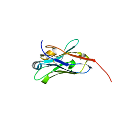
Crystal structure of human Cathepsin L in complex with covalently bound GC376
Descriptor: CHLORIDE ION, Cathepsin L, DI(HYDROXYETHYL)ETHER, ...
Authors:Reinke, P.Y.A, Falke, S, Lieske, J, Ewert, W, Loboda, J, Rahmani Mashhour, A, Hauser, M, Karnicar, K, Usenik, A, Lindic, N, Lach, M, Boehler, H, Beck, T, Cox, R, Chapman, H.N, Hinrichs, W, Turk, D, Guenther, S, Meents, A.
Deposit date:2021-12-17
Release date:2022-12-28
Last modified:2024-01-31
Method:X-RAY DIFFRACTION (1.8 Å)
Cite:Calpeptin is a potent cathepsin inhibitor and drug candidate for SARS-CoV-2 infections.
Commun Biol, 6, 2023
7QKA
DownloadVisualize
BU of 7qka by Molmil
Crystal structure of SARS-CoV-2 Main Protease in complex with covalently bound GC376
Descriptor: 3C-like proteinase nsp5, DIMETHYL SULFOXIDE, N~2~-[(benzyloxy)carbonyl]-N-{(2S)-1-hydroxy-3-[(3S)-2-oxopyrrolidin-3-yl]propan-2-yl}-L-leucinamide
Authors:Reinke, P.Y.A, Falke, S, Lieske, J, Ewert, W, Loboda, J, Rahmani Mashhour, A, Hauser, M, Karnicar, K, Usenik, A, Lindic, N, Lach, M, Boehler, H, Beck, T, Cox, R, Chapman, H.N, Hinrichs, W, Turk, D, Guenther, S, Meents, A.
Deposit date:2021-12-17
Release date:2022-12-28
Last modified:2024-01-31
Method:X-RAY DIFFRACTION (1.8 Å)
Cite:Calpeptin is a potent cathepsin inhibitor and drug candidate for SARS-CoV-2 infections.
Commun Biol, 6, 2023
8DTA
DownloadVisualize
BU of 8dta by Molmil
Metal sensitive GFP (mseGFP) complexed with phenylarsine oxide.
Descriptor: CALCIUM ION, DI(HYDROXYETHYL)ETHER, Green fluorescent protein, ...
Authors:Rosenbaum, J.C, Carlson, A.E.
Deposit date:2022-07-25
Release date:2023-07-05
Last modified:2024-01-31
Method:X-RAY DIFFRACTION (1.81 Å)
Cite:Glutathione modulates metal binding to proteins.
TBD
8EGK
DownloadVisualize
BU of 8egk by Molmil
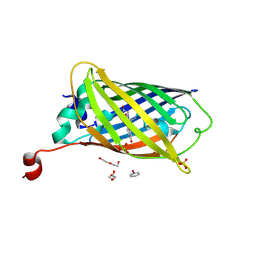
Re-refinement of Crystal Structure of NosGet3d, the All4481 protein from Nostoc sp. PCC 7120
Descriptor: AZIDE ION, All4481 protein
Authors:Barlow, A.N, Manu, M.S, Ramasamy, S, Clemons Jr, W.M.
Deposit date:2022-09-12
Release date:2023-05-24
Last modified:2023-11-15
Method:X-RAY DIFFRACTION (1.98 Å)
Cite:Structures of Get3d reveal a distinct architecture associated with the emergence of photosynthesis.
J.Biol.Chem., 299, 2023
7S8W
DownloadVisualize
BU of 7s8w by Molmil
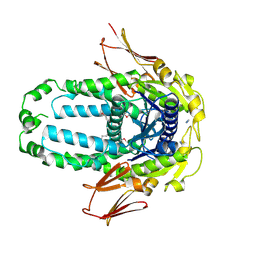
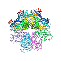
Amycolatopsis sp. T-1-60 N-succinylamino acid racemase/o-succinylbenzoate synthase R266Q mutant in complex with N-succinylphenylglycine
Descriptor: MAGNESIUM ION, N-succinyl-L-phenylglycine, N-succinylamino acid racemase/O-succinylbenzoate synthase, ...
Authors:Truong, D.P, Rousseau, S, Sacchettini, J.C, Glasner, M.E.
Deposit date:2021-09-20
Release date:2021-10-06
Last modified:2023-10-18
Method:X-RAY DIFFRACTION (2.9 Å)
Cite:Second-Shell Amino Acid R266 Helps Determine N -Succinylamino Acid Racemase Reaction Specificity in Promiscuous N -Succinylamino Acid Racemase/ o -Succinylbenzoate Synthase Enzymes.
Biochemistry, 60, 2021
8EB9
DownloadVisualize
BU of 8eb9 by Molmil
Crystal structure of SIX8 from Fusarium oxysporum f. sp. lycopersici
Descriptor: SULFATE ION, Secreted in xylem Six8
Authors:Yu, D.S, Ericsson, D.J, Williams, S.J.
Deposit date:2022-08-30
Release date:2023-06-21
Last modified:2024-04-03
Method:X-RAY DIFFRACTION (1.28 Å)
Cite:The structural repertoire of Fusarium oxysporum f. sp. lycopersici effectors revealed by experimental and computational studies.
Elife, 12, 2024
8EBB
DownloadVisualize
BU of 8ebb by Molmil
Crystal structure of SIX6 from Fusarium oxysporum f. sp. lycopersici
Descriptor: D(-)-TARTARIC ACID, Secreted in xylem Six6
Authors:Yu, D.S, Ericsson, D.J, Williams, S.J.
Deposit date:2022-08-30
Release date:2023-06-21
Last modified:2024-04-03
Method:X-RAY DIFFRACTION (1.88 Å)
Cite:The structural repertoire of Fusarium oxysporum f. sp. lycopersici effectors revealed by experimental and computational studies.
Elife, 12, 2024
8EE2
DownloadVisualize
BU of 8ee2 by Molmil
Crystal Structure of Nanobody VHH219 Bound to Its Antigen PA14 Cif
Descriptor: CFTR inhibitory factor, Nanobody VHH219
Authors:Simard, A.R, Madden, D.R.
Deposit date:2022-09-06
Release date:2023-09-13
Method:X-RAY DIFFRACTION (2.4 Å)
Cite:Crystal Structure of Nanobody VHH219 Bound to Its Antigen PA14 Cif
To Be Published
8EGZ
DownloadVisualize
BU of 8egz by Molmil
Engineered tyrosine synthase (TmTyrS1) derived from T. maritima TrpB with Ser bound as the amino-acrylate intermediate
Descriptor: 1,2-ETHANEDIOL, 2-{[(E)-{3-hydroxy-2-methyl-5-[(phosphonooxy)methyl]pyridin-4-yl}methylidene]amino}prop-2-enoic acid, POTASSIUM ION, ...
Authors:Porter, N.J, Almhjell, P.J, Arnold, F.H.
Deposit date:2022-09-13
Release date:2023-10-04
Method:X-RAY DIFFRACTION (1.9 Å)
Cite:Noncanonical L-tyrosine synthesis by evolved tryptophan synthases
To Be Published
8E2N
DownloadVisualize
BU of 8e2n by Molmil
Crystal Structure of Nanobody VHH113 Specific for PA14 Cif
Descriptor: Nanobody VHH113
Authors:Simard, A.R, Madden, D.R.
Deposit date:2022-08-15
Release date:2023-10-04
Method:X-RAY DIFFRACTION (1.1 Å)
Cite:Crystal Structure of Nanobody VHH113 Specific for PA14 Cif
To Be Published

219515

数据于2024-05-08公开中

PDB statisticsPDBj update infoContact PDBjnumon