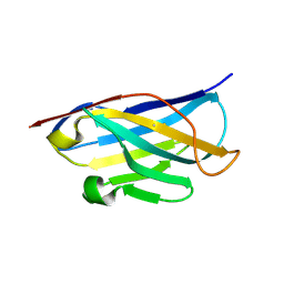
2LAL
 
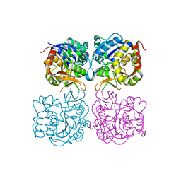
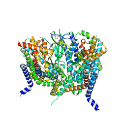
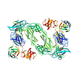
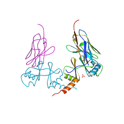
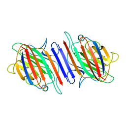 | | CRYSTAL STRUCTURE DETERMINATION AND REFINEMENT AT 2.3 ANGSTROMS RESOLUTION OF THE LENTIL LECTIN | | 分子名称: | CALCIUM ION, LENTIL LECTIN (ALPHA CHAIN), LENTIL LECTIN (BETA CHAIN), ... | | 著者 | Loris, R, Steyaert, J, Maes, D, Lisgarten, J, Pickersgill, R, Wyns, L. | | 登録日 | 1993-06-10 | | 公開日 | 1993-10-31 | | 最終更新日 | 2024-02-21 | | 実験手法 | X-RAY DIFFRACTION (1.8 Å) | | 主引用文献 | Structural analysis of two crystal forms of lentil lectin at 1.8 A resolution.
Proteins, 20, 1994
|
|
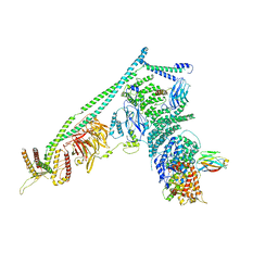
8E0G
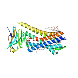 | | Re-refined model of active mu-opioid receptor (PDB 5c1m) as an adduct with BU72 | | 分子名称: | (2R)-2,3-dihydroxypropyl (9Z)-octadec-9-enoate, (2R,3S,3aR,5aR,6R,11bR,11cS)-3a-methoxy-3,14-dimethyl-2-phenyl-2,3,3a,6,7,11c-hexahydro-1H-6,11b-(epiminoethano)-3,5a-methanonaphtho[2,1-g]indol-10-ol, CHOLESTEROL, ... | | 著者 | Munro, T.A. | | 登録日 | 2022-08-09 | | 公開日 | 2023-10-18 | | 最終更新日 | 2023-11-15 | | 実験手法 | X-RAY DIFFRACTION (2.1 Å) | | 主引用文献 | Reanalysis of a mu opioid receptor crystal structure reveals a covalent adduct with BU72.
Bmc Biol., 21, 2023
|
|
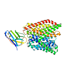
5N7O
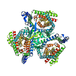
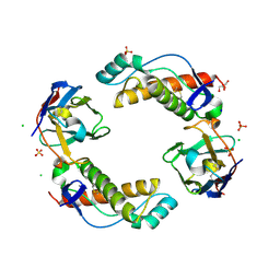
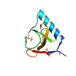
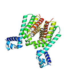 | | EthR2 in complex with SMARt-420 compound | | 分子名称: | 4,4,4-trifluoro-1-(3-phenyl-1-oxa-2,8-diazaspiro[4.5]dec-2-en-8-yl)butan-1-one, Probable transcriptional regulatory protein | | 著者 | Wohlkonig, A, Wintjens, R. | | 登録日 | 2017-02-21 | | 公開日 | 2017-04-26 | | 最終更新日 | 2024-01-17 | | 実験手法 | X-RAY DIFFRACTION (2.3 Å) | | 主引用文献 | Structural analysis of the interaction between spiroisoxazoline SMARt-420 and the Mycobacterium tuberculosis repressor EthR2.
Biochem. Biophys. Res. Commun., 487, 2017
|
|
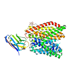
6GS7
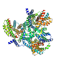
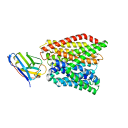 | | Crystal structure of peptide transporter DtpA-nanobody in glycine buffer | | 分子名称: | Dipeptide and tripeptide permease A, nanobody | | 著者 | Ural-Blimke, Y, Flayhan, A, Quistgaard, E.M, Loew, C. | | 登録日 | 2018-06-13 | | 公開日 | 2019-01-30 | | 最終更新日 | 2024-01-17 | | 実験手法 | X-RAY DIFFRACTION (3.3 Å) | | 主引用文献 | Structure of Prototypic Peptide Transporter DtpA from E. coli in Complex with Valganciclovir Provides Insights into Drug Binding of Human PepT1.
J. Am. Chem. Soc., 141, 2019
|
|
6GS1
 | | Crystal structure of peptide transporter DtpA-nanobody in MES buffer | | 分子名称: | Dipeptide and tripeptide permease A, Nanobody 00 | | 著者 | Ural-Blimke, Y, Flayhan, A, Loew, C, Quistgaard, E.M. | | 登録日 | 2018-06-13 | | 公開日 | 2019-01-30 | | 最終更新日 | 2024-01-17 | | 実験手法 | X-RAY DIFFRACTION (3.29 Å) | | 主引用文献 | Structure of Prototypic Peptide Transporter DtpA from E. coli in Complex with Valganciclovir Provides Insights into Drug Binding of Human PepT1.
J. Am. Chem. Soc., 141, 2019
|
|
6GS4
 | | Crystal structure of peptide transporter DtpA-nanobody in complex with valganciclovir | | 分子名称: | DODECYL-BETA-D-MALTOSIDE, Dipeptide and tripeptide permease A, [(2~{S})-2-[(2-azanyl-6-oxidanylidene-3~{H}-purin-9-yl)methoxy]-3-oxidanyl-propyl] (2~{S})-2-azanyl-3-methyl-butanoate, ... | | 著者 | Ural-Blimke, Y, Flayhan, A, Quistgaard, E.M, Loew, C. | | 登録日 | 2018-06-13 | | 公開日 | 2019-01-30 | | 最終更新日 | 2024-01-17 | | 実験手法 | X-RAY DIFFRACTION (2.645 Å) | | 主引用文献 | Structure of Prototypic Peptide Transporter DtpA from E. coli in Complex with Valganciclovir Provides Insights into Drug Binding of Human PepT1.
J. Am. Chem. Soc., 141, 2019
|
|
5DA0
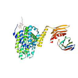 | | Structure of the the SLC26 transporter SLC26Dg in complex with a nanobody | | 分子名称: | DECYL-BETA-D-MALTOPYRANOSIDE, Nanobody, Sulphate transporter | | 著者 | Dutzler, R, Geertsma, E.R, Chang, Y, Shaik, F.R. | | 登録日 | 2015-08-19 | | 公開日 | 2015-09-09 | | 最終更新日 | 2015-10-14 | | 実験手法 | X-RAY DIFFRACTION (3.2 Å) | | 主引用文献 | Structure of a prokaryotic fumarate transporter reveals the architecture of the SLC26 family.
Nat.Struct.Mol.Biol., 22, 2015
|
|
4AQ1
 | |
5DA4
 | |
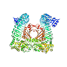
5DFZ
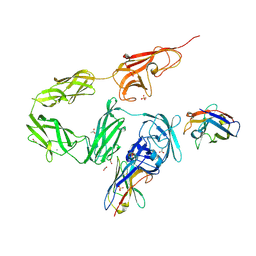
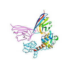
 | | Structure of Vps34 complex II from S. cerevisiae. | | 分子名称: | Nanobody binding S. cerevisiae Vps34, Phosphatidylinositol 3-kinase VPS34, Putative N-terminal domain of S. cerevisiae Vps30, ... | | 著者 | Rostislavleva, K, Soler, N, Ohashi, Y, Zhang, L, Williams, R.L. | | 登録日 | 2015-08-27 | | 公開日 | 2015-10-07 | | 最終更新日 | 2024-05-08 | | 実験手法 | X-RAY DIFFRACTION (4.4 Å) | | 主引用文献 | Structure and flexibility of the endosomal Vps34 complex reveals the basis of its function on membranes.
Science, 350, 2015
|
|
6HLU
 | |
5M94
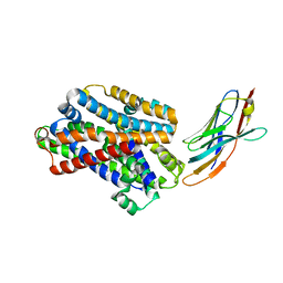 | |
5MJ7
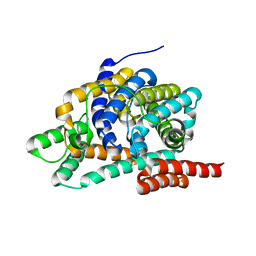
 | | Structure of the C. elegans nucleoside hydrolase | | 分子名称: | 2-AMINO-2-HYDROXYMETHYL-PROPANE-1,3-DIOL, CALCIUM ION, Uncharacterized protein | | 著者 | Versees, W, Singh, R.K. | | 登録日 | 2016-11-30 | | 公開日 | 2017-03-08 | | 最終更新日 | 2024-01-17 | | 実験手法 | X-RAY DIFFRACTION (1.65 Å) | | 主引用文献 | Structural and biochemical characterization of the nucleoside hydrolase from C. elegans reveals the role of two active site cysteine residues in catalysis.
Protein Sci., 26, 2017
|
|
8OPR
 | |
8OUJ
 | |
8OUI
 | | Complex of ASCT2 with Suppressyn | | 分子名称: | ALANINE, Neutral amino acid transporter B(0), Suppressyn | | 著者 | Khare, S, Kumar, A, Reyes, N. | | 登録日 | 2023-04-23 | | 公開日 | 2024-05-01 | | 最終更新日 | 2024-05-15 | | 実験手法 | ELECTRON MICROSCOPY (3.39 Å) | | 主引用文献 | Receptor-recognition and antiviral mechanisms of retrovirus-derived human proteins.
Nat.Struct.Mol.Biol., 2024
|
|
8OUH
 | | Complex of human ASCT2 with Syncytin-1 | | 分子名称: | ALANINE, Neutral amino acid transporter B(0), Syncytin-1 | | 著者 | Khare, S, Reyes, N. | | 登録日 | 2023-04-23 | | 公開日 | 2024-05-01 | | 最終更新日 | 2024-05-15 | | 実験手法 | ELECTRON MICROSCOPY (2.62 Å) | | 主引用文献 | Receptor-recognition and antiviral mechanisms of retrovirus-derived human proteins.
Nat.Struct.Mol.Biol., 2024
|
|
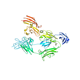
6HER
 | | Mouse prion protein in complex with Nanobody 484 | | 分子名称: | GLYCEROL, Major prion protein, Nanobody 484, ... | | 著者 | Soror, S.H, Abskharon, R, Wohlkonig, A. | | 登録日 | 2018-08-20 | | 公開日 | 2019-12-04 | | 最終更新日 | 2024-01-17 | | 実験手法 | X-RAY DIFFRACTION (1.199 Å) | | 主引用文献 | Structural evidence for the critical role of the prion protein hydrophobic region in forming an infectious prion.
Plos Pathog., 15, 2019
|
|
6HHD
 | | Mouse Prion Protein in complex with Nanobody 484 | | 分子名称: | CHLORIDE ION, GLYCEROL, Major prion protein, ... | | 著者 | Soror, S, Abskharon, R, Wohlkonig, A. | | 登録日 | 2018-08-28 | | 公開日 | 2019-12-11 | | 最終更新日 | 2024-01-17 | | 実験手法 | X-RAY DIFFRACTION (2.102 Å) | | 主引用文献 | Structural evidence for the critical role of the prion protein hydrophobic region in forming an infectious prion.
Plos Pathog., 15, 2019
|
|
6QX4
 | |
3GSP
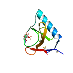 | | RIBONUCLEASE T1 COMPLEXED WITH 2',3'-CGPS + 3'-GMP, 4 DAYS | | 分子名称: | CALCIUM ION, GUANOSINE-2',3'-CYCLOPHOSPHOROTHIOATE, GUANOSINE-3'-MONOPHOSPHATE, ... | | 著者 | Zegers, I, Wyns, L. | | 登録日 | 1997-12-02 | | 公開日 | 1998-08-12 | | 最終更新日 | 2023-08-09 | | 実験手法 | X-RAY DIFFRACTION (1.9 Å) | | 主引用文献 | Hydrolysis of a slow cyclic thiophosphate substrate of RNase T1 analyzed by time-resolved crystallography.
Nat.Struct.Biol., 5, 1998
|
|
5G5R
 | |
5GSP
 | | RIBONUCLEASE T1/3'-GMP, 9 WEEKS | | 分子名称: | CALCIUM ION, GUANOSINE-3'-MONOPHOSPHATE, RIBONUCLEASE T1 | | 著者 | Zegers, I, Wyns, L. | | 登録日 | 1997-12-09 | | 公開日 | 1998-03-18 | | 最終更新日 | 2023-08-09 | | 実験手法 | X-RAY DIFFRACTION (1.8 Å) | | 主引用文献 | Hydrolysis of a slow cyclic thiophosphate substrate of RNase T1 analyzed by time-resolved crystallography.
Nat.Struct.Biol., 5, 1998
|
|
5G5X
 | |
5IOF
 | |


