Loading
PDBj
MenuPDBj@FacebookPDBj@TwitterPDBj@YouTubewwPDB FoundationwwPDB
RCSB PDBPDBeBMRBAdv. SearchSearch help
Search by PDB author
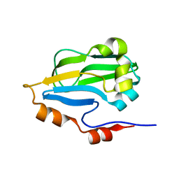
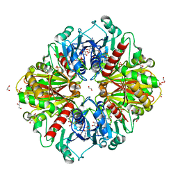
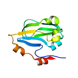
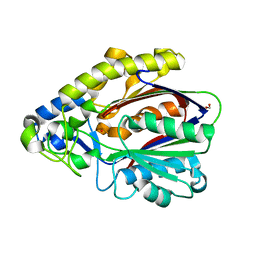
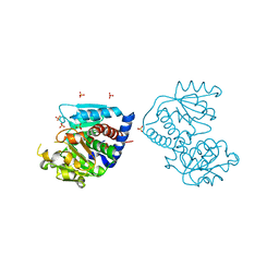
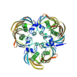
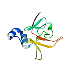
3CEZ
DownloadVisualize
BU of 3cez by Molmil
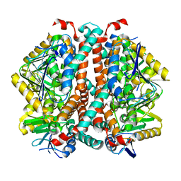
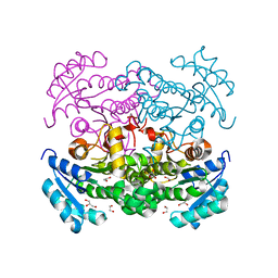
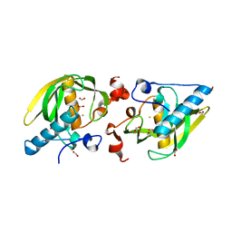
Crystal structure of methionine-R-sulfoxide reductase from Burkholderia pseudomallei
Descriptor: ACETIC ACID, Methionine-R-sulfoxide reductase, ZINC ION
Authors:Staker, B, Napuli, A, Nakazawa, S.H, Castaneda, L, Alkafeef, S, Vanvoorhis, W, Stewart, L, Myler, P, Seattle Structural Genomics Center for Infectious Disease (SSGCID)
Deposit date:2008-02-29
Release date:2008-03-18
Last modified:2023-08-30
Method:X-RAY DIFFRACTION (2.1 Å)
Cite:Methionine-R-sulfoxide reductase from Burkholderia pseudomallei.
To be Published
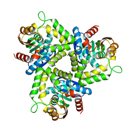
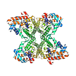
8SOY
DownloadVisualize
BU of 8soy by Molmil
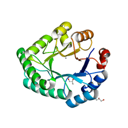
Cryo-EM structure of Enoyl-CoA hydratase from Mycobacterium smegmatis
Descriptor: Enoyl-CoA hydratase EchA21
Authors:Shek, R, Quispe, J, Staker, B, Seattle Structural Genomics Center for Infectious Disease (SSGCID)
Deposit date:2023-04-30
Release date:2023-05-10
Method:ELECTRON MICROSCOPY (2.85 Å)
Cite:Cryo-EM structure of Enoyl-CoA hydratase from Mycobacterium smegmatis
To Be Published
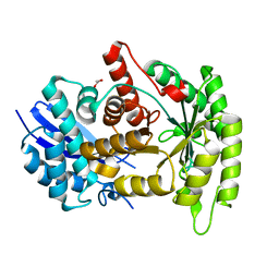
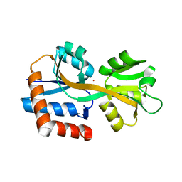
1R42
DownloadVisualize
BU of 1r42 by Molmil
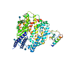
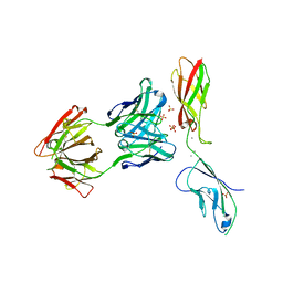
Native Human Angiotensin Converting Enzyme-Related Carboxypeptidase (ACE2)
Descriptor: 2-acetamido-2-deoxy-beta-D-glucopyranose, CHLORIDE ION, ZINC ION, ...
Authors:Towler, P, Staker, B, Prasad, S.G, Menon, S, Ryan, D, Tang, J, Parsons, T, Fisher, M, Williams, D, Dales, N.A, Patane, M.A, Pantoliano, M.W.
Deposit date:2003-10-07
Release date:2004-02-03
Last modified:2020-07-29
Method:X-RAY DIFFRACTION (2.2 Å)
Cite:ACE2 X-ray structures reveal a large hinge-bending motion important for inhibitor binding and catalysis.
J.Biol.Chem., 279, 2004
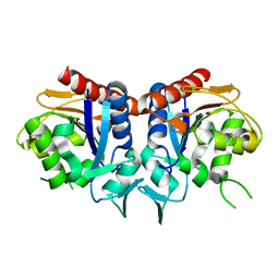
1R4L
DownloadVisualize
BU of 1r4l by Molmil
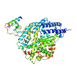
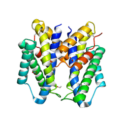
Inhibitor Bound Human Angiotensin Converting Enzyme-Related Carboxypeptidase (ACE2)
Descriptor: (S,S)-2-{1-CARBOXY-2-[3-(3,5-DICHLORO-BENZYL)-3H-IMIDAZOL-4-YL]-ETHYLAMINO}-4-METHYL-PENTANOIC ACID, 2-acetamido-2-deoxy-beta-D-glucopyranose, CHLORIDE ION, ...
Authors:Towler, P, Staker, B, Prasad, S.G, Menon, S, Ryan, D, Tang, J, Parsons, T, Fisher, M, Williams, D, Dales, N.A, Patane, M.A, Pantoliano, M.W.
Deposit date:2003-10-07
Release date:2004-02-03
Last modified:2023-08-23
Method:X-RAY DIFFRACTION (3 Å)
Cite:ACE2 X-ray structures reveal a large hinge-bending motion important for inhibitor binding and catalysis.
J.Biol.Chem., 279, 2004
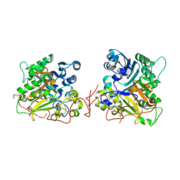
8FBP
DownloadVisualize
BU of 8fbp by Molmil
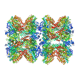
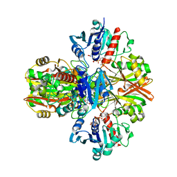
Glutamine synthetase from Pseudomonas aeruginosa, filament double-unit in compressed conformation
Descriptor: Glutamine synthetase
Authors:Phan, I.Q, Staker, B, Shek, R, Moser, T.H, Evans, J.E, van Voorhis, W.C, Myler, P.J, Seattle Structural Genomics Center for Infectious Disease (SSGCID)
Deposit date:2022-11-29
Release date:2024-02-14
Method:ELECTRON MICROSCOPY (2.8 Å)
Cite:Glutamine synthetase from Pseudomonas aeruginosa, filament double-unit in compressed conformation
To Be Published
1QZQ
DownloadVisualize
BU of 1qzq by Molmil
human Tyrosyl DNA phosphodiesterase
Descriptor: tyrosyl-DNA phosphodiesterase 1
Authors:Raymond, A.C, Rideout, M.C, Staker, B, Hjerrild, K, Burgin Jr, A.B.
Deposit date:2003-09-17
Release date:2004-05-11
Last modified:2024-02-14
Method:X-RAY DIFFRACTION (2.4 Å)
Cite:Analysis of Human Tyrosyl-DNA Phosphodiesterase I Catalytic Residues.
J.Mol.Biol., 338, 2004
3IFT
DownloadVisualize
BU of 3ift by Molmil
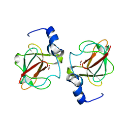
Crystal structure of glycine cleavage system protein H from Mycobacterium tuberculosis, using X-rays from the Compact Light Source.
Descriptor: Glycine cleavage system H protein
Authors:Edwards, T.E, Abendroth, J, Staker, B, Mayer, C, Phan, I, Kelley, A, Analau, E, Leibly, D, Rifkin, J, Loewen, R, Ruth, R.D, Stewart, L.J, Accelerated Technologies Center for Gene to 3D Structure (ATCG3D)
Deposit date:2009-07-25
Release date:2009-08-11
Last modified:2023-09-06
Method:X-RAY DIFFRACTION (2 Å)
Cite:X-ray structure determination of the glycine cleavage system protein H of Mycobacterium tuberculosis using an inverse Compton synchrotron X-ray source.
J.Struct.Funct.Genom., 11, 2010
4DGQ
DownloadVisualize
BU of 4dgq by Molmil
Crystal structure of Non-heme chloroperoxidase from Burkholderia cenocepacia
Descriptor: 1,2-ETHANEDIOL, Non-heme chloroperoxidase
Authors:Gardberg, A.S, Edwards, T.E, Abendroth, J.A, Staker, B, Stewart, L, Seattle Structural Genomics Center for Infectious Disease (SSGCID)
Deposit date:2012-01-26
Release date:2012-03-07
Last modified:2023-09-13
Method:X-RAY DIFFRACTION (1.85 Å)
Cite:Crystal structure of Non-heme chloroperoxidase from Burkholderia cenocepacia
To be Published
4G7F
DownloadVisualize
BU of 4g7f by Molmil
Crystal Structure of Enolase from Trypanosoma Cruzi
Descriptor: 1,2-ETHANEDIOL, Enolase, MAGNESIUM ION
Authors:Craig, T.K, Edwards, T.E, Staker, B, Seattle Structural Genomics Center for Infectious Disease (SSGCID)
Deposit date:2012-07-20
Release date:2012-08-29
Last modified:2023-09-13
Method:X-RAY DIFFRACTION (2.4 Å)
Cite:Crystal Structure of Enolase from Trypanosoma Cruzi
To be Published
3K2E
DownloadVisualize
BU of 3k2e by Molmil
Crystal structure of enoyl-(acyl-carrier-protein) reductase from Anaplasma phagocytophilum at 1.9A resolution
Descriptor: 1,2-ETHANEDIOL, Enoyl-(Acyl-carrier-protein) reductase, GLYCEROL
Authors:Abendroth, J, Staker, B, Seattle Structural Genomics Center for Infectious Disease (SSGCID)
Deposit date:2009-09-30
Release date:2009-10-13
Last modified:2023-09-06
Method:X-RAY DIFFRACTION (1.9 Å)
Cite:Crystal structure of enoyl-(acyl-carrier-protein) reductase from Anaplasma phagocytophilum at 1.9A resolution
To be Published
3NGF
DownloadVisualize
BU of 3ngf by Molmil
Crystal structure of AP endonuclease, family 2 from Brucella melitensis
Descriptor: AP endonuclease, family 2, GLYCEROL, ...
Authors:Seattle Structural Genomics Center for Infectious Disease (SSGCID)
Deposit date:2010-06-11
Release date:2010-07-07
Last modified:2023-09-06
Method:X-RAY DIFFRACTION (1.8 Å)
Cite:Crystal structure of AP endonuclease, family 2 from Brucella melitensis
To be Published
6VEL
DownloadVisualize
BU of 6vel by Molmil
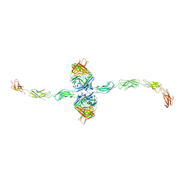
Crystal Structure of Human E-cadherin bound by mouse monoclonal antibody 66E8Fab
Descriptor: 66E8 Fab Heavy Chain, 66E8 Fab Light Chain, CALCIUM ION, ...
Authors:Seattle Structural Genomics Center for Infectious Disease, Seattle Structural Genomics Center for Infectious Disease (SSGCID)
Deposit date:2020-01-02
Release date:2020-01-29
Last modified:2023-10-25
Method:X-RAY DIFFRACTION (2.65 Å)
Cite:Regulation of multiple dimeric states of E-cadherin by adhesion activating antibodies revealed through Cryo-EM and X-ray crystallography.
Pnas Nexus, 1, 2022
6CNZ
DownloadVisualize
BU of 6cnz by Molmil
Crystal Structure of Chorismate Mutase from Burkholderia thailandensis
Descriptor: 1,2-ETHANEDIOL, Chorismate mutase, NITRATE ION
Authors:Seattle Structural Genomics Center for Infectious Disease (SSGCID)
Deposit date:2018-03-09
Release date:2018-03-21
Last modified:2023-10-04
Method:X-RAY DIFFRACTION (2.15 Å)
Cite:Crystal structure of chorismate mutase from Burkholderia thailandensis.
Acta Crystallogr F Struct Biol Commun, 74, 2018
7STZ
DownloadVisualize
BU of 7stz by Molmil
Crystal Structure of Human E-cadherin EC1-5 bound by mouse monoclonal antibody Fab mAb-1_19A11
Descriptor: 1,2-ETHANEDIOL, 2-acetamido-2-deoxy-beta-D-glucopyranose, CALCIUM ION, ...
Authors:Seattle Structural Genomics Center for Infectious Disease, Seattle Structural Genomics Center for Infectious Disease (SSGCID)
Deposit date:2021-11-15
Release date:2022-01-26
Last modified:2023-10-25
Method:X-RAY DIFFRACTION (2.95 Å)
Cite:Regulation of multiple dimeric states of E-cadherin by adhesion activating antibodies revealed through Cryo-EM and X-ray crystallography.
Pnas Nexus, 1, 2022
5VMT
DownloadVisualize
BU of 5vmt by Molmil
Crystal structure of a glyceraldehyde-3-phosphate dehydrogenase from Neisseria gonorrhoeae bound to NAD
Descriptor: CHLORIDE ION, Glyceraldehyde-3-phosphate dehydrogenase, NICOTINAMIDE-ADENINE-DINUCLEOTIDE
Authors:Seattle Structural Genomics Center for Infectious Disease (SSGCID)
Deposit date:2017-04-28
Release date:2017-05-10
Last modified:2023-10-04
Method:X-RAY DIFFRACTION (2.5 Å)
Cite:Structures of glyceraldehyde 3-phosphate dehydrogenase in Neisseria gonorrhoeae and Chlamydia trachomatis.
Protein Sci., 29, 2020
5KOB
DownloadVisualize
BU of 5kob by Molmil
Crystal structure of a peptide deformylase from Burkholderia xenovorans
Descriptor: 1,2-ETHANEDIOL, FE (II) ION, FORMIC ACID, ...
Authors:SSGCID, Seattle Structural Genomics Center for Infectious Disease (SSGCID)
Deposit date:2016-06-30
Release date:2016-07-13
Last modified:2023-10-04
Method:X-RAY DIFFRACTION (1.6 Å)
Cite:Crystal structure of a peptide deformylase from Burkholderia xenovorans
to be published
3CXK
DownloadVisualize
BU of 3cxk by Molmil
1.7 A Crystal structure of methionine-R-sulfoxide reductase from Burkholderia pseudomallei: crystallization in a microfluidic crystal card.
Descriptor: ACETATE ION, Methionine-R-sulfoxide reductase, ZINC ION
Authors:Lovell, S, Gerdts, C, Staker, B, Craigen, D, Stewart, L, Accelerated Technologies Center for Gene to 3D Structure (ATCG3D), Seattle Structural Genomics Center for Infectious Disease (SSGCID)
Deposit date:2008-04-24
Release date:2008-05-27
Last modified:2023-08-30
Method:X-RAY DIFFRACTION (1.7 Å)
Cite:The plug-based nanovolume Microcapillary Protein Crystallization System (MPCS).
Acta Crystallogr.,Sect.D, 64, 2008
6OK4
DownloadVisualize
BU of 6ok4 by Molmil
Crystal Structure of Glyceraldehyde-3-phosphate dehydrogenase (GAPDH) from Chlamydia trachomatis with bound NAD
Descriptor: 1,2-ETHANEDIOL, CHLORIDE ION, DI(HYDROXYETHYL)ETHER, ...
Authors:Seattle Structural Genomics Center for Infectious Disease, Seattle Structural Genomics Center for Infectious Disease (SSGCID)
Deposit date:2019-04-12
Release date:2019-04-24
Last modified:2023-10-11
Method:X-RAY DIFFRACTION (2.4 Å)
Cite:Structures of glyceraldehyde 3-phosphate dehydrogenase in Neisseria gonorrhoeae and Chlamydia trachomatis.
Protein Sci., 29, 2020
3HGB
DownloadVisualize
BU of 3hgb by Molmil
Crystal structure of glycine cleavage system protein H from Mycobacterium tuberculosis
Descriptor: Glycine cleavage system H protein
Authors:Seattle Structural Genomics Center for Infectious Disease (SSGCID)
Deposit date:2009-05-13
Release date:2009-05-26
Last modified:2023-09-06
Method:X-RAY DIFFRACTION (1.75 Å)
Cite:X-ray structure determination of the glycine cleavage system protein H of Mycobacterium tuberculosis using an inverse Compton synchrotron X-ray source.
J.Struct.Funct.Genom., 11, 2010
4EFI
DownloadVisualize
BU of 4efi by Molmil
Crystal Structure of 3-oxoacyl-(Acyl-carrier protein) Synthase from Burkholderia Xenovorans LB400
Descriptor: 3-oxoacyl-(Acyl-carrier protein) synthase, CHLORIDE ION, FORMIC ACID, ...
Authors:Seattle Structural Genomics Center for Infectious Disease, Craig, T.K, Abendroth, J, Staker, B, Stewart, L, Seattle Structural Genomics Center for Infectious Disease (SSGCID)
Deposit date:2012-03-29
Release date:2012-04-11
Last modified:2023-09-13
Method:X-RAY DIFFRACTION (1.35 Å)
Cite:Combining functional and structural genomics to sample the essential Burkholderia structome.
Plos One, 8, 2013
4F2G
DownloadVisualize
BU of 4f2g by Molmil
The Crystal Structure of Ornithine carbamoyltransferase from Burkholderia thailandensis E264
Descriptor: 1,2-ETHANEDIOL, Ornithine carbamoyltransferase 1, PHOSPHATE ION
Authors:Craig, T.K, Fox, D, Staker, B, Stewart, L, Seattle Structural Genomics Center for Infectious Disease (SSGCID)
Deposit date:2012-05-07
Release date:2012-05-30
Last modified:2023-09-13
Method:X-RAY DIFFRACTION (2.1 Å)
Cite:Combining functional and structural genomics to sample the essential Burkholderia structome.
Plos One, 8, 2013
4H4G
DownloadVisualize
BU of 4h4g by Molmil
Crystal Structure of (3R)-hydroxymyristoyl-[acyl-carrier-protein] dehydratase from Burkholderia thailandensis E264
Descriptor: (3R)-hydroxymyristoyl-[acyl-carrier-protein] dehydratase
Authors:Craig, T.K, Edwards, T.E, Staker, B, Stewart, L, Lorimer, D, Seattle Structural Genomics Center for Infectious Disease (SSGCID)
Deposit date:2012-09-17
Release date:2012-10-03
Last modified:2023-09-20
Method:X-RAY DIFFRACTION (2.65 Å)
Cite:Combining functional and structural genomics to sample the essential Burkholderia structome.
Plos One, 8, 2013
3MMT
DownloadVisualize
BU of 3mmt by Molmil
Crystal structure of fructose bisphosphate aldolase from Bartonella henselae, bound to fructose bisphosphate
Descriptor: 1,6-FRUCTOSE DIPHOSPHATE (LINEAR FORM), Fructose-bisphosphate aldolase
Authors:Seattle Structural Genomics Center for Infectious Disease (SSGCID)
Deposit date:2010-04-20
Release date:2010-05-19
Last modified:2023-09-06
Method:X-RAY DIFFRACTION (2.35 Å)
Cite:Structure of fructose bisphosphate aldolase from Bartonella henselae bound to fructose 1,6-bisphosphate.
Acta Crystallogr.,Sect.F, 67, 2011
4F3P
DownloadVisualize
BU of 4f3p by Molmil
Crystal structure of a Glutamine-binding periplasmic protein from Burkholderia pseudomallei in complex with glutamine
Descriptor: Glutamine-binding periplasmic protein
Authors:Seattle Structural Genomics Center for Infectious Disease (SSGCID)
Deposit date:2012-05-09
Release date:2012-05-23
Last modified:2023-09-13
Method:X-RAY DIFFRACTION (2.4 Å)
Cite:Crystal structure of a Glutamine-binding periplasmic protein from Burkholderia pseudomallei in complex with glutamine
To be Published
3L3B
DownloadVisualize
BU of 3l3b by Molmil
Crystal structure of isoprenoid biosynthesis protein with amidotransferase-like domain from Ehrlichia Chaffeensis at 1.90A resolution
Descriptor: Es1 family protein
Authors:Seattle Structural Genomics Center for Infectious Disease (SSGCID)
Deposit date:2009-12-16
Release date:2010-01-19
Last modified:2023-09-06
Method:X-RAY DIFFRACTION (1.9 Å)
Cite:Crystal structure of isoprenoid biosynthesis protein with amidotransferase-like domain from Ehrlichia Chaffeensis at 1.90A resolution
To be Published


1234>

220113

PDB entries from 2024-05-22

PDB statisticsPDBj update infoContact PDBjnumon