Loading
PDBj
MenuPDBj@FacebookPDBj@TwitterPDBj@YouTubewwPDB FoundationwwPDB
RCSB PDBPDBeBMRBAdv. SearchSearch help
Search by PDB author
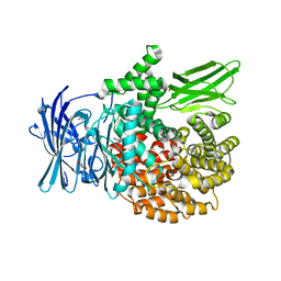
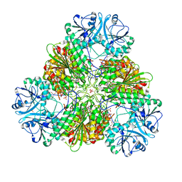
4R7M
DownloadVisualize
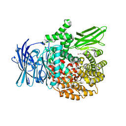
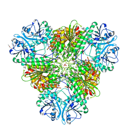
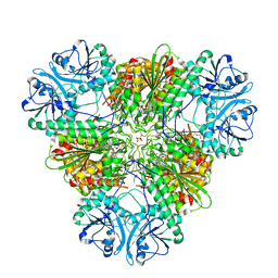
BU of 4r7m by Molmil
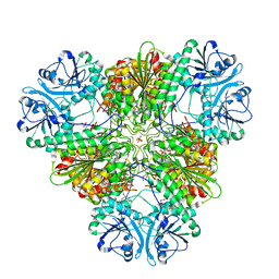
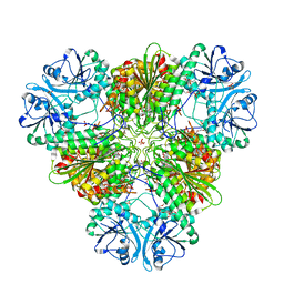
Structure of the m17 leucyl aminopeptidase from malaria complexed with a hydroxamic acid-based inhibitor
Descriptor: 4-amino-N-{(1R)-2-(hydroxyamino)-2-oxo-1-[4-(1H-pyrazol-1-yl)phenyl]ethyl}benzamide, CARBONATE ION, DIMETHYL SULFOXIDE, ...
Authors:Drinkwater, N, Mcgowan, S.
Deposit date:2014-08-28
Release date:2014-10-29
Last modified:2023-09-20
Method:X-RAY DIFFRACTION (2.85 Å)
Cite:Two-Pronged Attack: Dual Inhibition of Plasmodium falciparum M1 and M17 Metalloaminopeptidases by a Novel Series of Hydroxamic Acid-Based Inhibitors.
J.Med.Chem., 57, 2014
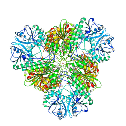
4R5V
DownloadVisualize
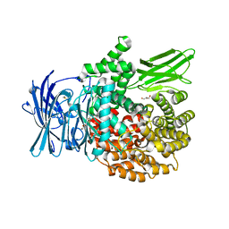
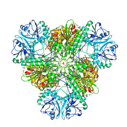
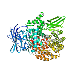
BU of 4r5v by Molmil
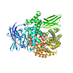
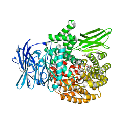
Structure of the m1 alanylaminopeptidase from malaria complexed with a hydroxamic acid-based inhibitor
Descriptor: GLYCEROL, M1 family aminopeptidase, N-{(1R)-2-(hydroxyamino)-2-oxo-1-[4-(1H-pyrazol-1-yl)phenyl]ethyl}-2,2-dimethylpropanamide, ...
Authors:Drinkwater, N, Mcgowan, S.
Deposit date:2014-08-22
Release date:2014-10-29
Last modified:2023-09-20
Method:X-RAY DIFFRACTION (2.1 Å)
Cite:Two-Pronged Attack: Dual Inhibition of Plasmodium falciparum M1 and M17 Metalloaminopeptidases by a Novel Series of Hydroxamic Acid-Based Inhibitors.
J.Med.Chem., 57, 2014
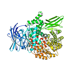
4R1B
DownloadVisualize
BU of 4r1b by Molmil
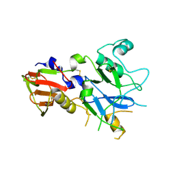
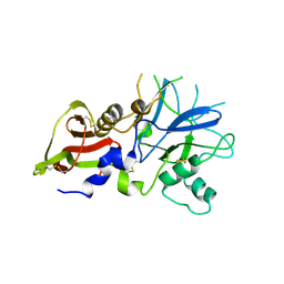
Crystal Structure of 3D7 strain Plasmodium falciparum AMA1
Descriptor: Apical membrane antigen 1, AMA1
Authors:Lim, S.S, Norton, R.S, McGowan, S.
Deposit date:2014-08-04
Release date:2015-01-14
Last modified:2023-09-20
Method:X-RAY DIFFRACTION (1.6 Å)
Cite:Structure and Dynamics of Apical Membrane Antigen 1 from Plasmodium falciparum FVO.
Biochemistry, 53, 2014
4R5X
DownloadVisualize
BU of 4r5x by Molmil
Structure of the m1 alanylaminopeptidase from malaria complexed with a hydroxamic acid-based inhibitor
Descriptor: 3-amino-N-{(1R)-2-(hydroxyamino)-2-oxo-1-[4-(1H-pyrazol-1-yl)phenyl]ethyl}benzamide, DIMETHYL SULFOXIDE, GLYCEROL, ...
Authors:Drinkwater, N, Mcgowan, S.
Deposit date:2014-08-22
Release date:2014-10-29
Last modified:2024-02-28
Method:X-RAY DIFFRACTION (1.85 Å)
Cite:Two-Pronged Attack: Dual Inhibition of Plasmodium falciparum M1 and M17 Metalloaminopeptidases by a Novel Series of Hydroxamic Acid-Based Inhibitors.
J.Med.Chem., 57, 2014
4R76
DownloadVisualize
BU of 4r76 by Molmil
Structure of the m17 leucyl aminopeptidase from malaria complexed with a hydroxamic acid-based inhibitor
Descriptor: 3-amino-N-{(1R)-2-(hydroxyamino)-2-oxo-1-[4-(1H-pyrazol-1-yl)phenyl]ethyl}benzamide, CARBONATE ION, M17 family aminopeptidase, ...
Authors:Drinkwater, N, Mcgowan, S.
Deposit date:2014-08-27
Release date:2014-10-29
Last modified:2023-09-20
Method:X-RAY DIFFRACTION (2.5 Å)
Cite:Two-Pronged Attack: Dual Inhibition of Plasmodium falciparum M1 and M17 Metalloaminopeptidases by a Novel Series of Hydroxamic Acid-Based Inhibitors.
J.Med.Chem., 57, 2014
4R1A
DownloadVisualize
BU of 4r1a by Molmil
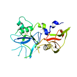
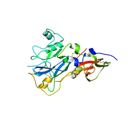
Crystal Structure of FVO strain Plasmodium falciparum AMA1
Descriptor: Apical membrane antigen 1
Authors:Lim, S.S, Norton, R.S, McGowan, S.
Deposit date:2014-08-04
Release date:2015-01-14
Last modified:2023-09-20
Method:X-RAY DIFFRACTION (2 Å)
Cite:Structure and Dynamics of Apical Membrane Antigen 1 from Plasmodium falciparum FVO.
Biochemistry, 53, 2014
4R1C
DownloadVisualize
BU of 4r1c by Molmil
Crystal Structure of 3D7 strain Plasmodium falciparum AMA1
Descriptor: Apical membrane antigen 1, AMA1
Authors:Lim, S.S, Norton, R.S, McGowan, S.
Deposit date:2014-08-04
Release date:2015-01-14
Last modified:2023-09-20
Method:X-RAY DIFFRACTION (2 Å)
Cite:Structure and Dynamics of Apical Membrane Antigen 1 from Plasmodium falciparum FVO.
Biochemistry, 53, 2014
4R5T
DownloadVisualize
BU of 4r5t by Molmil
Structure of the m1 alanylaminopeptidase from malaria complexed with a hydroxamic acid-based inhibitor
Descriptor: DIMETHYL SULFOXIDE, GLYCEROL, M1 family aminopeptidase, ...
Authors:Drinkwater, N, Mcgowan, S.
Deposit date:2014-08-22
Release date:2014-10-29
Last modified:2023-09-20
Method:X-RAY DIFFRACTION (1.98 Å)
Cite:Two-Pronged Attack: Dual Inhibition of Plasmodium falciparum M1 and M17 Metalloaminopeptidases by a Novel Series of Hydroxamic Acid-Based Inhibitors.
J.Med.Chem., 57, 2014
4R6T
DownloadVisualize
BU of 4r6t by Molmil
Structure of the m17 leucyl aminopeptidase from malaria complexed with a hydroxamic acid-based inhibitor
Descriptor: CARBONATE ION, DIMETHYL SULFOXIDE, M17 leucyl aminopeptidase, ...
Authors:Drinkwater, N, Mcgowan, S.
Deposit date:2014-08-26
Release date:2014-10-29
Last modified:2023-09-20
Method:X-RAY DIFFRACTION (2.6 Å)
Cite:Two-Pronged Attack: Dual Inhibition of Plasmodium falciparum M1 and M17 Metalloaminopeptidases by a Novel Series of Hydroxamic Acid-Based Inhibitors.
J.Med.Chem., 57, 2014
4R19
DownloadVisualize
BU of 4r19 by Molmil
Crystal Structure of 3D7 strain Plasmodium falciparum AMA1
Descriptor: Apical membrane antigen 1, AMA1
Authors:Lim, S.S, Norton, R.S, McGowan, S.
Deposit date:2014-08-04
Release date:2015-01-14
Last modified:2023-09-20
Method:X-RAY DIFFRACTION (1.8 Å)
Cite:Structure and Dynamics of Apical Membrane Antigen 1 from Plasmodium falciparum FVO.
Biochemistry, 53, 2014
4ZW8
DownloadVisualize
BU of 4zw8 by Molmil
X-ray crystal structure of PfA-M1 in complex with hydroxamic acid-based inhibitor 9r
Descriptor: DIMETHYL SULFOXIDE, GLYCEROL, M1 family aminopeptidase, ...
Authors:Drinkwater, N, McGowan, S.
Deposit date:2015-05-19
Release date:2016-03-30
Last modified:2023-09-27
Method:X-RAY DIFFRACTION (2 Å)
Cite:Potent dual inhibitors of Plasmodium falciparum M1 and M17 aminopeptidases through optimization of S1 pocket interactions.
Eur.J.Med.Chem., 110, 2016
4ZY0
DownloadVisualize
BU of 4zy0 by Molmil
X-ray crystal structure of PfA-M17 in complex with hydroxamic acid-based inhibitor 10q
Descriptor: CARBONATE ION, GLYCEROL, N-{(1R)-2-(hydroxyamino)-2-oxo-1-[4-(thiophen-3-yl)phenyl]ethyl}-2,2-dimethylpropanamide, ...
Authors:Drinkwater, N, McGowan, S.
Deposit date:2015-05-21
Release date:2016-03-30
Last modified:2023-09-27
Method:X-RAY DIFFRACTION (2.2 Å)
Cite:Potent dual inhibitors of Plasmodium falciparum M1 and M17 aminopeptidases through optimization of S1 pocket interactions.
Eur.J.Med.Chem., 110, 2016
4ZW7
DownloadVisualize
BU of 4zw7 by Molmil
X-ray crystal structure of PfA-M1 in complex with hydroxamic acid-based inhibitor 9m
Descriptor: DIMETHYL SULFOXIDE, GLYCEROL, M1 family aminopeptidase, ...
Authors:Drinkwater, N, McGowan, S.
Deposit date:2015-05-19
Release date:2016-03-30
Last modified:2023-09-27
Method:X-RAY DIFFRACTION (1.95 Å)
Cite:Potent dual inhibitors of Plasmodium falciparum M1 and M17 aminopeptidases through optimization of S1 pocket interactions.
Eur.J.Med.Chem., 110, 2016
4ZYQ
DownloadVisualize
BU of 4zyq by Molmil
X-ray crystal structure of PfA-M17 in complex with hydroxamic acid-based inhibitor 10s
Descriptor: CARBONATE ION, N-{(1R)-2-(hydroxyamino)-1-[3'-(N'-hydroxycarbamimidoyl)biphenyl-4-yl]-2-oxoethyl}-2,2-dimethylpropanamide, PENTAETHYLENE GLYCOL, ...
Authors:Drinkwater, N, McGowan, S.
Deposit date:2015-05-22
Release date:2016-03-30
Last modified:2023-09-27
Method:X-RAY DIFFRACTION (2.6 Å)
Cite:Potent dual inhibitors of Plasmodium falciparum M1 and M17 aminopeptidases through optimization of S1 pocket interactions.
Eur.J.Med.Chem., 110, 2016
4ZX6
DownloadVisualize
BU of 4zx6 by Molmil
X-ray crystal structure of PfA-M1 in complex with hydroxamic acid-based inhibitor 10s
Descriptor: DIMETHYL SULFOXIDE, GLYCEROL, M1 family aminopeptidase, ...
Authors:Drinkwater, N, McGowan, S.
Deposit date:2015-05-20
Release date:2016-03-30
Last modified:2023-09-27
Method:X-RAY DIFFRACTION (2.05 Å)
Cite:Potent dual inhibitors of Plasmodium falciparum M1 and M17 aminopeptidases through optimization of S1 pocket interactions.
Eur.J.Med.Chem., 110, 2016
4ZY2
DownloadVisualize
BU of 4zy2 by Molmil
X-ray crystal structure of PfA-M17 in complex with hydroxamic acid-based inhibitor 10o
Descriptor: CARBONATE ION, DIMETHYL SULFOXIDE, N-[(1R)-2-(hydroxyamino)-2-oxo-1-(3',4',5'-trifluorobiphenyl-4-yl)ethyl]-2,2-dimethylpropanamide, ...
Authors:Drinkwater, N, McGowan, S.
Deposit date:2015-05-21
Release date:2016-03-30
Last modified:2023-09-27
Method:X-RAY DIFFRACTION (2.1 Å)
Cite:Potent dual inhibitors of Plasmodium falciparum M1 and M17 aminopeptidases through optimization of S1 pocket interactions.
Eur.J.Med.Chem., 110, 2016
4ZX9
DownloadVisualize
BU of 4zx9 by Molmil
X-ray crystal structure of PfA-M17 in complex with hydroxamic acid-based inhibitor 10b
Descriptor: CARBONATE ION, DIMETHYL SULFOXIDE, GLYCEROL, ...
Authors:Drinkwater, N, McGowan, S.
Deposit date:2015-05-20
Release date:2016-03-30
Last modified:2023-09-27
Method:X-RAY DIFFRACTION (2.6 Å)
Cite:Potent dual inhibitors of Plasmodium falciparum M1 and M17 aminopeptidases through optimization of S1 pocket interactions.
Eur.J.Med.Chem., 110, 2016
4ZY1
DownloadVisualize
BU of 4zy1 by Molmil
X-ray crystal structure of PfA-M17 in complex with hydroxamic acid-based inhibitor 10r
Descriptor: CARBONATE ION, GLYCEROL, N-{(1R)-2-(hydroxyamino)-1-[4-(1-methyl-1H-pyrazol-4-yl)phenyl]-2-oxoethyl}-2,2-dimethylpropanamide, ...
Authors:Drinkwater, N, McGowan, S.
Deposit date:2015-05-21
Release date:2016-03-30
Last modified:2023-09-27
Method:X-RAY DIFFRACTION (2.5 Å)
Cite:Potent dual inhibitors of Plasmodium falciparum M1 and M17 aminopeptidases through optimization of S1 pocket interactions.
Eur.J.Med.Chem., 110, 2016
4ZX3
DownloadVisualize
BU of 4zx3 by Molmil
X-ray crystal structure of PfA-M1 in complex with hydroxamic acid-based inhibitor 10b
Descriptor: DIMETHYL SULFOXIDE, M1 family aminopeptidase, MAGNESIUM ION, ...
Authors:Drinkwater, N, McGowan, S.
Deposit date:2015-05-20
Release date:2016-03-30
Last modified:2023-09-27
Method:X-RAY DIFFRACTION (2 Å)
Cite:Potent dual inhibitors of Plasmodium falciparum M1 and M17 aminopeptidases through optimization of S1 pocket interactions.
Eur.J.Med.Chem., 110, 2016
5QKC
DownloadVisualize
BU of 5qkc by Molmil
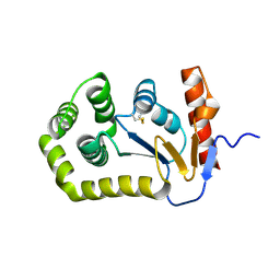
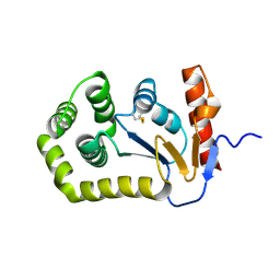
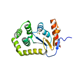
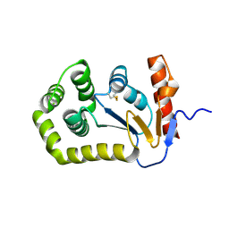
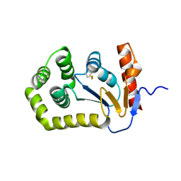
Group deposition of library data - Crystal Structure of EcDsbA after initial refinement with no ligand modelled (structure A1_1)
Descriptor: Thiol:disulfide interchange protein
Authors:Ilyichova, O.V, Bentley, M.R, Doak, B.C, Scanlon, M.J.
Deposit date:2019-01-27
Release date:2020-02-05
Last modified:2024-04-24
Method:X-RAY DIFFRACTION (2.146 Å)
Cite:Rapid Elaboration of Fragments into Leads by X-ray Crystallographic Screening of Parallel Chemical Libraries (REFiLX).
J.Med.Chem., 63, 2020
5QKS
DownloadVisualize
BU of 5qks by Molmil
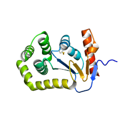
Group deposition of library data - Crystal Structure of EcDsbA after initial refinement with no ligand modelled (structure A8_1)
Descriptor: Thiol:disulfide interchange protein
Authors:Ilyichova, O.V, Bentley, M.R, Doak, B.C, Scanlon, M.J.
Deposit date:2019-01-27
Release date:2020-02-05
Last modified:2020-07-22
Method:X-RAY DIFFRACTION (2.118 Å)
Cite:Rapid Elaboration of Fragments into Leads by X-ray Crystallographic Screening of Parallel Chemical Libraries (REFiLX).
J.Med.Chem., 63, 2020
5QL9
DownloadVisualize
BU of 5ql9 by Molmil
Group deposition of library data - Crystal Structure of EcDsbA after initial refinement with no ligand modelled (structure B9_2)
Descriptor: Thiol:disulfide interchange protein
Authors:Ilyichova, O.V, Bentley, M.R, Doak, B.C, Scanlon, M.J.
Deposit date:2019-01-27
Release date:2020-02-05
Last modified:2020-07-22
Method:X-RAY DIFFRACTION (2.151 Å)
Cite:Rapid Elaboration of Fragments into Leads by X-ray Crystallographic Screening of Parallel Chemical Libraries (REFiLX).
J.Med.Chem., 63, 2020
5QLS
DownloadVisualize
BU of 5qls by Molmil
Group deposition of library data - Crystal Structure of EcDsbA after initial refinement with no ligand modelled (structure D11_1)
Descriptor: Thiol:disulfide interchange protein
Authors:Ilyichova, O.V, Bentley, M.R, Doak, B.C, Scanlon, M.J.
Deposit date:2019-01-27
Release date:2020-02-05
Last modified:2020-07-22
Method:X-RAY DIFFRACTION (2.007 Å)
Cite:Rapid Elaboration of Fragments into Leads by X-ray Crystallographic Screening of Parallel Chemical Libraries (REFiLX).
J.Med.Chem., 63, 2020
5QM7
DownloadVisualize
BU of 5qm7 by Molmil
Group deposition of library data - Crystal Structure of EcDsbA after initial refinement with no ligand modelled (structure E2_1)
Descriptor: Thiol:disulfide interchange protein
Authors:Ilyichova, O.V, Bentley, M.R, Doak, B.C, Scanlon, M.J.
Deposit date:2019-01-27
Release date:2020-02-05
Last modified:2020-07-22
Method:X-RAY DIFFRACTION (2.037 Å)
Cite:Rapid Elaboration of Fragments into Leads by X-ray Crystallographic Screening of Parallel Chemical Libraries (REFiLX).
J.Med.Chem., 63, 2020
5QKI
DownloadVisualize
BU of 5qki by Molmil
Group deposition of library data - Crystal Structure of EcDsbA after initial refinement with no ligand modelled (structure A2_2)
Descriptor: Thiol:disulfide interchange protein
Authors:Ilyichova, O.V, Bentley, M.R, Doak, B.C, Scanlon, M.J.
Deposit date:2019-01-27
Release date:2020-02-05
Last modified:2020-07-22
Method:X-RAY DIFFRACTION (2.304 Å)
Cite:Rapid Elaboration of Fragments into Leads by X-ray Crystallographic Screening of Parallel Chemical Libraries (REFiLX).
J.Med.Chem., 63, 2020

220113

PDB entries from 2024-05-22

PDB statisticsPDBj update infoContact PDBjnumon