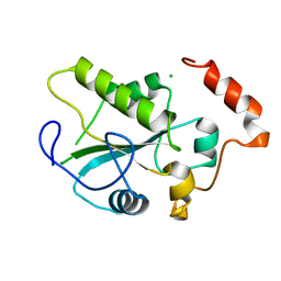
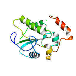
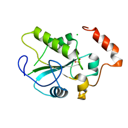
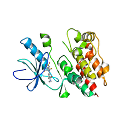
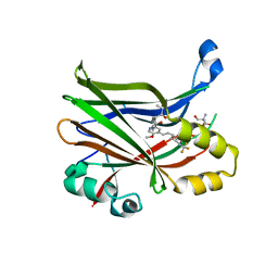
7SCZ
 
 | |
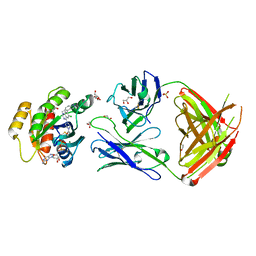
2IFD
 | |
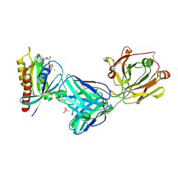
2IFV
 | |
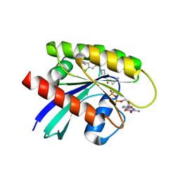
6W3B
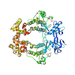
 | | Structure of apo unphosphorylated IRE1 | | 分子名称: | Serine/threonine-protein kinase/endoribonuclease IRE1 | | 著者 | Wallweber, H, Mortara, K, Ferri, E, Rudolph, J, Wang, W. | | 登録日 | 2020-03-09 | | 公開日 | 2020-12-09 | | 最終更新日 | 2023-10-18 | | 実験手法 | X-RAY DIFFRACTION (2.57 Å) | | 主引用文献 | Activation of the IRE1 RNase through remodeling of the kinase front pocket by ATP-competitive ligands.
Nat Commun, 11, 2020
|
|
6W3C
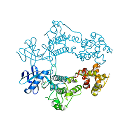
 | | Structure of phosphorylated apo IRE1 | | 分子名称: | Serine/threonine-protein kinase/endoribonuclease IRE1 | | 著者 | Wallweber, H, Mortara, K, Ferri, E, Wang, W, Rudolph, J. | | 登録日 | 2020-03-09 | | 公開日 | 2020-12-09 | | 最終更新日 | 2023-10-18 | | 実験手法 | X-RAY DIFFRACTION (2.3 Å) | | 主引用文献 | Activation of the IRE1 RNase through remodeling of the kinase front pocket by ATP-competitive ligands.
Nat Commun, 11, 2020
|
|
6W3K
 | | Structure of unphosphorylated human IRE1 bound to G-9807 | | 分子名称: | 4-[5-[2,6-bis(fluoranyl)phenyl]-2~{H}-pyrazolo[3,4-b]pyridin-3-yl]-2-(4-oxidanylpiperidin-1-yl)-1~{H}-pyrimidin-6-one, Serine/threonine-protein kinase/endoribonuclease IRE1 | | 著者 | Lammens, A, Wang, W, Ferri, E, Rudolph, J. | | 登録日 | 2020-03-09 | | 公開日 | 2020-12-09 | | 最終更新日 | 2023-10-18 | | 実験手法 | X-RAY DIFFRACTION (2.08 Å) | | 主引用文献 | Activation of the IRE1 RNase through remodeling of the kinase front pocket by ATP-competitive ligands.
Nat Commun, 11, 2020
|
|
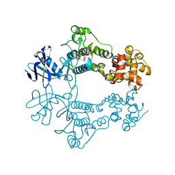
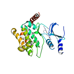
6OPK
 | | Phosphorylated ERK2 with Vertex-11e | | 分子名称: | 4-{2-[(2-chloro-4-fluorophenyl)amino]-5-methylpyrimidin-4-yl}-N-[(1S)-1-(3-chlorophenyl)-2-hydroxyethyl]-1H-pyrrole-2-carboxamide, Mitogen-activated protein kinase 1 | | 著者 | Vigers, G.P, Rudolph, J. | | 登録日 | 2019-04-25 | | 公開日 | 2019-07-31 | | 最終更新日 | 2023-11-15 | | 実験手法 | X-RAY DIFFRACTION (2.54 Å) | | 主引用文献 | Activation loop dynamics are controlled by conformation-selective inhibitors of ERK2.
Proc.Natl.Acad.Sci.USA, 116, 2019
|
|
2A2K
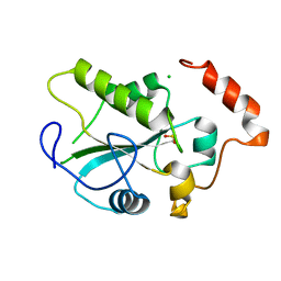
 | | Crystal Structure of an active site mutant, C473S, of Cdc25B Phosphatase Catalytic Domain | | 分子名称: | CHLORIDE ION, M-phase inducer phosphatase 2, SULFATE ION | | 著者 | Sohn, J, Parks, J, Buhrman, G, Brown, P, Kristjansdottir, K, Safi, A, Yang, W, Edelsbrunner, H, Rudolph, J. | | 登録日 | 2005-06-22 | | 公開日 | 2006-01-03 | | 最終更新日 | 2023-08-23 | | 実験手法 | X-RAY DIFFRACTION (1.52 Å) | | 主引用文献 | Experimental Validation of the Docking Orientation of Cdc25 with Its Cdk2-CycA Protein Substrate.
Biochemistry, 44, 2005
|
|
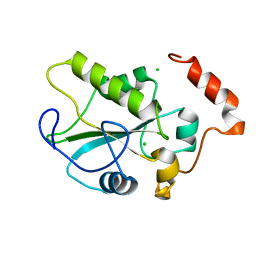
1YM9
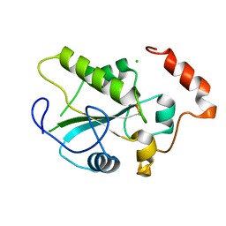
 | | Crystal structure of the CDC25B phosphatase catalytic domain with the active site cysteine in the sulfinic form | | 分子名称: | CHLORIDE ION, M-phase inducer phosphatase 2 | | 著者 | Buhrman, G.K, Parker, B, Sohn, J, Rudolph, J, Mattos, C. | | 登録日 | 2005-01-20 | | 公開日 | 2005-04-12 | | 最終更新日 | 2023-08-23 | | 実験手法 | X-RAY DIFFRACTION (2 Å) | | 主引用文献 | Structural Mechanism of Oxidative Regulation of the Phosphatase Cdc25B via an Intramolecular Disulfide Bond
Biochemistry, 44, 2005
|
|
1YML
 | | Crystal Structure of the CDC25B phosphatase catalytic domain with the active site cysteine in the sulfenic form | | 分子名称: | CHLORIDE ION, M-phase inducer phosphatase 2 | | 著者 | Buhrman, G.K, Parker, B, Sohn, J, Rudolph, J, Mattos, C. | | 登録日 | 2005-01-21 | | 公開日 | 2005-04-12 | | 最終更新日 | 2023-11-15 | | 実験手法 | X-RAY DIFFRACTION (1.7 Å) | | 主引用文献 | Structural Mechanism of Oxidative Regulation of the Phosphatase Cdc25B via an Intramolecular Disulfide Bond
Biochemistry, 44, 2005
|
|
1YMK
 | | Crystal Structure of the CDC25B phosphatase catalytic domain in the apo form | | 分子名称: | CHLORIDE ION, M-phase inducer phosphatase 2 | | 著者 | Buhrman, G.K, Parker, B, Sohn, J, Rudolph, J, Mattos, C. | | 登録日 | 2005-01-21 | | 公開日 | 2005-04-12 | | 最終更新日 | 2023-08-23 | | 実験手法 | X-RAY DIFFRACTION (1.7 Å) | | 主引用文献 | Structural Mechanism of Oxidative Regulation of the Phosphatase Cdc25B via an Intramolecular Disulfide Bond
Biochemistry, 44, 2005
|
|
1YS0
 | | Crystal Structure of the CDC25B phosphatase catalytic domain with the active site cysteine in the disulfide form | | 分子名称: | CHLORIDE ION, M-phase inducer phosphatase 2 | | 著者 | Buhrman, G.K, Parker, B, Sohn, J, Rudolph, J, Mattos, C. | | 登録日 | 2005-02-05 | | 公開日 | 2005-04-12 | | 最終更新日 | 2023-08-23 | | 実験手法 | X-RAY DIFFRACTION (2 Å) | | 主引用文献 | Structural Mechanism of Oxidative Regulation of the Phosphatase Cdc25B via an Intramolecular Disulfide Bond
Biochemistry, 44, 2005
|
|
1YMD
 | | Crystal Structure of the CDC25B phosphatase catalytic domain with the active site cysteine in the sulfonic form | | 分子名称: | CHLORIDE ION, M-phase inducer phosphatase 2 | | 著者 | Buhrman, G.K, Parker, B, Sohn, J, Rudolph, J, Mattos, C. | | 登録日 | 2005-01-20 | | 公開日 | 2005-04-12 | | 最終更新日 | 2023-11-15 | | 実験手法 | X-RAY DIFFRACTION (1.7 Å) | | 主引用文献 | Structural Mechanism of Oxidative Regulation of the Phosphatase Cdc25B via an Intramolecular Disulfide Bond
Biochemistry, 44, 2005
|
|
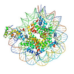
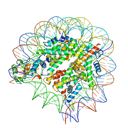
1XXE
 | | RDC refined solution structure of the AaLpxC/TU-514 complex | | 分子名称: | 1,5-ANHYDRO-2-C-(CARBOXYMETHYL-N-HYDROXYAMIDE)-2-DEOXY-3-O-MYRISTOYL-D-GLUCITOL, UDP-3-O-[3-hydroxymyristoyl] N-acetylglucosamine deacetylase, ZINC ION | | 著者 | Coggins, B.E, McClerren, A.L, Jiang, L, Li, X, Rudolph, J, Hindsgaul, O, Raetz, C.R.H, Zhou, P. | | 登録日 | 2004-11-04 | | 公開日 | 2004-11-23 | | 最終更新日 | 2022-03-02 | | 実験手法 | SOLUTION NMR | | 主引用文献 | Refined Solution Structure of the LpxC-TU-514 Complex and pK(a) Analysis of an Active Site Histidine: Insights into the Mechanism and Inhibitor Design
Biochemistry, 44, 2005
|
|
4PP7
 | | Highly Potent and Selective 3-N-methylquinazoline-4(3H)-one Based Inhibitors of B-RafV600E Kinase | | 分子名称: | N-{2,4-difluoro-3-[methyl(3-methyl-4-oxo-3,4-dihydroquinazolin-6-yl)amino]phenyl}propane-1-sulfonamide, Serine/threonine-protein kinase B-raf | | 著者 | Wenglowsky, S, Ren, L, Grina, J, Hansen, J.D, Laird, E.R, Moreno, D, Dinkel, V, Gloor, S.L, Hastings, G, Rana, S, Rasor, K, Sturgis, H.L, Voegtli, W.C, Vigers, G.P.A, Willis, B, Mathieu, S, Rudolph, J. | | 登録日 | 2014-02-26 | | 公開日 | 2014-04-09 | | 最終更新日 | 2024-02-28 | | 実験手法 | X-RAY DIFFRACTION (3.4 Å) | | 主引用文献 | Highly potent and selective 3-N-methylquinazoline-4(3H)-one based inhibitors of B-Raf(V600E) kinase.
Bioorg.Med.Chem.Lett., 24, 2014
|
|
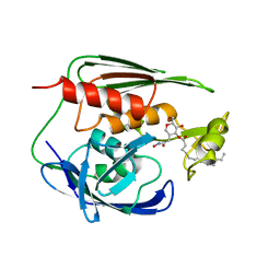
7SCY
 | |
5IME
 | | Crystal structure of P21-activated kinase 1 (PAK1) in complex with compound 9 | | 分子名称: | 8-(3-aminopropyl)-6-[2-chloro-4-(3-methyl-2-oxopyrazin-1(2H)-yl)phenyl]-2-(methylamino)pyrido[2,3-d]pyrimidin-7(8H)-one, Serine/threonine-protein kinase PAK 1 | | 著者 | Li, D, Wang, W. | | 登録日 | 2016-03-06 | | 公開日 | 2016-05-25 | | 最終更新日 | 2023-09-27 | | 実験手法 | X-RAY DIFFRACTION (2.217 Å) | | 主引用文献 | Chemically Diverse Group I p21-Activated Kinase (PAK) Inhibitors Impart Acute Cardiovascular Toxicity with a Narrow Therapeutic Window.
J.Med.Chem., 59, 2016
|
|
7MDP
 | | KRas G12C in complex with G-2897 | | 分子名称: | 1,2-ETHANEDIOL, 2,5,8,11,14,17-HEXAOXANONADECAN-19-OL, 4-(trifluoromethyl)-1,3-benzothiazol-2-amine, ... | | 著者 | Oh, A, Frank, Y, Wang, W. | | 登録日 | 2021-04-05 | | 公開日 | 2022-03-16 | | 最終更新日 | 2023-10-18 | | 実験手法 | X-RAY DIFFRACTION (1.96 Å) | | 主引用文献 | Conformation-locking antibodies for the discovery and characterization of KRAS inhibitors.
Nat.Biotechnol., 40, 2022
|
|
8E1O
 | | Crystal structure of hTEAD2 bound to a methoxypyridine lipid pocket binder | | 分子名称: | 2-AMINO-2-HYDROXYMETHYL-PROPANE-1,3-DIOL, 5-methoxy-N-({3-[2-(methylamino)-2-oxoethyl]phenyl}methyl)-4-{(E)-2-[trans-4-(trifluoromethyl)cyclohexyl]ethenyl}pyridine-2-carboxamide, Transcriptional enhancer factor TEF-4 | | 著者 | Noland, C.L, Dey, A, Zbieg, J, Crawford, J. | | 登録日 | 2022-08-10 | | 公開日 | 2023-08-16 | | 最終更新日 | 2024-02-28 | | 実験手法 | X-RAY DIFFRACTION (2.25 Å) | | 主引用文献 | Targeting the Hippo pathway in cancers via ubiquitination dependent TEAD degradation
Biorxiv, 2024
|
|
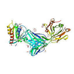
7RP3
 | | Crystal structure of GNE-1952 alkylated KRAS G12C in complex with 2H11 CLAMP | | 分子名称: | 1,2-ETHANEDIOL, 1-[4-[6-chloranyl-7-(5-methyl-1~{H}-indazol-4-yl)quinazolin-4-yl]piperazin-1-yl]propan-1-one, GLYCEROL, ... | | 著者 | Oh, A, Tam, C, Wang, W. | | 登録日 | 2021-08-03 | | 公開日 | 2022-03-16 | | 最終更新日 | 2023-10-18 | | 実験手法 | X-RAY DIFFRACTION (2 Å) | | 主引用文献 | Conformation-locking antibodies for the discovery and characterization of KRAS inhibitors.
Nat.Biotechnol., 40, 2022
|
|
7RP2
 | | Crystal structure of Kas G12C in complex with 2H11 CLAMP | | 分子名称: | 1,2-ETHANEDIOL, CACODYLATE ION, GTPase KRas, ... | | 著者 | Oh, A, Tam, C, Wang, W. | | 登録日 | 2021-08-03 | | 公開日 | 2022-03-16 | | 最終更新日 | 2023-10-18 | | 実験手法 | X-RAY DIFFRACTION (2.2 Å) | | 主引用文献 | Conformation-locking antibodies for the discovery and characterization of KRAS inhibitors.
Nat.Biotechnol., 40, 2022
|
|
7RP4
 | | Crystal structure of KRAS G12C in complex with GNE-1952 | | 分子名称: | 1-[4-[6-chloranyl-7-(5-methyl-1~{H}-indazol-4-yl)quinazolin-4-yl]piperazin-1-yl]propan-1-one, GLYCEROL, GUANOSINE-5'-DIPHOSPHATE, ... | | 著者 | Oh, A, Tam, C, Wang, W. | | 登録日 | 2021-08-03 | | 公開日 | 2022-03-16 | | 最終更新日 | 2023-10-18 | | 実験手法 | X-RAY DIFFRACTION (2.15 Å) | | 主引用文献 | Conformation-locking antibodies for the discovery and characterization of KRAS inhibitors.
Nat.Biotechnol., 40, 2022
|
|
4ZY4
 | |
4ZY6
 | |
4ZY5
 | |


