Loading
PDBj
MenuPDBj@FacebookPDBj@TwitterPDBj@YouTubewwPDB FoundationwwPDB
RCSB PDBPDBeBMRBAdv. SearchSearch help
Search by PDB author
6PHL
DownloadVisualize
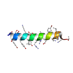
BU of 6phl by Molmil
Crystal structure of glucagon analog with mono-stereoinversion at position 23 (D-Val23) in space group I41 at 1.44 A resolution
Descriptor: Glucagon
Authors:Mroz, P.A, Gonzalez-Gutierrez, G, DiMarchi, R.D.
Deposit date:2019-06-25
Release date:2020-07-01
Last modified:2023-10-11
Method:X-RAY DIFFRACTION (1.443 Å)
Cite:High resolution X-ray structure of glucagon and selected stereo-inversed analogs in novel crystallographic packing arrangement.
To Be Published
6PHQ
DownloadVisualize
BU of 6phq by Molmil
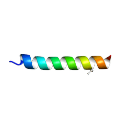
Crystal structure of glucagon analog fully composed of D-amino acids with 4-bromo-D-phenylalanine substitutions at position 6 and 22 in space group I41 at 1.1 A resolution
Descriptor: D-glucagon D-BrPhe 6,22
Authors:Mroz, P.A, Gonzalez-Gutierrez, G, DiMarchi, R.D.
Deposit date:2019-06-25
Release date:2020-07-01
Last modified:2023-11-15
Method:X-RAY DIFFRACTION (1.32 Å)
Cite:High resolution X-ray structure of glucagon and selected stereo-inversed analogs in novel crystallographic packing arrangement.
To Be Published
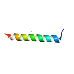
6PHI
DownloadVisualize
BU of 6phi by Molmil
Crystal structure of native glucagon in space group I41 at 1.1 A resolution
Descriptor: CHLORIDE ION, Glucagon
Authors:Mroz, P.A, Gonzalez-Gutierrez, G, DiMarchi, R.D.
Deposit date:2019-06-25
Release date:2020-07-01
Last modified:2023-10-11
Method:X-RAY DIFFRACTION (1.1 Å)
Cite:High resolution X-ray structure of glucagon and selected stereo-inversed analogs in novel crystallographic packing arrangement.
To Be Published
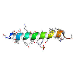
6PHM
DownloadVisualize
BU of 6phm by Molmil
Crystal structure of glucagon analog fully composed of D-amino acids in space group I41 at 1.1 A resolution
Descriptor: D-glucagon, SULFATE ION
Authors:Mroz, P.A, Gonzalez-Gutierrez, G, DiMarchi, R.D.
Deposit date:2019-06-25
Release date:2020-07-01
Last modified:2023-11-15
Method:X-RAY DIFFRACTION (1.1 Å)
Cite:High resolution X-ray structure of glucagon and selected stereo-inversed analogs in novel crystallographic packing arrangement.
To Be Published
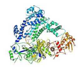
6P7M
DownloadVisualize
BU of 6p7m by Molmil
Cryo-EM structure of LbCas12a-crRNA: AcrVA4 (1:2 complex)
Descriptor: Cas12a, MAGNESIUM ION, anti-CRISPR VA4, ...
Authors:Knott, G.J, Liu, J.J, Doudna, J.A.
Deposit date:2019-06-06
Release date:2019-08-21
Last modified:2024-03-20
Method:ELECTRON MICROSCOPY (3 Å)
Cite:Structural basis for AcrVA4 inhibition of specific CRISPR-Cas12a.
Elife, 8, 2019
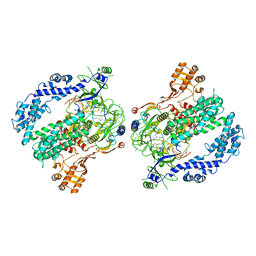
6P7N
DownloadVisualize
BU of 6p7n by Molmil
Cryo-EM structure of LbCas12a-crRNA: AcrVA4 (2:2 complex)
Descriptor: Cas12a, MAGNESIUM ION, anti-CRISPR VA4, ...
Authors:Knott, G.J, Liu, J.J, Doudna, J.A.
Deposit date:2019-06-06
Release date:2019-08-21
Last modified:2023-08-16
Method:ELECTRON MICROSCOPY (4.9 Å)
Cite:Structural basis for AcrVA4 inhibition of specific CRISPR-Cas12a.
Elife, 8, 2019
6PW8
DownloadVisualize
BU of 6pw8 by Molmil
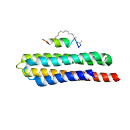
Hydrocarbon-Stapled Paxillin Peptide Bound to the Focal Adhesion Targeting (FAT) Domain of the Focal Adhesion Kinase (FAK)
Descriptor: CHLORIDE ION, Focal adhesion kinase 1, SP3, ...
Authors:Thifault, D.G, Fromme, P, Martin-Garcia, J.M.
Deposit date:2019-07-22
Release date:2020-07-22
Last modified:2023-11-15
Method:X-RAY DIFFRACTION (1.95 Å)
Cite:Stapled Peptide Ligand Bound to the Focal Adhesion Targeting (FAT) Domain of the Focal Adhesion Kinase (FAK)
To Be Published
6RPP
DownloadVisualize
BU of 6rpp by Molmil
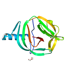
Crystal structure of PabCDC21-1 intein
Descriptor: ACETATE ION, Cell division control protein, DI(HYDROXYETHYL)ETHER
Authors:Mikula, K.M, Beyer, H.M, Iwai, H.
Deposit date:2019-05-14
Release date:2019-08-14
Last modified:2024-01-24
Method:X-RAY DIFFRACTION (1.6 Å)
Cite:Crystal structures of CDC21-1 inteins from hyperthermophilic archaea reveal the selection mechanism for the highly conserved homing endonuclease insertion site.
Extremophiles, 23, 2019
6RPQ
DownloadVisualize
BU of 6rpq by Molmil
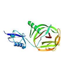
Crystal structure of PhoCDC21-1 intein
Descriptor: Ubiquitin-like protein SMT3,1108aa long hypothetical cell division control protein
Authors:Beyer, H.M, Mikula, K.M, Iwai, H.
Deposit date:2019-05-14
Release date:2019-08-14
Last modified:2024-01-24
Method:X-RAY DIFFRACTION (2.654 Å)
Cite:Crystal structures of CDC21-1 inteins from hyperthermophilic archaea reveal the selection mechanism for the highly conserved homing endonuclease insertion site.
Extremophiles, 23, 2019
7ZIE
DownloadVisualize
BU of 7zie by Molmil
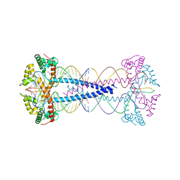
Gcf1p, multimerizes and bridges the mitochondrial DNA from Candida albicans by a specific mechanism.
Descriptor: 20-mer DNA, GLYCEROL, Gcf1p
Authors:Tarres-Sole, A, Sola, M.
Deposit date:2022-04-07
Release date:2023-06-14
Last modified:2023-07-19
Method:SOLUTION SCATTERING (2.9 Å), X-RAY DIFFRACTION
Cite:Gcf1p tetramerizes upon DNA binding, revealing a novel compaction method for mitochondrial DNA in Candida albicans.
Nucleic Acids Res., 2023
7ZJV
DownloadVisualize
BU of 7zjv by Molmil
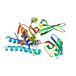
Structure of human USPL1 in covalent complex with DeltaN-SUMO2/3-PA probe
Descriptor: CHLORIDE ION, SUMO-specific isopeptidase USPL1, Small ubiquitin-related modifier 2, ...
Authors:Zhao, Z, Gersch, M.
Deposit date:2022-04-12
Release date:2023-09-27
Last modified:2023-11-15
Method:X-RAY DIFFRACTION (2.4 Å)
Cite:Native Semisynthesis of Isopeptide-Linked Substrates for Specificity Analysis of Deubiquitinases and Ubl Proteases.
J.Am.Chem.Soc., 145, 2023
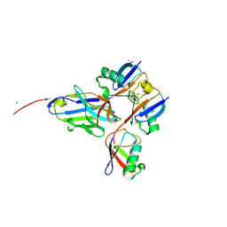
7ZJU
DownloadVisualize
BU of 7zju by Molmil
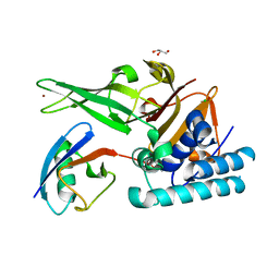
Structure of human USPL1 in covalent complex with SUMO3-2Br probe
Descriptor: 2-bromanylethanamine, CALCIUM ION, CHLORIDE ION, ...
Authors:Zhao, Z, Gersch, M.
Deposit date:2022-04-12
Release date:2023-09-27
Last modified:2023-11-15
Method:X-RAY DIFFRACTION (2.17 Å)
Cite:Native Semisynthesis of Isopeptide-Linked Substrates for Specificity Analysis of Deubiquitinases and Ubl Proteases.
J.Am.Chem.Soc., 145, 2023
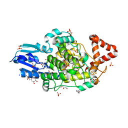
8A8L
DownloadVisualize
BU of 8a8l by Molmil
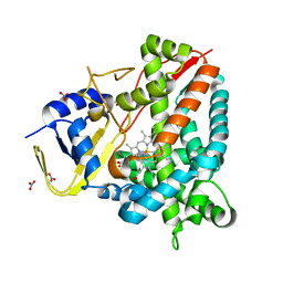
Crystal structure of a staphylococcal orthologue of CYP134A1 (CYPX) in complex with a heme-coordinated fragment
Descriptor: 6-methoxy-2,3,4,9-tetrahydro-1H-pyrido[3,4-b]indole, Cytochrome P450 protein, GLYCEROL, ...
Authors:Snee, M, Katariya, M, Levy, C.
Deposit date:2022-06-23
Release date:2023-07-05
Last modified:2024-02-07
Method:X-RAY DIFFRACTION (1.88 Å)
Cite:Crystal structure of a staphylococcal orthologue of CYP134A1 (CYPX) in complex with a heme-coordinated fragment
To Be Published
8A6W
DownloadVisualize
BU of 8a6w by Molmil
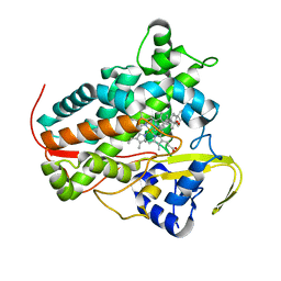
Crystal structure of CYP142 from Mycobacterium tuberculosis in complex with cholestenone
Descriptor: (8ALPHA,9BETA)-CHOLEST-4-EN-3-ONE, PROTOPORPHYRIN IX CONTAINING FE, Steroid C26-monooxygenase
Authors:Snee, M, Amadi, C, Levy, C.
Deposit date:2022-06-20
Release date:2023-07-05
Last modified:2024-02-07
Method:X-RAY DIFFRACTION (2.09 Å)
Cite:Crystal structure of CYP142 from Mycobacterium tuberculosis in complex with cholestenone
To Be Published
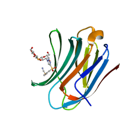
8PFF
DownloadVisualize
BU of 8pff by Molmil
Galectin-3C in complex with a triazolesulfone derivative
Descriptor: (2~{R},3~{S},4~{S},5~{R},6~{S})-2-(hydroxymethyl)-6-[(2~{S},3~{R},4~{S},5~{R},6~{R})-6-(hydroxymethyl)-3,5-bis(oxidanyl)-4-[4-(phenylsulfonyl)-1,2,3-triazol-1-yl]oxan-2-yl]sulfanyl-oxane-3,4,5-triol, Galectin-3, MAGNESIUM ION, ...
Authors:Kumar, R, Mahanti, M, Nilsson, U.J, Logan, D.T.
Deposit date:2023-06-15
Release date:2023-11-08
Last modified:2023-11-22
Method:X-RAY DIFFRACTION (1.08 Å)
Cite:Ligand Sulfur Oxidation State Progressively Alters Galectin-3-Ligand Complex Conformations To Induce Affinity-Influencing Hydrogen Bonds.
J.Med.Chem., 66, 2023
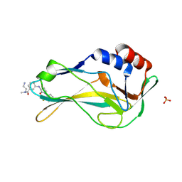
8A7N
DownloadVisualize
BU of 8a7n by Molmil
Crystal Structure of human Brachyury G177D variant in complex with (S)-N-(3-aminopropyl)-3-((1-(2-fluorophenyl)-2-oxopyrrolidin-3-yl)amino)-N-methylbenzamide (CF-2-125)
Descriptor: N-(3-azanylpropyl)-3-[[(3S)-1-(2-fluorophenyl)-2-oxidanylidene-pyrrolidin-3-yl]amino]-N-methyl-benzamide, PHOSPHATE ION, T-box transcription factor T
Authors:Newman, J.A, Gavard, A, Aitkenhead, H, Imprachim, N, Sherestha, L, Burgess-Brown, N.A, von Delft, F, Bountra, C, Gileadi, O.
Deposit date:2022-06-21
Release date:2022-10-05
Last modified:2024-01-31
Method:X-RAY DIFFRACTION (1.9 Å)
Cite:Crystal Structure of human Brachyury G177D variant in complex with (S)-N-(3-aminopropyl)-3-((1-(2-fluorophenyl)-2-oxopyrrolidin-3-yl)amino)-N-methylbenzamide (CF-2-125)
To Be Published
8A1H
DownloadVisualize
BU of 8a1h by Molmil
Bacterial 6-4 photolyase from Vibrio cholerase
Descriptor: 1-deoxy-1-(6,7-dimethyl-2,4-dioxo-3,4-dihydropteridin-8(2H)-yl)-D-ribitol, 4-(2-HYDROXYETHYL)-1-PIPERAZINE ETHANESULFONIC ACID, 6-4 photolyase (FeS-BCP, ...
Authors:Essen, L.-O, Emmerich, H.J.
Deposit date:2022-06-01
Release date:2023-02-08
Last modified:2024-02-07
Method:X-RAY DIFFRACTION (1.65 Å)
Cite:Structural and Functional Analysis of a Prokaryotic (6-4) Photolyase from the Aquatic Pathogen Vibrio Cholerae † .
Photochem.Photobiol., 99, 2023
8A67
DownloadVisualize
BU of 8a67 by Molmil
Branched Lys48- and Lys63-linked tri-ubiquitin (K48-K63-Ub3) in complex with matured synthetic nanobody NbSL3.3Q (3rd generation)
Descriptor: CHLORIDE ION, GLYCEROL, ISOPROPYL ALCOHOL, ...
Authors:Lange, S.M, Kulathu, Y.
Deposit date:2022-06-16
Release date:2023-02-15
Last modified:2024-02-07
Method:X-RAY DIFFRACTION (1.86 Å)
Cite:Comprehensive approach to study branched ubiquitin chains reveals roles for K48-K63 branches in VCP/p97-related processes
Biorxiv, 2023
8A58
DownloadVisualize
BU of 8a58 by Molmil
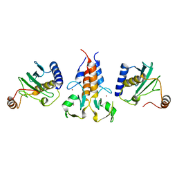
X-ray structure of TRIM21 RING E3 ligase in complex with E2 enzyme Ube2W
Descriptor: E3 ubiquitin-protein ligase TRIM21, Ubiquitin-conjugating enzyme E2 W, ZINC ION
Authors:James, L.C, Kiss, L.
Deposit date:2022-06-14
Release date:2023-04-26
Last modified:2024-02-07
Method:X-RAY DIFFRACTION (2.25 Å)
Cite:Trim-Away ubiquitinates and degrades lysine-less and N-terminally acetylated substrates.
Nat Commun, 14, 2023
8QI7
DownloadVisualize
BU of 8qi7 by Molmil
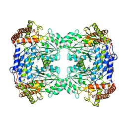
Cryo-EM Structure of Human Serine Hydroxymethyltransferase, isoform 2 (SHMT2)
Descriptor: Serine hydroxymethyltransferase, mitochondrial
Authors:Rutkiewicz, M, Tran, L.H, Ruszkowski, M.
Deposit date:2023-09-11
Release date:2023-09-20
Last modified:2023-11-15
Method:ELECTRON MICROSCOPY (2.9 Å)
Cite:New Structural Models of SHMT2
To Be Published
8R61
DownloadVisualize
BU of 8r61 by Molmil
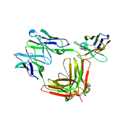
Structure of IgE delta epsilon 3-4 in complex with a kappa binding nanobody
Descriptor: HMM5 IgE light chain, Immunoglobulin E VH-Ceps1-Ceps1, kappa binding nanobody
Authors:Andersen, G.R, Gandini, R.
Deposit date:2023-11-20
Release date:2023-11-29
Method:X-RAY DIFFRACTION (3.1 Å)
Cite:Structure of IgE delta epsilon 3-4 in complex with a kappa binding nanobody
To Be Published
8B23
DownloadVisualize
BU of 8b23 by Molmil
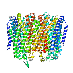
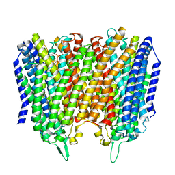
Time-resolved structure of K+-dependent Na+-PPase from Thermotoga maritima 600-seconds post reaction initiation with Na+
Descriptor: DIPHOSPHATE, K(+)-stimulated pyrophosphate-energized sodium pump, MAGNESIUM ION
Authors:Strauss, J, Vidilaseris, K, Goldman, A.
Deposit date:2022-09-12
Release date:2024-01-17
Last modified:2024-04-10
Method:X-RAY DIFFRACTION (3.84 Å)
Cite:Functional and structural asymmetry suggest a unifying principle for catalysis in membrane-bound pyrophosphatases.
Embo Rep., 25, 2024
8B22
DownloadVisualize
BU of 8b22 by Molmil
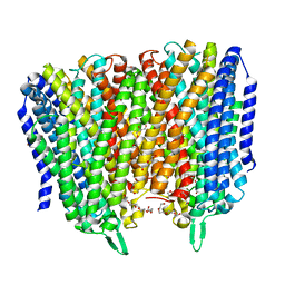
Time-resolved structure of K+-dependent Na+-PPase from Thermotoga maritima 300-seconds post reaction initiation with Na+
Descriptor: DIPHOSPHATE, K(+)-stimulated pyrophosphate-energized sodium pump, MAGNESIUM ION
Authors:Strauss, J, Vidilaseris, K, Goldman, A.
Deposit date:2022-09-12
Release date:2024-01-17
Last modified:2024-04-10
Method:X-RAY DIFFRACTION (3.98 Å)
Cite:Functional and structural asymmetry suggest a unifying principle for catalysis in membrane-bound pyrophosphatases.
Embo Rep., 25, 2024
8B21
DownloadVisualize
BU of 8b21 by Molmil
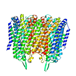
Time-resolved structure of K+-dependent Na+-PPase from Thermotoga maritima 0-60-seconds post reaction initiation with Na+
Descriptor: DI(HYDROXYETHYL)ETHER, DODECYL-BETA-D-MALTOSIDE, K(+)-stimulated pyrophosphate-energized sodium pump, ...
Authors:Strauss, J, Vidilaseris, K, Goldman, A.
Deposit date:2022-09-12
Release date:2024-01-17
Last modified:2024-04-10
Method:X-RAY DIFFRACTION (2.59 Å)
Cite:Functional and structural asymmetry suggest a unifying principle for catalysis in membrane-bound pyrophosphatases.
Embo Rep., 25, 2024
8B24
DownloadVisualize
BU of 8b24 by Molmil
Time-resolved structure of K+-dependent Na+-PPase from Thermotoga maritima 3600-seconds post reaction initiation with Na+
Descriptor: DIPHOSPHATE, K(+)-stimulated pyrophosphate-energized sodium pump, MAGNESIUM ION, ...
Authors:Strauss, J, Vidilaseris, K, Goldman, A.
Deposit date:2022-09-12
Release date:2024-01-17
Last modified:2024-04-10
Method:X-RAY DIFFRACTION (4.53 Å)
Cite:Functional and structural asymmetry suggest a unifying principle for catalysis in membrane-bound pyrophosphatases.
Embo Rep., 25, 2024

219869

건을2024-05-15부터공개중

PDB statisticsPDBj update infoContact PDBjnumon