6PHL
 
 | |
6PHQ
 | |
6PHI
 | |
6PHM
 | |
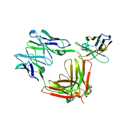
6P7M
 | | Cryo-EM structure of LbCas12a-crRNA: AcrVA4 (1:2 complex) | | 分子名称: | Cas12a, MAGNESIUM ION, anti-CRISPR VA4, ... | | 著者 | Knott, G.J, Liu, J.J, Doudna, J.A. | | 登録日 | 2019-06-06 | | 公開日 | 2019-08-21 | | 最終更新日 | 2024-03-20 | | 実験手法 | ELECTRON MICROSCOPY (3 Å) | | 主引用文献 | Structural basis for AcrVA4 inhibition of specific CRISPR-Cas12a.
Elife, 8, 2019
|
|
6P7N
 | | Cryo-EM structure of LbCas12a-crRNA: AcrVA4 (2:2 complex) | | 分子名称: | Cas12a, MAGNESIUM ION, anti-CRISPR VA4, ... | | 著者 | Knott, G.J, Liu, J.J, Doudna, J.A. | | 登録日 | 2019-06-06 | | 公開日 | 2019-08-21 | | 最終更新日 | 2023-08-16 | | 実験手法 | ELECTRON MICROSCOPY (4.9 Å) | | 主引用文献 | Structural basis for AcrVA4 inhibition of specific CRISPR-Cas12a.
Elife, 8, 2019
|
|
6PW8
 | |
6RPP
 | | Crystal structure of PabCDC21-1 intein | | 分子名称: | ACETATE ION, Cell division control protein, DI(HYDROXYETHYL)ETHER | | 著者 | Mikula, K.M, Beyer, H.M, Iwai, H. | | 登録日 | 2019-05-14 | | 公開日 | 2019-08-14 | | 最終更新日 | 2024-01-24 | | 実験手法 | X-RAY DIFFRACTION (1.6 Å) | | 主引用文献 | Crystal structures of CDC21-1 inteins from hyperthermophilic archaea reveal the selection mechanism for the highly conserved homing endonuclease insertion site.
Extremophiles, 23, 2019
|
|
6RPQ
 | | Crystal structure of PhoCDC21-1 intein | | 分子名称: | Ubiquitin-like protein SMT3,1108aa long hypothetical cell division control protein | | 著者 | Beyer, H.M, Mikula, K.M, Iwai, H. | | 登録日 | 2019-05-14 | | 公開日 | 2019-08-14 | | 最終更新日 | 2024-01-24 | | 実験手法 | X-RAY DIFFRACTION (2.654 Å) | | 主引用文献 | Crystal structures of CDC21-1 inteins from hyperthermophilic archaea reveal the selection mechanism for the highly conserved homing endonuclease insertion site.
Extremophiles, 23, 2019
|
|
7ZIE
 | |
7ZJV
 | |
7ZJU
 | |
8A8L
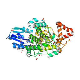
 | | Crystal structure of a staphylococcal orthologue of CYP134A1 (CYPX) in complex with a heme-coordinated fragment | | 分子名称: | 6-methoxy-2,3,4,9-tetrahydro-1H-pyrido[3,4-b]indole, Cytochrome P450 protein, GLYCEROL, ... | | 著者 | Snee, M, Katariya, M, Levy, C. | | 登録日 | 2022-06-23 | | 公開日 | 2023-07-05 | | 最終更新日 | 2024-02-07 | | 実験手法 | X-RAY DIFFRACTION (1.88 Å) | | 主引用文献 | Crystal structure of a staphylococcal orthologue of CYP134A1 (CYPX) in complex with a heme-coordinated fragment
To Be Published
|
|
8A6W
 | |
8PFF
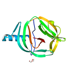
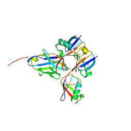
 | | Galectin-3C in complex with a triazolesulfone derivative | | 分子名称: | (2~{R},3~{S},4~{S},5~{R},6~{S})-2-(hydroxymethyl)-6-[(2~{S},3~{R},4~{S},5~{R},6~{R})-6-(hydroxymethyl)-3,5-bis(oxidanyl)-4-[4-(phenylsulfonyl)-1,2,3-triazol-1-yl]oxan-2-yl]sulfanyl-oxane-3,4,5-triol, Galectin-3, MAGNESIUM ION, ... | | 著者 | Kumar, R, Mahanti, M, Nilsson, U.J, Logan, D.T. | | 登録日 | 2023-06-15 | | 公開日 | 2023-11-08 | | 最終更新日 | 2023-11-22 | | 実験手法 | X-RAY DIFFRACTION (1.08 Å) | | 主引用文献 | Ligand Sulfur Oxidation State Progressively Alters Galectin-3-Ligand Complex Conformations To Induce Affinity-Influencing Hydrogen Bonds.
J.Med.Chem., 66, 2023
|
|
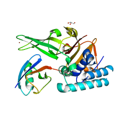
8A7N
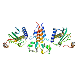
 | | Crystal Structure of human Brachyury G177D variant in complex with (S)-N-(3-aminopropyl)-3-((1-(2-fluorophenyl)-2-oxopyrrolidin-3-yl)amino)-N-methylbenzamide (CF-2-125) | | 分子名称: | N-(3-azanylpropyl)-3-[[(3S)-1-(2-fluorophenyl)-2-oxidanylidene-pyrrolidin-3-yl]amino]-N-methyl-benzamide, PHOSPHATE ION, T-box transcription factor T | | 著者 | Newman, J.A, Gavard, A, Aitkenhead, H, Imprachim, N, Sherestha, L, Burgess-Brown, N.A, von Delft, F, Bountra, C, Gileadi, O. | | 登録日 | 2022-06-21 | | 公開日 | 2022-10-05 | | 最終更新日 | 2024-01-31 | | 実験手法 | X-RAY DIFFRACTION (1.9 Å) | | 主引用文献 | Crystal Structure of human Brachyury G177D variant in complex with (S)-N-(3-aminopropyl)-3-((1-(2-fluorophenyl)-2-oxopyrrolidin-3-yl)amino)-N-methylbenzamide (CF-2-125)
To Be Published
|
|
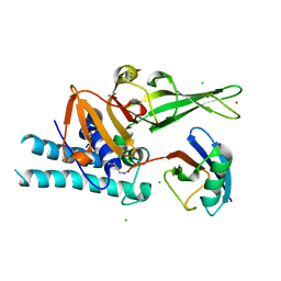
8A1H
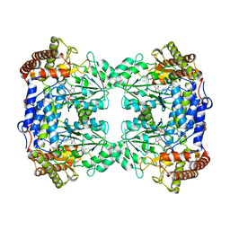
 | | Bacterial 6-4 photolyase from Vibrio cholerase | | 分子名称: | 1-deoxy-1-(6,7-dimethyl-2,4-dioxo-3,4-dihydropteridin-8(2H)-yl)-D-ribitol, 4-(2-HYDROXYETHYL)-1-PIPERAZINE ETHANESULFONIC ACID, 6-4 photolyase (FeS-BCP, ... | | 著者 | Essen, L.-O, Emmerich, H.J. | | 登録日 | 2022-06-01 | | 公開日 | 2023-02-08 | | 最終更新日 | 2024-02-07 | | 実験手法 | X-RAY DIFFRACTION (1.65 Å) | | 主引用文献 | Structural and Functional Analysis of a Prokaryotic (6-4) Photolyase from the Aquatic Pathogen Vibrio Cholerae † .
Photochem.Photobiol., 99, 2023
|
|
8A67
 | |
8A58
 | |
8QI7
 | |
8R61
 | |
8B23
 | |
8B22
 | |
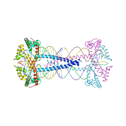
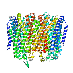
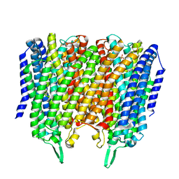
8B21
 | | Time-resolved structure of K+-dependent Na+-PPase from Thermotoga maritima 0-60-seconds post reaction initiation with Na+ | | 分子名称: | DI(HYDROXYETHYL)ETHER, DODECYL-BETA-D-MALTOSIDE, K(+)-stimulated pyrophosphate-energized sodium pump, ... | | 著者 | Strauss, J, Vidilaseris, K, Goldman, A. | | 登録日 | 2022-09-12 | | 公開日 | 2024-01-17 | | 最終更新日 | 2024-04-10 | | 実験手法 | X-RAY DIFFRACTION (2.59 Å) | | 主引用文献 | Functional and structural asymmetry suggest a unifying principle for catalysis in membrane-bound pyrophosphatases.
Embo Rep., 25, 2024
|
|
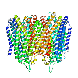
8B24
 | | Time-resolved structure of K+-dependent Na+-PPase from Thermotoga maritima 3600-seconds post reaction initiation with Na+ | | 分子名称: | DIPHOSPHATE, K(+)-stimulated pyrophosphate-energized sodium pump, MAGNESIUM ION, ... | | 著者 | Strauss, J, Vidilaseris, K, Goldman, A. | | 登録日 | 2022-09-12 | | 公開日 | 2024-01-17 | | 最終更新日 | 2024-04-10 | | 実験手法 | X-RAY DIFFRACTION (4.53 Å) | | 主引用文献 | Functional and structural asymmetry suggest a unifying principle for catalysis in membrane-bound pyrophosphatases.
Embo Rep., 25, 2024
|
|


