3OTM
 
 | |
3OUP
 | |
3OYS
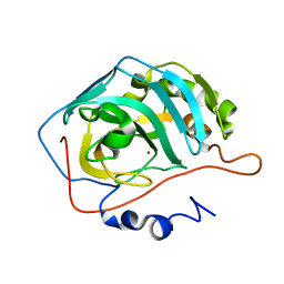
 | | Human Carbonic Anhydrase II complexed with 2-{[4-AMINO-3-(3-HYDROXYPROP-1-YN-1-YL)-1H-PYRAZOLO[3,4-D]PYRIMIDIN-1-YL]METHYL}-5-METHYL-3-(2-METHYLPHENYL)QUINAZOLIN-4(3H)-ONE | | 分子名称: | 2-phenyl-N-(4-sulfamoylphenyl)acetamide, Carbonic anhydrase 2, DIMETHYL SULFOXIDE, ... | | 著者 | Aggarwal, M, McKenna, R. | | 登録日 | 2010-09-23 | | 公開日 | 2011-08-10 | | 最終更新日 | 2024-02-21 | | 実験手法 | X-RAY DIFFRACTION (1.538 Å) | | 主引用文献 | Anticonvulsant 4-aminobenzenesulfonamide derivatives with branched-alkylamide moieties: X-ray crystallography and inhibition studies of human carbonic anhydrase isoforms I, II, VII, and XIV.
J.Med.Chem., 54, 2011
|
|
3QYK
 | |
3R17
 | |
3PWE
 | | Crystal structure of the E. coli beta clamp mutant R103C, I305C, C260S, C333S at 2.2A resolution | | 分子名称: | DNA polymerase III subunit beta | | 著者 | Marzahn, M.R, Robbins, A.H, McKenna, R, Bloom, L.B. | | 登録日 | 2010-12-08 | | 公開日 | 2011-10-19 | | 最終更新日 | 2023-09-13 | | 実験手法 | X-RAY DIFFRACTION (2.199 Å) | | 主引用文献 | The E. coli clamp loader can actively pry open the beta-sliding clamp
J.Biol.Chem., 286, 2011
|
|
3OW5
 | |
3OU9
 | |
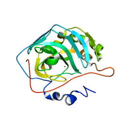
3R16
 | | Human CAII bound to N-(4-sulfamoylphenyl)-2-(thiophen-2-yl) acetamide | | 分子名称: | Carbonic anhydrase 2, DIMETHYL SULFOXIDE, GLYCEROL, ... | | 著者 | Biswas, S, McKenna, R, Supuran, C.T. | | 登録日 | 2011-03-09 | | 公開日 | 2011-05-25 | | 最終更新日 | 2024-02-21 | | 実験手法 | X-RAY DIFFRACTION (1.6 Å) | | 主引用文献 | Conformational variability of different sulfonamide inhibitors with thienyl-acetamido moieties attributes to differential binding in the active site of cytosolic human carbonic anhydrase isoforms.
Bioorg.Med.Chem., 19, 2011
|
|
1GFF
 | |
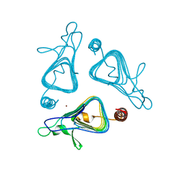
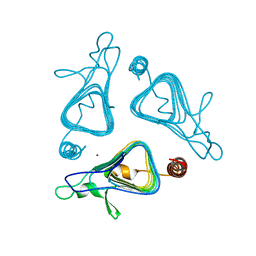
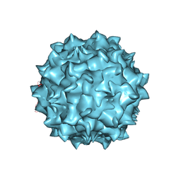
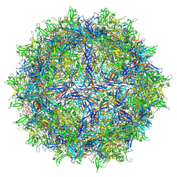
1AL0
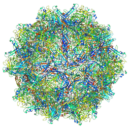
 | | PROCAPSID OF BACTERIOPHAGE PHIX174 | | 分子名称: | CAPSID PROTEIN GPF, SCAFFOLDING PROTEIN GPB, SCAFFOLDING PROTEIN GPD, ... | | 著者 | Rossmann, M.G, Dokland, T. | | 登録日 | 1997-06-06 | | 公開日 | 1998-01-28 | | 最終更新日 | 2024-04-03 | | 実験手法 | X-RAY DIFFRACTION (3.5 Å) | | 主引用文献 | Structure of a viral procapsid with molecular scaffolding.
Nature, 389, 1997
|
|
5EGC
 | |
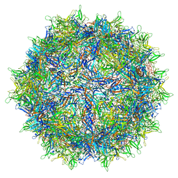
1MVM
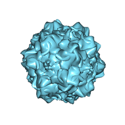 | | MVM(STRAIN I), COMPLEX(VIRAL COAT/DNA), VP2, PH=7.5, T=4 DEGREES C | | 分子名称: | DNA (5'-D(*CP*AP*AP*A)-3'), DNA (5'-D(*CP*CP*AP*CP*CP*CP*CP*AP*AP*CP*A)-3'), DNA (5'-D(P*A)-3'), ... | | 著者 | Llamas-Saiz, A.L, Agbandje-McKenna, M, Rossmann, M.G. | | 登録日 | 1996-06-21 | | 公開日 | 1998-02-25 | | 最終更新日 | 2024-04-03 | | 実験手法 | X-RAY DIFFRACTION (3.5 Å) | | 主引用文献 | Structure determination of minute virus of mice.
Acta Crystallogr.,Sect.D, 53, 1997
|
|
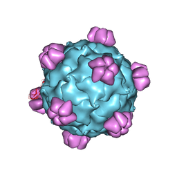
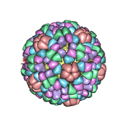
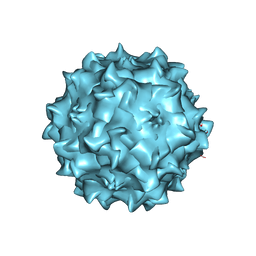
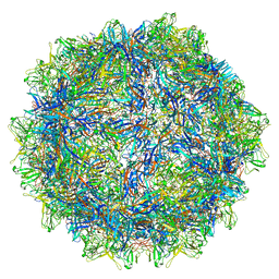
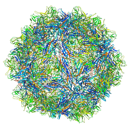
7LTM
 | | Hum8 capsid | | 分子名称: | 2'-DEOXYADENOSINE-5'-MONOPHOSPHATE, Capsid protein | | 著者 | Mietzsch, M, Agbandje-McKenna, M. | | 登録日 | 2021-02-19 | | 公開日 | 2021-07-07 | | 最終更新日 | 2021-09-22 | | 実験手法 | ELECTRON MICROSCOPY (2.49 Å) | | 主引用文献 | Receptor Switching in Newly Evolved Adeno-associated Viruses.
J.Virol., 95, 2021
|
|
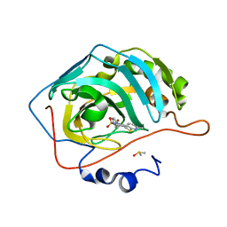
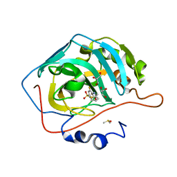
2AX2
 | | Production and X-ray crystallographic analysis of fully deuterated human carbonic anhydrase II | | 分子名称: | Carbonic anhydrase II, ZINC ION | | 著者 | Budayova-Spano, M, Fisher, S.Z, Dauvergne, M.T, Silverman, D.N, Myles, D.A.A, McKenna, R.M. | | 登録日 | 2005-09-02 | | 公開日 | 2006-01-03 | | 最終更新日 | 2023-08-23 | | 実験手法 | X-RAY DIFFRACTION (1.5 Å) | | 主引用文献 | Production and X-ray crystallographic analysis of fully deuterated human carbonic anhydrase II.
Acta Crystallogr.,Sect.F, 62, 2006
|
|
1IJJ
 | | THE X-RAY CRYSTAL STRUCTURE OF THE COMPLEX BETWEEN RABBIT SKELETAL MUSCLE ACTIN AND LATRUNCULIN A AT 2.85 A RESOLUTION | | 分子名称: | ACTIN, ALPHA SKELETAL MUSCLE, ADENOSINE-5'-TRIPHOSPHATE, ... | | 著者 | Vorobiev, S.M, Bubb, M.R, Almo, S.C. | | 登録日 | 2001-04-26 | | 公開日 | 2002-04-15 | | 最終更新日 | 2023-08-16 | | 実験手法 | X-RAY DIFFRACTION (2.85 Å) | | 主引用文献 | Polylysine induces an antiparallel actin dimer that nucleates filament assembly: crystal structure at 3.5-A resolution
J.Biol.Chem., 277, 2002
|
|
2QA0
 | |
8TXO
 | | E. coli DNA-directed RNA polymerase transcription elongation complex bound to the unnatural dZ-PTP base pair in the active site | | 分子名称: | DNA-directed RNA polymerase subunit alpha, DNA-directed RNA polymerase subunit beta, DNA-directed RNA polymerase subunit beta', ... | | 著者 | Aiyer, S, Shan, Z, Oh, J, Wang, D, Tan, Y.Z, Lyumkis, D. | | 登録日 | 2023-08-23 | | 公開日 | 2024-01-24 | | 実験手法 | ELECTRON MICROSCOPY (3.1 Å) | | 主引用文献 | Overcoming resolution attenuation during tilted cryo-EM data collection.
Nat Commun, 15, 2024
|
|
6X2K
 | |
6O9R
 | |
6WFT
 | |
6X2I
 | |
6WFU
 | |
4Y0J
 | |
3TMJ
 | |


