3BHX
 
 | |
3BXM
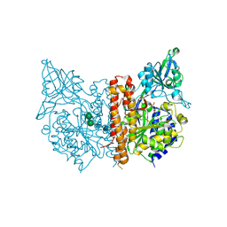
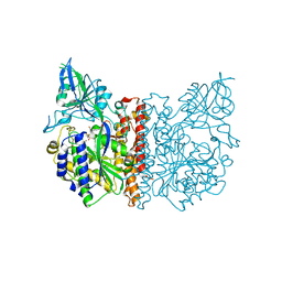
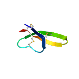
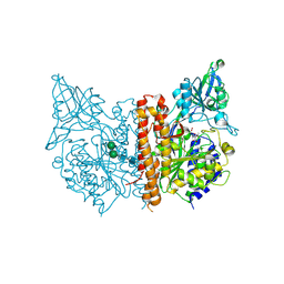
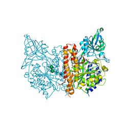
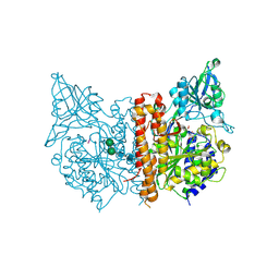 | | Structure of an inactive mutant of human glutamate carboxypeptidase II [GCPII(E424A)] in complex with N-acetyl-Asp-Glu (NAAG) | | 分子名称: | 2-acetamido-2-deoxy-beta-D-glucopyranose, 2-acetamido-2-deoxy-beta-D-glucopyranose-(1-4)-2-acetamido-2-deoxy-beta-D-glucopyranose, CALCIUM ION, ... | | 著者 | Lubkowski, J, Barinka, C. | | 登録日 | 2008-01-14 | | 公開日 | 2009-01-27 | | 最終更新日 | 2023-08-30 | | 実験手法 | X-RAY DIFFRACTION (1.71 Å) | | 主引用文献 | Reaction mechanism of glutamate carboxypeptidase II revealed by mutagenesis, X-ray crystallography, and computational methods.
Biochemistry, 48, 2009
|
|
3FF3
 | |
3FEE
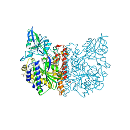
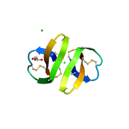
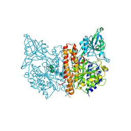
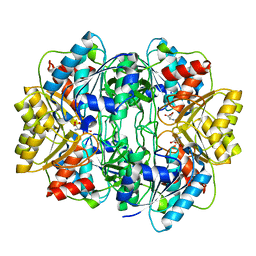
 | | The high resolution structure of human glutamate carboxypeptidase III (GCPIII/NAALADase II) in complex with quisqualic acid | | 分子名称: | (S)-2-AMINO-3-(3,5-DIOXO-[1,2,4]OXADIAZOLIDIN-2-YL)-PROPIONIC ACID, 2-acetamido-2-deoxy-beta-D-glucopyranose, 2-acetamido-2-deoxy-beta-D-glucopyranose-(1-4)-2-acetamido-2-deoxy-beta-D-glucopyranose, ... | | 著者 | Lubkowski, J, Barinka, C, Hlouchova, K. | | 登録日 | 2008-11-28 | | 公開日 | 2009-08-25 | | 最終更新日 | 2023-09-06 | | 実験手法 | X-RAY DIFFRACTION (1.56 Å) | | 主引用文献 | Structural insight into the evolutionary and pharmacologic homology of glutamate carboxypeptidases II and III
Febs J., 276, 2009
|
|
3FED
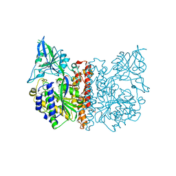
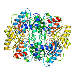
 | | The high resolution structure of human glutamate carboxypeptidase III (GCPIII/NAALADase II) in complex with a transition state analog of Glu-Glu | | 分子名称: | (2S)-2-{[(S)-[(3S)-3-amino-3-carboxypropyl](hydroxy)phosphoryl]methyl}pentanedioic acid, 2-acetamido-2-deoxy-beta-D-glucopyranose, 2-acetamido-2-deoxy-beta-D-glucopyranose-(1-4)-2-acetamido-2-deoxy-beta-D-glucopyranose, ... | | 著者 | Lubkowski, J, Barinka, C, Hlouchova, K. | | 登録日 | 2008-11-28 | | 公開日 | 2009-08-25 | | 最終更新日 | 2023-09-06 | | 実験手法 | X-RAY DIFFRACTION (1.29 Å) | | 主引用文献 | Structural insight into the evolutionary and pharmacologic homology of glutamate carboxypeptidases II and III
Febs J., 276, 2009
|
|
3HJ2
 | |
3HJD
 | |
2PM4
 | |
2PLZ
 | |
2RA4
 | |
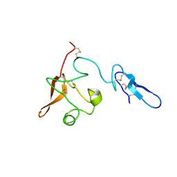
1ZMH
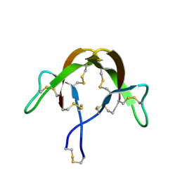
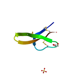
 | | Crystal structure of human neutrophil peptide 2, HNP-2 (variant Gly16-> D-Ala) | | 分子名称: | GLYCEROL, HEXAETHYLENE GLYCOL, Neutrophil defensin 2, ... | | 著者 | Lubkowski, J, Prahl, A, Lu, W. | | 登録日 | 2005-05-10 | | 公開日 | 2005-08-16 | | 最終更新日 | 2023-08-23 | | 実験手法 | X-RAY DIFFRACTION (1.5 Å) | | 主引用文献 | Reconstruction of the conserved beta-bulge in mammalian defensins using D-amino acids.
J.Biol.Chem., 280, 2005
|
|
1ZMI
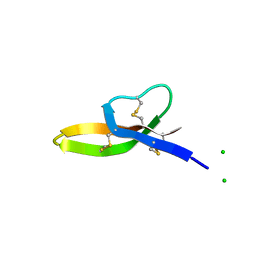
 | | Crystal structure of human alpha_defensin-2 (variant GLY16->D-ALA), P 32 2 1 space group ) | | 分子名称: | GLYCEROL, Neutrophil defensin 2, SULFATE ION | | 著者 | Lubkowski, J, Prahl, A, Lu, W. | | 登録日 | 2005-05-10 | | 公開日 | 2005-08-16 | | 最終更新日 | 2023-08-23 | | 実験手法 | X-RAY DIFFRACTION (1.15 Å) | | 主引用文献 | Reconstruction of the conserved beta-bulge in mammalian defensins using D-amino acids.
J.Biol.Chem., 280, 2005
|
|
1ZMM
 | |
1ZMK
 | | Crystal structure of human alpha-defensin-2 (variant Gly16-> D-ALA), P 42 21 2 space group | | 分子名称: | CHLORIDE ION, GLYCEROL, Neutrophil defensin 2 | | 著者 | Lubkowski, J, Prahl, A, Lu, W. | | 登録日 | 2005-05-10 | | 公開日 | 2005-08-16 | | 最終更新日 | 2023-08-23 | | 実験手法 | X-RAY DIFFRACTION (1.3 Å) | | 主引用文献 | Reconstruction of the conserved beta-bulge in mammalian defensins using D-amino acids.
J.Biol.Chem., 280, 2005
|
|
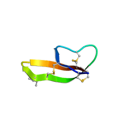
1ZMP
 | | Crystal structure of human defensin-5 | | 分子名称: | CHLORIDE ION, Defensin 5, GLYCEROL, ... | | 著者 | Lubkowski, J, Szyk, A, Lu, W. | | 登録日 | 2005-05-10 | | 公開日 | 2006-05-30 | | 最終更新日 | 2024-04-03 | | 実験手法 | X-RAY DIFFRACTION (1.65 Å) | | 主引用文献 | Crystal structures of human {alpha}-defensins HNP4, HD5, and HD6.
Protein Sci., 15, 2006
|
|
1ZMQ
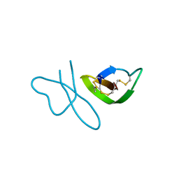
 | | Crystal structure of human alpha-defensin-6 | | 分子名称: | CHLORIDE ION, Defensin 6 | | 著者 | Lubkowski, J, Szyk, A, Lu, W. | | 登録日 | 2005-05-10 | | 公開日 | 2006-05-30 | | 最終更新日 | 2024-04-03 | | 実験手法 | X-RAY DIFFRACTION (2.1 Å) | | 主引用文献 | Crystal structures of human {alpha}-defensins HNP4, HD5, and HD6.
Protein Sci., 15, 2006
|
|
2I9A
 | |
2I9B
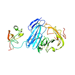 | | Crystal structure of ATF-urokinase receptor complex | | 分子名称: | 2-acetamido-2-deoxy-beta-D-glucopyranose-(1-4)-2-acetamido-2-deoxy-beta-D-glucopyranose, SULFATE ION, Urokinase plasminogen activator surface receptor, ... | | 著者 | Lubkowski, J, Barinka, C. | | 登録日 | 2006-09-05 | | 公開日 | 2007-01-02 | | 最終更新日 | 2023-08-30 | | 実験手法 | X-RAY DIFFRACTION (2.8 Å) | | 主引用文献 | Structural basis of interaction between urokinase-type plasminogen activator and its receptor.
J.Mol.Biol., 363, 2006
|
|
3D7G
 | |
3D7F
 | |
3D7H
 | |
3D7D
 | |
1HG1
 | |
1HFK
 | | Asparaginase from Erwinia chrysanthemi, hexagonal form with weak sulfate | | 分子名称: | L-ASPARAGINE AMIDOHYDROLASE, SULFATE ION | | 著者 | Lubkowski, J, Palm, G.J, Kozak, M, Jaskolski, M, Wlodawer, A. | | 登録日 | 2000-12-05 | | 公開日 | 2000-12-07 | | 最終更新日 | 2023-12-13 | | 実験手法 | X-RAY DIFFRACTION (2.17 Å) | | 主引用文献 | Structures of Two Highly Homologous Bacterial L-Asparaginases: A Case of Enantiomorphic Space Groups
Acta Crystallogr.,Sect.D, 57, 2001
|
|
1HFW
 | |


