2J1Y
 
 | |
2J1X
 | |
2J1W
 | |
2J20
 | |
2J1Z
 | |
2J21
 | |
1HL2
 | |
2BIN
 | |
2BIP
 | |
2BIO
 | |
2BIQ
 | |
2BIM
 | |
5AB9
 | |
5ABA
 | |
1UOL
 | |
5AOL
 | |
5AOM
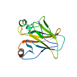
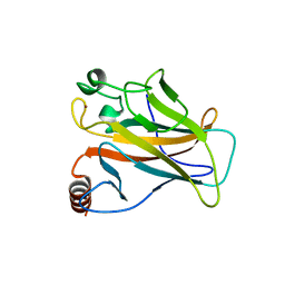
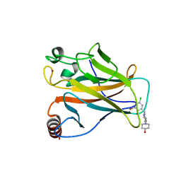
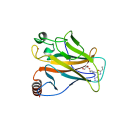
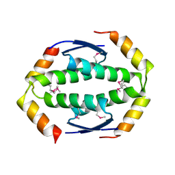
 | | Structure of the p53 cancer mutant Y220C with bound small molecule PhiKan883 | | 分子名称: | CELLULAR TUMOR ANTIGEN P53, GLYCEROL, N-(5-chloranyl-2-oxidanyl-phenyl)piperidine-4-carboxamide, ... | | 著者 | Joerger, A.C, Boeckler, F.M, Wilcken, R. | | 登録日 | 2015-09-10 | | 公開日 | 2015-12-16 | | 最終更新日 | 2024-01-10 | | 実験手法 | X-RAY DIFFRACTION (1.74 Å) | | 主引用文献 | Exploiting Transient Protein States for the Design of Small-Molecule Stabilizers of Mutant P53.
Structure, 23, 2015
|
|
5AOJ
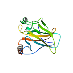
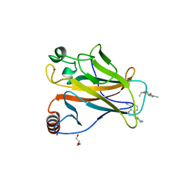
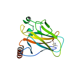
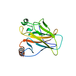
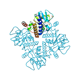
 | | Structure of the p53 cancer mutant Y220C in complex with 2-hydroxy-3, 5-diiodo-4-(1H-pyrrol-1-yl)benzoic acid | | 分子名称: | 2-hydroxy-3,5-diiodo-4-(1H-pyrrol-1-yl)benzoic acid, CELLULAR TUMOR ANTIGEN P53, DI(HYDROXYETHYL)ETHER, ... | | 著者 | Joerger, A.C, Baud, M.G, Bauer, M.R, Fersht, A.R. | | 登録日 | 2015-09-10 | | 公開日 | 2015-12-16 | | 最終更新日 | 2024-01-10 | | 実験手法 | X-RAY DIFFRACTION (1.47 Å) | | 主引用文献 | Exploiting Transient Protein States for the Design of Small-Molecule Stabilizers of Mutant P53.
Structure, 23, 2015
|
|
5AOK
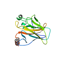
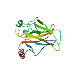
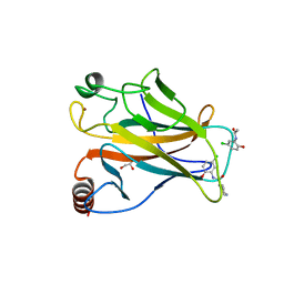
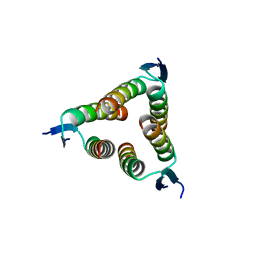
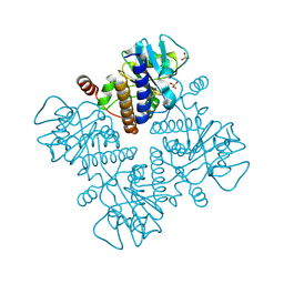
 | | Structure of the p53 cancer mutant Y220C with bound small molecule PhiKan7099 | | 分子名称: | 5-[2-cyclopropyl-5-(1H-pyrrol-1-yl)-1,3-oxazol-4-yl]-1H-1,2,3,4-tetrazole, CELLULAR TUMOR ANTIGEN P53, DI(HYDROXYETHYL)ETHER, ... | | 著者 | Joerger, A.C. | | 登録日 | 2015-09-10 | | 公開日 | 2015-12-16 | | 最終更新日 | 2024-01-10 | | 実験手法 | X-RAY DIFFRACTION (1.35 Å) | | 主引用文献 | Exploiting Transient Protein States for the Design of Small-Molecule Stabilizers of Mutant P53.
Structure, 23, 2015
|
|
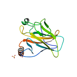
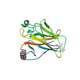
5AOI
 | |
2WQJ
 | |
2WQI
 | |
2WTT
 | |
1E4A
 | |
1E47
 | |


