3LBX
 
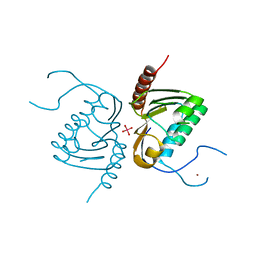
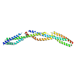 | | Crystal Structure of the Erythrocyte Spectrin Tetramerization Domain Complex | | 分子名称: | Spectrin alpha chain, erythrocyte, Spectrin beta chain | | 著者 | Ipsaro, J.J, Harper, S.L, Messick, T.E, Marmorstein, R, Mondragon, A, Speicher, D.W. | | 登録日 | 2010-01-08 | | 公開日 | 2010-03-09 | | 最終更新日 | 2024-02-21 | | 実験手法 | X-RAY DIFFRACTION (2.8 Å) | | 主引用文献 | Crystal structure and functional interpretation of the erythrocyte spectrin tetramerization domain complex.
Blood, 115, 2010
|
|
6X46
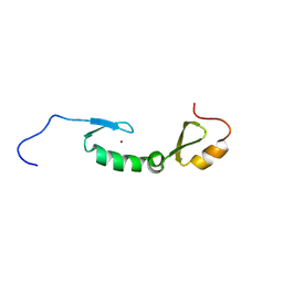 | | NMR solution structure of Asterix/Gtsf1 from mouse (CHHC zinc finger domains) | | 分子名称: | Gametocyte-specific factor 1, ZINC ION | | 著者 | Ipsaro, J.J, O'Brien, P.A, Bhattacharya, S, Palmer III, A.G, Joshua-Tor, L. | | 登録日 | 2020-05-22 | | 公開日 | 2021-03-03 | | 最終更新日 | 2021-04-14 | | 実験手法 | SOLUTION NMR | | 主引用文献 | Asterix/Gtsf1 links tRNAs and piRNA silencing of retrotransposons.
Cell Rep, 34, 2021
|
|
3KBT
 | |
3KBU
 | |
3F57
 | |
3F59
 | |
4GGJ
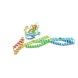
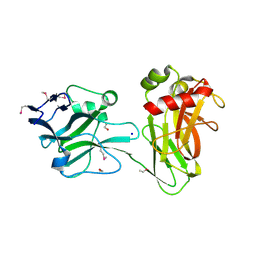
 | | Crystal structure of Zucchini from mouse (mZuc / PLD6 / MitoPLD) | | 分子名称: | Mitochondrial cardiolipin hydrolase, ZINC ION | | 著者 | Ipsaro, J.J, Haase, A.D, Hannon, G.J, Joshua-Tor, L. | | 登録日 | 2012-08-06 | | 公開日 | 2012-10-10 | | 最終更新日 | 2023-09-13 | | 実験手法 | X-RAY DIFFRACTION (1.75 Å) | | 主引用文献 | The structural biochemistry of Zucchini implicates it as a nuclease in piRNA biogenesis.
Nature, 491, 2012
|
|
4GGK
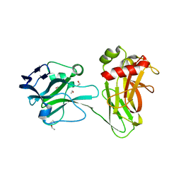
 | | Crystal structure of Zucchini from mouse (mZuc / PLD6 / MitoPLD) bound to tungstate | | 分子名称: | Mitochondrial cardiolipin hydrolase, TUNGSTATE(VI)ION, ZINC ION | | 著者 | Ipsaro, J.J, Haase, A.D, Hannon, G.J, Joshua-Tor, L. | | 登録日 | 2012-08-06 | | 公開日 | 2012-10-10 | | 最終更新日 | 2023-09-13 | | 実験手法 | X-RAY DIFFRACTION (2.1 Å) | | 主引用文献 | The structural biochemistry of Zucchini implicates it as a nuclease in piRNA biogenesis.
Nature, 491, 2012
|
|
7UX9
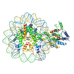 | | Arabidopsis DDM1 bound to nucleosome (H2A.W, H2B, H3.3, H4, with 147 bp DNA) | | 分子名称: | ATP-dependent DNA helicase DDM1, DNA (antisense strand), DNA (sense strand), ... | | 著者 | Ipsaro, J.J, Adams, D.W, Joshua-Tor, L. | | 登録日 | 2022-05-05 | | 公開日 | 2023-08-09 | | 最終更新日 | 2023-09-27 | | 実験手法 | ELECTRON MICROSCOPY (3.2 Å) | | 主引用文献 | Chromatin remodeling of histone H3 variants by DDM1 underlies epigenetic inheritance of DNA methylation.
Cell, 186, 2023
|
|
3UD2
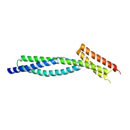
 | | Crystal structure of Selenomethionine ZU5A-ZU5B protein domains of human erythrocyte ankyrin | | 分子名称: | Ankyrin-1, CHLORIDE ION, ETHANOL, ... | | 著者 | Yasunaga, M, Ipsaro, J.J, Mondragon, A. | | 登録日 | 2011-10-27 | | 公開日 | 2012-02-22 | | 最終更新日 | 2019-12-25 | | 実験手法 | X-RAY DIFFRACTION (2.21 Å) | | 主引用文献 | Structurally Similar but Functionally Diverse ZU5 Domains in Human Erythrocyte Ankyrin.
J.Mol.Biol., 417, 2012
|
|
3UD1
 | |


