Loading
PDBj
MenuPDBj@FacebookPDBj@X(formerly Twitter)PDBj@BlueSkyPDBj@YouTubewwPDB FoundationwwPDB
RCSB PDBPDBeBMRBAdv. SearchSearch help
Search by PDB author
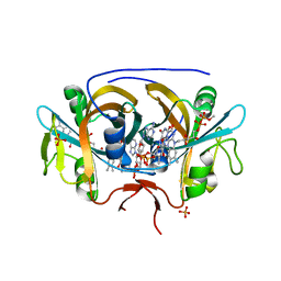
8CT0
DownloadVisualize
BU of 8ct0 by Molmil
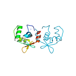
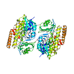
Crystal structure of FAD reductase CtcQ from Kitasatospora aureofaciens in complex with FAD and NAD
Descriptor: 1,4-DIHYDRONICOTINAMIDE ADENINE DINUCLEOTIDE, FLAVIN-ADENINE DINUCLEOTIDE, Flavin reductase (NADH), ...
Authors:Hou, C, Tsodikov, O.V.
Deposit date:2022-05-13
Release date:2023-05-17
Last modified:2023-10-25
Method:X-RAY DIFFRACTION (2.45 Å)
Cite:Crystal structures and complex formation of halogenase CtcP and FAD reductase CtcQ from the chlortetracycline biosynthetic pathway
To Be Published
6DY5
DownloadVisualize
BU of 6dy5 by Molmil
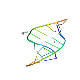
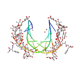
Crystal structure of double-stranded DNA AGGGATCCCT in complex with Zn2+
Descriptor: DNA (5'-D(*AP*GP*GP*GP*AP*TP*CP*CP*CP*T)-3'), SPERMIDINE, ZINC ION
Authors:Hou, C, Tsodikov, O.V.
Deposit date:2018-07-01
Release date:2019-01-23
Last modified:2024-03-13
Method:X-RAY DIFFRACTION (1.26 Å)
Cite:Utilizing guanine coordinated Zn2+ to determine DNA crystal structures by single-wavelength anomalous diffraction
Acta Crystallogr.,Sect.D, 75, 2019
6DWT
DownloadVisualize
BU of 6dwt by Molmil
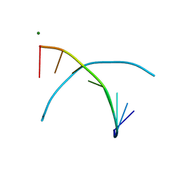
Crystal structure of double-stranded DNA GAGGCCTC, crystals grown in Mg2+
Descriptor: DNA (5'-D(*GP*AP*GP*GP*CP*CP*TP*C)-3'), MAGNESIUM ION
Authors:Hou, C, Tsodikov, O.V.
Deposit date:2018-06-27
Release date:2019-01-23
Last modified:2023-10-11
Method:X-RAY DIFFRACTION (1.6 Å)
Cite:Utilizing guanine coordinated Zn2+ to determine DNA crystal structures by single-wavelength anomalous diffraction
Acta Crystallogr.,Sect.D, 75, 2019
6DXJ
DownloadVisualize
BU of 6dxj by Molmil
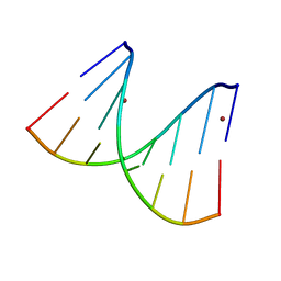
Crystal structure of double-stranded DNA GAGGCCTC; crystals grown in Zn2+
Descriptor: DNA (5'-D(*GP*AP*GP*GP*CP*CP*TP*C)-3'), ZINC ION
Authors:Hou, C, Tsodikov, O.V.
Deposit date:2018-06-29
Release date:2019-01-23
Last modified:2024-03-13
Method:X-RAY DIFFRACTION (1.65 Å)
Cite:Utilizing guanine coordinated Zn2+ to determine DNA crystal structures by single-wavelength anomalous diffraction
Acta Crystallogr.,Sect.D, 75, 2019
6DY9
DownloadVisualize
BU of 6dy9 by Molmil
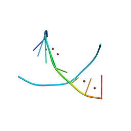
Crystal structure of double-stranded DNA GGGATCCC; crystals grown in Zn2+
Descriptor: DNA (5'-D(*GP*GP*GP*AP*TP*CP*CP*C)-3'), ZINC ION
Authors:Hou, C, Tsodikov, O.V.
Deposit date:2018-07-01
Release date:2019-01-23
Last modified:2024-03-13
Method:X-RAY DIFFRACTION (2.3 Å)
Cite:Utilizing guanine coordinated Zn2+ to determine DNA crystal structures by single-wavelength anomalous diffraction
Acta Crystallogr.,Sect.D, 75, 2019
7JSL
DownloadVisualize
BU of 7jsl by Molmil
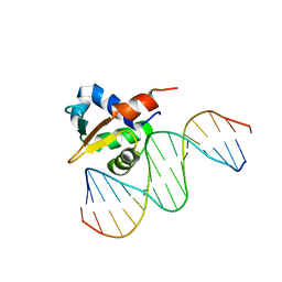
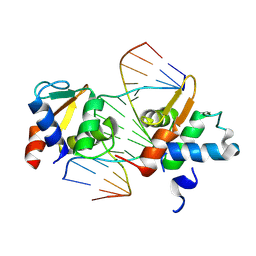
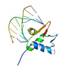
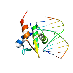
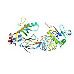
Crystal structure of the DNA binding domain of human transcription factor ERF in the oxidized form, in complex with double-stranded DNA ACCGGAAGTG
Descriptor: DNA (5'-D(*AP*CP*CP*GP*GP*AP*AP*GP*TP*G)-3'), DNA (5'-D(*CP*AP*CP*TP*TP*CP*CP*GP*GP*T)-3'), ETS domain-containing transcription factor ERF
Authors:Hou, C, Tsodikov, O.V.
Deposit date:2020-08-14
Release date:2020-11-25
Last modified:2024-10-09
Method:X-RAY DIFFRACTION (4.51 Å)
Cite:Structural Insight into the DNA Binding Function of Transcription Factor ERF.
Biochemistry, 2020
7JSA
DownloadVisualize
BU of 7jsa by Molmil
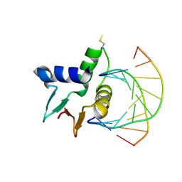
Crystal structure of the DNA binding domain of human transcription factor ERF in the reduced form, in complex with double-stranded DNA ACCGGAAGTG
Descriptor: DNA (5'-D(*AP*CP*CP*GP*GP*AP*AP*GP*TP*G)-3'), DNA (5'-D(*CP*AP*CP*TP*TP*CP*CP*GP*GP*T)-3'), ETS domain-containing transcription factor ERF
Authors:Hou, C, Tsodikov, O.V.
Deposit date:2020-08-14
Release date:2020-11-25
Last modified:2023-10-18
Method:X-RAY DIFFRACTION (2.85 Å)
Cite:Structural Insight into the DNA Binding Function of Transcription Factor ERF.
Biochemistry, 2020
6VGD
DownloadVisualize
BU of 6vgd by Molmil
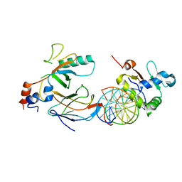
Crystal structure of the DNA binding domain (DBD) of human FLI1 and the complex of the DBD of human Runx2 with core binding factor beta (Cbfb), in complex with 16mer DNA CAGAGGATGTGGCTTC
Descriptor: Core-binding factor subunit beta, DNA (5'-D(P*CP*AP*GP*AP*GP*GP*AP*TP*GP*TP*GP*GP*CP*TP*TP*C)-3'), DNA (5'-D(P*GP*AP*AP*GP*CP*CP*AP*CP*AP*TP*CP*CP*TP*CP*TP*G)-3'), ...
Authors:Hou, C, Tsodikov, O.V.
Deposit date:2020-01-07
Release date:2020-11-25
Last modified:2023-10-11
Method:X-RAY DIFFRACTION (4.2 Å)
Cite:Allosteric interference in oncogenic FLI1 and ERG transactions by mithramycins.
Structure, 29, 2021
6VG2
DownloadVisualize
BU of 6vg2 by Molmil
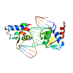
Crystal structure of the DNA binding domain of human transcription factor FLI1 in complex with 16-mer DNA CAGAGGATGTGGCTTC
Descriptor: DNA (5'-D(*GP*AP*AP*GP*CP*CP*AP*CP*TP*TP*CP*CP*GP*GP*TP*C)-3'), DNA (5'-D(*GP*AP*CP*CP*GP*GP*AP*AP*GP*TP*GP*GP*CP*TP*TP*C)-3'), Friend leukemia integration 1 transcription factor
Authors:Hou, C, Tsodikov, O.V.
Deposit date:2020-01-07
Release date:2020-11-25
Last modified:2023-10-11
Method:X-RAY DIFFRACTION (3.9 Å)
Cite:Allosteric interference in oncogenic FLI1 and ERG transactions by mithramycins.
Structure, 29, 2021
6VGG
DownloadVisualize
BU of 6vgg by Molmil
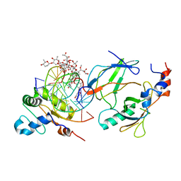
Crystal structure of the DNA binding domains of human transcription factor ERG, human Runx2 bound to core binding factor beta (Cbfb), and mithramycin, in complex with 16mer DNA CAGAGGATGTGGCTTC
Descriptor: Core-binding factor subunit beta, DNA (5'-D(P*CP*AP*GP*AP*GP*GP*AP*TP*GP*TP*GP*GP*CP*TP*TP*C)-3'), DNA (5'-D(P*GP*AP*AP*GP*CP*CP*AP*CP*AP*TP*CP*CP*TP*CP*TP*G)-3'), ...
Authors:Hou, C, Rohr, J, Tsodikov, O.V.
Deposit date:2020-01-08
Release date:2020-11-25
Last modified:2023-10-11
Method:X-RAY DIFFRACTION (4.31 Å)
Cite:Allosteric interference in oncogenic FLI1 and ERG transactions by mithramycins.
Structure, 29, 2021
6VG8
DownloadVisualize
BU of 6vg8 by Molmil
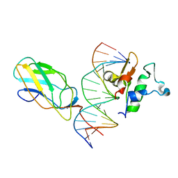
Crystal structure of the DNA binding domains of human FLI1 and Runx2 in complex with 16-mer DNA CAGAGGATGTGGCTTC
Descriptor: DNA (5'-D(P*CP*AP*GP*AP*GP*GP*AP*TP*GP*TP*GP*GP*CP*TP*TP*C)-3'), DNA (5'-D(P*GP*AP*AP*GP*CP*CP*AP*CP*AP*TP*CP*CP*TP*CP*TP*G)-3'), Friend leukemia integration 1 transcription factor, ...
Authors:Hou, C, Tsodikov, O.V.
Deposit date:2020-01-07
Release date:2020-11-25
Last modified:2023-10-11
Method:X-RAY DIFFRACTION (4.31 Å)
Cite:Allosteric interference in oncogenic FLI1 and ERG transactions by mithramycins.
Structure, 29, 2021
6VGE
DownloadVisualize
BU of 6vge by Molmil
Crystal structure of the DNA binding domains of human transcription factor ERG, human Runx2 bound to core binding factor beta (Cbfb), in complex with 16mer DNA CAGAGGATGTGGCTTC
Descriptor: Core-binding factor subunit beta, DNA (5'-D(P*CP*AP*GP*AP*GP*GP*AP*TP*GP*TP*GP*GP*CP*TP*TP*C)-3'), DNA (5'-D(P*GP*AP*AP*GP*CP*CP*AP*CP*AP*TP*CP*CP*TP*CP*TP*G)-3'), ...
Authors:Hou, C, Tsodikov, O.V.
Deposit date:2020-01-07
Release date:2020-11-25
Last modified:2023-10-11
Method:X-RAY DIFFRACTION (4.25 Å)
Cite:Allosteric interference in oncogenic FLI1 and ERG transactions by mithramycins.
Structure, 29, 2021
5JW0
DownloadVisualize
BU of 5jw0 by Molmil
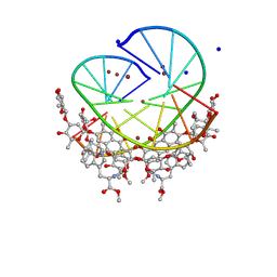
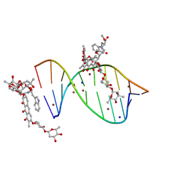
Crystal structure of mithramycin analogue MTM SA-Phe in complex with a 10-mer DNA AGGGTACCCT
Descriptor: DNA (5'-D(P*AP*GP*GP*GP*TP*AP*CP*CP*CP*T)-3'), Plicamycin, mithramycin analogue MTM SA-Phe, ...
Authors:Hou, C, Rohr, J, Tsodikov, O.V.
Deposit date:2016-05-11
Release date:2016-09-14
Last modified:2024-03-06
Method:X-RAY DIFFRACTION (2.4 Å)
Cite:Structures of mithramycin analogues bound to DNA and implications for targeting transcription factor FLI1.
Nucleic Acids Res., 44, 2016
5JW2
DownloadVisualize
BU of 5jw2 by Molmil
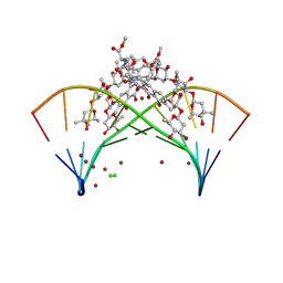
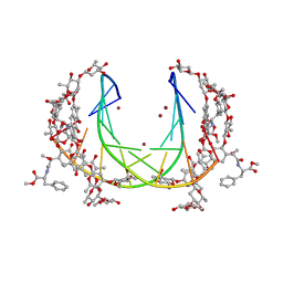
Crystal structure of mithramycin analogue MTM SA-Phe in complex with a 10-mer DNA AGGGATCCCT
Descriptor: DNA (5'-D(*AP*GP*GP*GP*AP*TP*CP*CP*CP*T)-3'), Plicamycin, mithramycin analogue MTM SA-Phe, ...
Authors:Hou, C, Rohr, J, Tsodikov, O.V.
Deposit date:2016-05-11
Release date:2016-09-14
Last modified:2024-03-06
Method:X-RAY DIFFRACTION (3.1 Å)
Cite:Structures of mithramycin analogues bound to DNA and implications for targeting transcription factor FLI1.
Nucleic Acids Res., 44, 2016
5JVT
DownloadVisualize
BU of 5jvt by Molmil
Crystal structure of the DNA binding domain of transcription factor FLI1 in complex with an 11-mer DNA GACCGGAAGTG
Descriptor: DNA (5'-D(*CP*AP*CP*TP*TP*CP*CP*GP*GP*TP*C)-3'), DNA (5'-D(*GP*AP*CP*CP*GP*GP*AP*AP*GP*TP*G)-3'), Friend leukemia integration 1 transcription factor, ...
Authors:Hou, C, Tsodikov, O.V.
Deposit date:2016-05-11
Release date:2016-09-14
Last modified:2023-09-27
Method:X-RAY DIFFRACTION (3.1 Å)
Cite:Structures of mithramycin analogues bound to DNA and implications for targeting transcription factor FLI1.
Nucleic Acids Res., 44, 2016
5JVW
DownloadVisualize
BU of 5jvw by Molmil
Crystal structure of mithramycin analogue MTM SA-Trp in complex with a 10-mer DNA AGAGGCCTCT.
Descriptor: DNA (5'-D(*AP*GP*AP*GP*GP*CP*CP*TP*CP*T)-3'), Plicamycin, mithramycin analogue MTM SA-Trp, ...
Authors:Hou, C, Rohr, J, Tsodikov, O.V.
Deposit date:2016-05-11
Release date:2016-09-14
Last modified:2024-03-06
Method:X-RAY DIFFRACTION (2 Å)
Cite:Structures of mithramycin analogues bound to DNA and implications for targeting transcription factor FLI1.
Nucleic Acids Res., 44, 2016
7V0B
DownloadVisualize
BU of 7v0b by Molmil
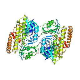
Crystal structure of halogenase CtcP from Kitasatospora aureofaciens in complex with FAD
Descriptor: 4-(2-HYDROXYETHYL)-1-PIPERAZINE ETHANESULFONIC ACID, CHLORIDE ION, FLAVIN-ADENINE DINUCLEOTIDE, ...
Authors:Hou, C, Tsodikov, O.V.
Deposit date:2022-05-10
Release date:2023-05-17
Last modified:2023-10-25
Method:X-RAY DIFFRACTION (2.15 Å)
Cite:Crystal structures and complex formation of halogenase CtcP and FAD reductase CtcQ from the chlortetracycline biosynthetic pathway
To Be Published
7V0D
DownloadVisualize
BU of 7v0d by Molmil
Crystal structure of halogenase CtcP from Kitasatospora aureofaciens
Descriptor: 4-(2-HYDROXYETHYL)-1-PIPERAZINE ETHANESULFONIC ACID, CHLORIDE ION, DI(HYDROXYETHYL)ETHER, ...
Authors:Hou, C, Tsodikov, O.V.
Deposit date:2022-05-10
Release date:2023-05-17
Last modified:2023-10-25
Method:X-RAY DIFFRACTION (2.6 Å)
Cite:Crystal structures and complex formation of halogenase CtcP and FAD reductase CtcQ from the chlortetracycline biosynthetic pathway
To Be Published
5E8G
DownloadVisualize
BU of 5e8g by Molmil
Crystal structure of the DNA binding domain of human transcription factor FLI1
Descriptor: COBALT (II) ION, Friend leukemia integration 1 transcription factor
Authors:Hou, C, Tsodikov, O.V.
Deposit date:2015-10-14
Release date:2015-12-09
Last modified:2023-09-27
Method:X-RAY DIFFRACTION (2.7 Å)
Cite:Structural Basis for Dimerization and DNA Binding of Transcription Factor FLI1.
Biochemistry, 54, 2015
5E8I
DownloadVisualize
BU of 5e8i by Molmil
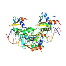
Crystal structure of the DNA binding domain of human transcription factor FLI1 in complex with a 10-mer DNA ACCGGAAGTG
Descriptor: CALCIUM ION, DNA (5'-D(*AP*CP*CP*GP*GP*AP*AP*GP*TP*G)-3'), DNA (5'-D(*CP*AP*CP*TP*TP*CP*CP*GP*GP*T)-3'), ...
Authors:Hou, C, Tsodikov, O.V.
Deposit date:2015-10-14
Release date:2015-12-09
Last modified:2023-09-27
Method:X-RAY DIFFRACTION (3.45 Å)
Cite:Structural Basis for Dimerization and DNA Binding of Transcription Factor FLI1.
Biochemistry, 54, 2015
5W36
DownloadVisualize
BU of 5w36 by Molmil
Crystal structure of the RNA polymerase domain (RPD) of Mycobacterium tuberculosis primase DnaG in complex with a double-stranded DNA oligomer with a 6-nucleotide overhang
Descriptor: DNA primase, STRONTIUM ION, synthetic DNA 5'-TATCGTCCCGCCTC
Authors:Hou, C, Tsodikov, O.V.
Deposit date:2017-06-07
Release date:2018-03-28
Last modified:2023-10-04
Method:X-RAY DIFFRACTION (2.46 Å)
Cite:Structures of the Catalytic Domain of Bacterial Primase DnaG in Complexes with DNA Provide Insight into Key Priming Events.
Biochemistry, 57, 2018
5W33
DownloadVisualize
BU of 5w33 by Molmil
Crystal structure of the RNA polymerase domain (RPD) of Mycobacterium tuberculosis primase DnaG
Descriptor: DNA primase, SULFATE ION
Authors:Hou, C, Biswas, T, Tsodikov, O.V.
Deposit date:2017-06-07
Release date:2018-03-28
Last modified:2023-10-04
Method:X-RAY DIFFRACTION (2.85 Å)
Cite:Structures of the Catalytic Domain of Bacterial Primase DnaG in Complexes with DNA Provide Insight into Key Priming Events.
Biochemistry, 57, 2018
5W34
DownloadVisualize
BU of 5w34 by Molmil
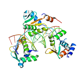
Crystal structure of the RNA polymerase domain (RPD) of Mycobacterium tuberculosis primase DnaG in complex with double-stranded DNA GACCGGAAGTGG
Descriptor: DNA oligomer 5'-CCACTTCCGGTC, DNA oligomer 5'-GACCGGAAGTGG, DNA primase
Authors:Hou, C, Tsodikov, O.V.
Deposit date:2017-06-07
Release date:2018-03-28
Last modified:2023-10-04
Method:X-RAY DIFFRACTION (2.95 Å)
Cite:Structures of the Catalytic Domain of Bacterial Primase DnaG in Complexes with DNA Provide Insight into Key Priming Events.
Biochemistry, 57, 2018
5W35
DownloadVisualize
BU of 5w35 by Molmil
Crystal structure of the RNA polymerase domain (RPD) of Mycobacterium tuberculosis primase DnaG in complex with a double-stranded DNA oligomer with a 1-nucleotide overhang
Descriptor: DNA primase, synthetic DNA oligomer 5'-CCACTTCCGGTC, synthetic DNA oligomer 5'-TGACCGGAAGTGG
Authors:Hou, C, Tsodikov, O.V.
Deposit date:2017-06-07
Release date:2018-03-28
Last modified:2023-10-04
Method:X-RAY DIFFRACTION (3.31 Å)
Cite:Structures of the Catalytic Domain of Bacterial Primase DnaG in Complexes with DNA Provide Insight into Key Priming Events.
Biochemistry, 57, 2018
7CH7
DownloadVisualize
BU of 7ch7 by Molmil
Cryo-EM structure of E.coli MlaFEB
Descriptor: Lipid asymmetry maintenance ABC transporter permease subunit MlaE, Lipid asymmetry maintenance protein MlaB, Phospholipid ABC transporter ATP-binding protein MlaF
Authors:Zhou, C, Shi, H, Huang, Y.
Deposit date:2020-07-05
Release date:2021-05-19
Last modified:2024-03-27
Method:ELECTRON MICROSCOPY (3.9 Å)
Cite:Structural Insight into Phospholipid Transport by the MlaFEBD Complex from P. aeruginosa.
J.Mol.Biol., 433, 2021

227561

PDB entries from 2024-11-20

PDB statisticsPDBj update infoContact PDBjnumon