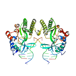
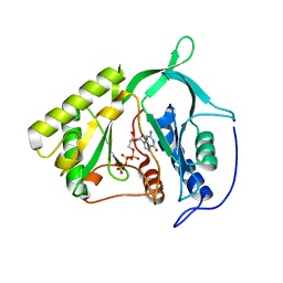
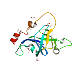
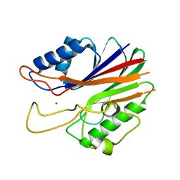
3PKY
 
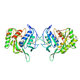 | | Polymerase Domain from Mycobacterium tuberculosis Ligase D in complex with DNA, UTP and Manganese. | | 分子名称: | DNA 5'-D(*G*CP*CP*GP*CP*AP*AP*CP*GP*CP*AP*CP*G)-3', DNA 5'-D(P*GP*CP*GP*GP*C)-3', MANGANESE (II) ION, ... | | 著者 | Brissett, N.C, Fox, G.C, Pitcher, R.S, Doherty, A.J. | | 登録日 | 2010-11-12 | | 公開日 | 2011-02-16 | | 最終更新日 | 2024-02-21 | | 実験手法 | X-RAY DIFFRACTION (3.1 Å) | | 主引用文献 | Structure of a Preternary Complex Involving a Prokaryotic NHEJ DNA Polymerase.
Mol.Cell, 41, 2011
|
|
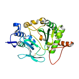
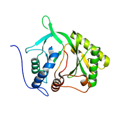
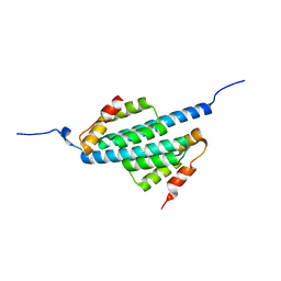
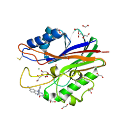
2R9L
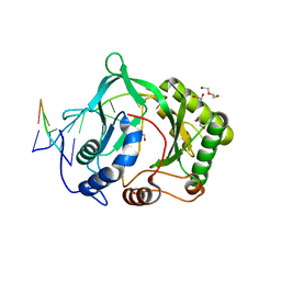 | | Polymerase Domain from Mycobacterium tuberculosis Ligase D in complex with DNA | | 分子名称: | 1,2-ETHANEDIOL, DI(HYDROXYETHYL)ETHER, DNA (5'-D(*DGP*DCP*DCP*DGP*DCP*DAP*DAP*DCP*DGP*DCP*DA)-3'), ... | | 著者 | Brissett, N.C, Fox, G.C, Pitcher, R.S, Doherty, A.J. | | 登録日 | 2007-09-13 | | 公開日 | 2008-01-08 | | 最終更新日 | 2023-08-30 | | 実験手法 | X-RAY DIFFRACTION (2.4 Å) | | 主引用文献 | Structure of a NHEJ polymerase-mediated DNA synaptic complex
Science, 318, 2007
|
|
6SA1
 | |
6SA0
 | |
4MKY
 | |
5OP0
 | |
5N85
 | |
5N8A
 | |
2IRX
 | |
2IRU
 | |
2IRY
 | |
4LIT
 | |
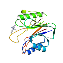
4LIU
 | | Structure of YcfD, a Ribosomal oxygenase from Escherichia coli. | | 分子名称: | 50S ribosomal protein L16 arginine hydroxylase, PHOSPHATE ION, TRIS(HYDROXYETHYL)AMINOMETHANE | | 著者 | Brissett, N.C, Doherty, A.J, Fox, G.C. | | 登録日 | 2013-07-03 | | 公開日 | 2014-05-14 | | 最終更新日 | 2014-07-09 | | 実験手法 | X-RAY DIFFRACTION (2.7 Å) | | 主引用文献 | Ribosomal oxygenases are structurally conserved from prokaryotes to humans.
Nature, 509, 2014
|
|
4LIV
 | |
5DMU
 | | Structure of the NHEJ polymerase from Methanocella paludicola | | 分子名称: | 1,2-ETHANEDIOL, GLYCEROL, NHEJ Polymerase, ... | | 著者 | Brissett, N.C, Bartlett, E.J, Doherty, A.J. | | 登録日 | 2015-09-09 | | 公開日 | 2015-10-07 | | 最終更新日 | 2024-01-10 | | 実験手法 | X-RAY DIFFRACTION (1.949 Å) | | 主引用文献 | Molecular basis for DNA strand displacement by NHEJ repair polymerases.
Nucleic Acids Res., 44, 2016
|
|
5DMP
 | | Structure of the Archaeal NHEJ Phosphoesterase from Methanocella paludicola. | | 分子名称: | 1,2-ETHANEDIOL, MAGNESIUM ION, Uncharacterized protein, ... | | 著者 | Brissett, N.C, Bartlett, E.J, Doherty, A.J. | | 登録日 | 2015-09-09 | | 公開日 | 2015-10-07 | | 最終更新日 | 2024-01-10 | | 実験手法 | X-RAY DIFFRACTION (1.793 Å) | | 主引用文献 | Molecular basis for DNA strand displacement by NHEJ repair polymerases.
Nucleic Acids Res., 44, 2016
|
|
2XDV
 | | Crystal Structure of the Catalytic Domain of FLJ14393 | | 分子名称: | 1,2-ETHANEDIOL, CADMIUM ION, MANGANESE (II) ION, ... | | 著者 | Krojer, T, Muniz, J.R.C, Ng, S.S, Pilka, E, Guo, K, Pike, A.C.W, Filippakopoulos, P, Knapp, S, Kavanagh, K.L, Gileadi, O, Bunkoczi, G, Yue, W.W, Niesen, F, Sobott, F, Fedorov, O, Savitsky, P, Kochan, G, Daniel, M, von Delft, F, Arrowsmith, C.H, Edwards, A.M, Weigelt, J, Bountra, C, Oppermann, U. | | 登録日 | 2010-05-07 | | 公開日 | 2010-05-26 | | 最終更新日 | 2018-02-21 | | 実験手法 | X-RAY DIFFRACTION (2.57 Å) | | 主引用文献 | Ribosomal oxygenases are structurally conserved from prokaryotes to humans.
Nature, 510, 2014
|
|
1UZ3
 | |
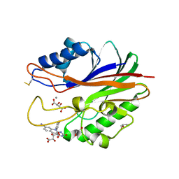
5J3S
 | | Crystal structure of the catalytic domain of human tyrosyl DNA phosphodiesterase 2 in complex with a small molecule inhibitor | | 分子名称: | 2,4-dioxo-10-[3-(1H-tetrazol-5-yl)phenyl]-2,3,4,10-tetrahydropyrimido[4,5-b]quinoline-8-carbonitrile, Tyrosyl-DNA phosphodiesterase 2 | | 著者 | Hornyak, P, Pearl, L.H, Caldecott, K.W, Oliver, A.W. | | 登録日 | 2016-03-31 | | 公開日 | 2016-05-04 | | 最終更新日 | 2024-01-10 | | 実験手法 | X-RAY DIFFRACTION (3.4 Å) | | 主引用文献 | Mode of action of DNA-competitive small molecule inhibitors of tyrosyl DNA phosphodiesterase 2.
Biochem.J., 473, 2016
|
|
5J42
 | | Crystal structure of m2hTDP2-CAT in complex with a small molecule inhibitor | | 分子名称: | 1,2-ETHANEDIOL, 10-(4-hydroxyphenyl)-2,4-dioxo-2,3,4,10-tetrahydropyrimido[4,5-b]quinoline-8-carbonitrile, GLYCEROL, ... | | 著者 | Hornyak, P, Pearl, L.H, Caldecott, K.W, Oliver, A.W. | | 登録日 | 2016-03-31 | | 公開日 | 2016-05-04 | | 最終更新日 | 2024-01-10 | | 実験手法 | X-RAY DIFFRACTION (1.7 Å) | | 主引用文献 | Mode of action of DNA-competitive small molecule inhibitors of tyrosyl DNA phosphodiesterase 2.
Biochem.J., 473, 2016
|
|
5J3P
 | | Crystal structure of the catalytic domain of human tyrosyl DNA phosphodiesterase 2 | | 分子名称: | GLYCEROL, MAGNESIUM ION, Tyrosyl-DNA phosphodiesterase 2 | | 著者 | Hornyak, P, Pearl, L.H, Caldecott, K.W, Oliver, A.W. | | 登録日 | 2016-03-31 | | 公開日 | 2016-05-04 | | 最終更新日 | 2024-01-10 | | 実験手法 | X-RAY DIFFRACTION (3.1 Å) | | 主引用文献 | Mode of action of DNA-competitive small molecule inhibitors of tyrosyl DNA phosphodiesterase 2.
Biochem.J., 473, 2016
|
|
5J3Z
 | | Crystal structure of m2hTDP2-CAT in complex with a small molecule inhibitor | | 分子名称: | 1,2-ETHANEDIOL, 2,4-dioxo-10-[3-(1H-tetrazol-5-yl)phenyl]-2,3,4,10-tetrahydropyrimido[4,5-b]quinoline-8-carbonitrile, ACETATE ION, ... | | 著者 | Hornyak, P, Pearl, L.H, Caldecott, K.W, Oliver, A.W. | | 登録日 | 2016-03-31 | | 公開日 | 2016-05-04 | | 最終更新日 | 2024-01-10 | | 実験手法 | X-RAY DIFFRACTION (1.8 Å) | | 主引用文献 | Mode of action of DNA-competitive small molecule inhibitors of tyrosyl DNA phosphodiesterase 2.
Biochem.J., 473, 2016
|
|
4BU2
 | | 60S ribosomal protein L27A histidine hydroxylase (MINA53) in complex with Ni(II) and 2-oxoglutarate (2OG) | | 分子名称: | 1,2-ETHANEDIOL, 2-OXOGLUTARIC ACID, BIFUNCTIONAL LYSINE-SPECIFIC DEMETHYLASE AND HISTIDYL-HYDROXYLASE MINA, ... | | 著者 | Chowdhury, R, Clifton, I.J, McDonough, M.A, Ng, S.S, Pilka, E, Oppermann, U, Schofield, C.J. | | 登録日 | 2013-06-19 | | 公開日 | 2014-05-14 | | 最終更新日 | 2019-02-06 | | 実験手法 | X-RAY DIFFRACTION (2.78 Å) | | 主引用文献 | Ribosomal oxygenases are structurally conserved from prokaryotes to humans.
Nature, 510, 2014
|
|
4CCL
 | | X-Ray structure of E. coli ycfD | | 分子名称: | 50S RIBOSOMAL PROTEIN L16 ARGININE HYDROXYLASE, MANGANESE (II) ION, SULFATE ION | | 著者 | McDonough, M.A, Ho, C.H, Kershaw, N.J, Schofield, C.J. | | 登録日 | 2013-10-23 | | 公開日 | 2014-04-30 | | 最終更新日 | 2020-06-03 | | 実験手法 | X-RAY DIFFRACTION (2.596 Å) | | 主引用文献 | Ribosomal oxygenases are structurally conserved from prokaryotes to humans.
Nature, 510, 2014
|
|
4BXF
 | |


