Loading
PDBj
MenuPDBj@FacebookPDBj@TwitterPDBj@YouTubewwPDB FoundationwwPDB
RCSB PDBPDBeBMRBAdv. SearchSearch help
Search by PDB author
3HAV
DownloadVisualize
BU of 3hav by Molmil
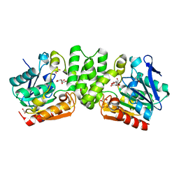
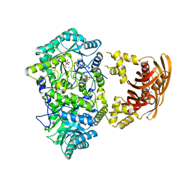
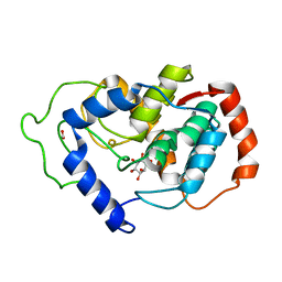
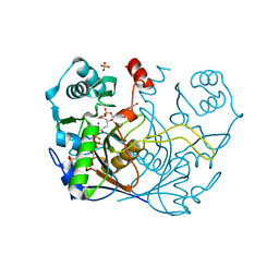
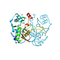
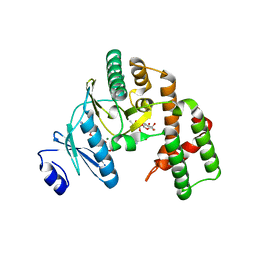
Structure of the streptomycin-ATP-APH(2")-IIa ternary complex
Descriptor: ADENOSINE-5'-TRIPHOSPHATE, Aminoglycoside phosphotransferase, MAGNESIUM ION, ...
Authors:Young, P.G, Baker, E.N, Vakulenko, S.B, Smith, C.A.
Deposit date:2009-05-02
Release date:2009-11-17
Last modified:2023-09-06
Method:X-RAY DIFFRACTION (2.45 Å)
Cite:The crystal structures of substrate and nucleotide complexes of Enterococcus faecium aminoglycoside-2''-phosphotransferase-IIa [APH(2'')-IIa] provide insights into substrate selectivity in the APH(2'') subfamily.
J.Bacteriol., 191, 2009
3GLD
DownloadVisualize
BU of 3gld by Molmil
Crystal Structure of the Major Pilin from Streptococcus pyogenes E117A mutant
Descriptor: Pilin
Authors:Kang, H.J, Baker, E.N.
Deposit date:2009-03-12
Release date:2009-06-02
Last modified:2023-11-01
Method:X-RAY DIFFRACTION (2.03 Å)
Cite:Intramolecular isopeptide bonds give thermodynamic and proteolytic stability to the major pilin protein of Streptococcus pyogenes
To be Published
3HAM
DownloadVisualize
BU of 3ham by Molmil
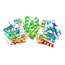
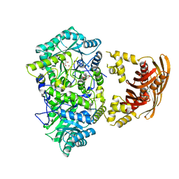
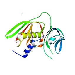
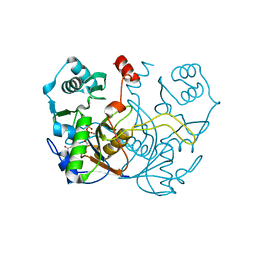
Structure of the gentamicin-APH(2")-IIa complex
Descriptor: (2R,3R,4R,5R)-2-((1S,2S,3R,4S,6R)-4,6-DIAMINO-3-((2R,3R,6S)-3-AMINO-6-(AMINOMETHYL)-TETRAHYDRO-2H-PYRAN-2-YLOXY)-2-HYDR OXYCYCLOHEXYLOXY)-5-METHYL-4-(METHYLAMINO)-TETRAHYDRO-2H-PYRAN-3,5-DIOL, Aminoglycoside phosphotransferase, GLYCEROL
Authors:Young, P.G, Baker, E.N, Vakulenko, S.B, Smith, C.A.
Deposit date:2009-05-02
Release date:2009-07-28
Last modified:2024-02-21
Method:X-RAY DIFFRACTION (2.5 Å)
Cite:The crystal structures of substrate and nucleotide complexes of Enterococcus faecium aminoglycoside-2''-phosphotransferase-IIa [APH(2'')-IIa] provide insights into substrate selectivity in the APH(2'') subfamily.
J.Bacteriol., 191, 2009
3GLE
DownloadVisualize
BU of 3gle by Molmil
Crystal Structure of the Major Pilin from Streptococcus pyogenes N168A mutant
Descriptor: Pilin
Authors:Kang, H.J, Baker, E.N.
Deposit date:2009-03-12
Release date:2009-06-02
Last modified:2023-11-01
Method:X-RAY DIFFRACTION (2.19 Å)
Cite:Intramolecular isopeptide bonds give thermodynamic and proteolytic stability to the major pilin protein of Streptococcus pyogenes
To be Published
3HYS
DownloadVisualize
BU of 3hys by Molmil
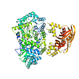
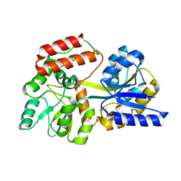
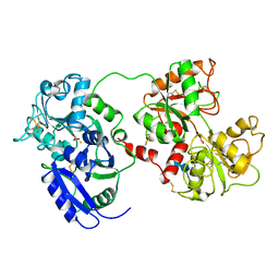
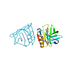
Structure of Rv0554 from Mycobacterium tuberculosis complexed with Malonic Acid
Descriptor: 1,2-ETHANEDIOL, 2-AMINO-2-HYDROXYMETHYL-PROPANE-1,3-DIOL, MALONIC ACID, ...
Authors:Johnston, J.M, Baker, E.N.
Deposit date:2009-06-23
Release date:2010-06-02
Last modified:2024-02-21
Method:X-RAY DIFFRACTION (2.3 Å)
Cite:Structural and functional analysis of Rv0554 from Mycobacterium tuberculosis: testing a putative role in menaquinone biosynthesis.
Acta Crystallogr.,Sect.D, 66, 2010
3HZO
DownloadVisualize
BU of 3hzo by Molmil
Rv0554 from Mycobacterium tuberculosis - the structure solved from the tetragonal crystal form
Descriptor: 1,2-ETHANEDIOL, MALONIC ACID, SODIUM ION, ...
Authors:Johnston, J.M, Baker, E.N.
Deposit date:2009-06-23
Release date:2010-06-02
Last modified:2024-02-21
Method:X-RAY DIFFRACTION (2.6 Å)
Cite:The structure of Rv0554 from Mycobacterium tuberculosis
To be Published
3HPS
DownloadVisualize
BU of 3hps by Molmil
Crystal structure of Mycobacterium tuberculosis LeuA complexed with ketoisocaproate (KIC)
Descriptor: 2-OXO-4-METHYLPENTANOIC ACID, 2-isopropylmalate synthase, GLYCEROL, ...
Authors:Koon, N, Squire, C.J, Baker, E.N.
Deposit date:2009-06-04
Release date:2010-05-19
Last modified:2023-09-06
Method:X-RAY DIFFRACTION (1.8 Å)
Cite:Probing the active site of M. tuberculosis LeuA
To be Published
3HSS
DownloadVisualize
BU of 3hss by Molmil
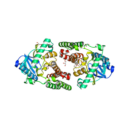
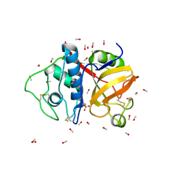
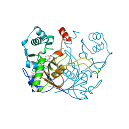
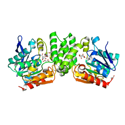
A higher resolution structure of Rv0554 from Mycobacterium tuberculosis complexed with malonic acid
Descriptor: 1,2-ETHANEDIOL, 2-AMINO-2-HYDROXYMETHYL-PROPANE-1,3-DIOL, ACETATE ION, ...
Authors:Johnston, J.M, Baker, E.N.
Deposit date:2009-06-10
Release date:2010-05-26
Last modified:2024-02-21
Method:X-RAY DIFFRACTION (1.9 Å)
Cite:Structural and functional analysis of Rv0554 from Mycobacterium tuberculosis: testing a putative role in menaquinone biosynthesis.
Acta Crystallogr.,Sect.D, 66, 2010
3HQ1
DownloadVisualize
BU of 3hq1 by Molmil
Crystal structure of Mycobacterium tuberculosis LeuA complexed with citrate and Mn2+
Descriptor: 2-isopropylmalate synthase, CHLORIDE ION, CITRATE ANION, ...
Authors:Koon, N, Squire, C.J, Baker, E.N.
Deposit date:2009-06-05
Release date:2010-05-19
Last modified:2023-09-06
Method:X-RAY DIFFRACTION (1.7 Å)
Cite:Probing the active site of M. tuberculosis LeuA
To be Published
3HPZ
DownloadVisualize
BU of 3hpz by Molmil
Crystal structure of Mycobacterium tuberculosis LeuA complexed with bromopyruvate
Descriptor: 2-isopropylmalate synthase, Bromopyruvate, CHLORIDE ION, ...
Authors:Koon, N, Squire, C.J, Baker, E.N.
Deposit date:2009-06-05
Release date:2010-05-19
Last modified:2023-09-06
Method:X-RAY DIFFRACTION (2.2 Å)
Cite:Probing the active site of M. tuberculosis LeuA
To be Published
3HST
DownloadVisualize
BU of 3hst by Molmil
N-Terminal RNASE H domain of rv2228c from mycobacterium tuberculosis as a fusion protein with maltose binding protein
Descriptor: 1,2-ETHANEDIOL, D(-)-TARTARIC ACID, Maltose-binding periplasmic protein, ...
Authors:Watkins, H.A, Baker, E.N.
Deposit date:2009-06-10
Release date:2010-04-28
Last modified:2024-02-21
Method:X-RAY DIFFRACTION (2.25 Å)
Cite:Structural and functional characterization of an RNase HI domain from the bifunctional protein Rv2228c from Mycobacterium tuberculosis.
J.Bacteriol., 192, 2010
3HTL
DownloadVisualize
BU of 3htl by Molmil
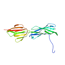
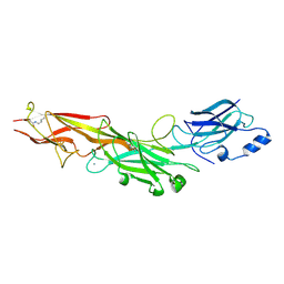
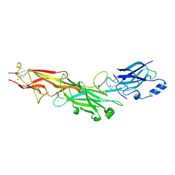
Structure of the Corynebacterium diphtheriae major pilin SpaA points to a modular pilus assembly with stabilizing isopeptide bonds
Descriptor: CALCIUM ION, Putative surface-anchored fimbrial subunit, SODIUM ION
Authors:Kang, H.J, Paterson, N.G, Gaspar, A.H, Ton-That, H, Baker, E.N.
Deposit date:2009-06-11
Release date:2009-09-15
Last modified:2011-07-13
Method:X-RAY DIFFRACTION (1.8 Å)
Cite:The Corynebacterium diphtheriae shaft pilin SpaA is built of tandem Ig-like modules with stabilizing isopeptide and disulfide bonds
Proc.Natl.Acad.Sci.USA, 106, 2009
3HR6
DownloadVisualize
BU of 3hr6 by Molmil
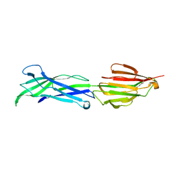
Structure of the Corynebacterium diphtheriae major pilin SpaA points to a modular pilus assembly stabilizing isopeptide bonds
Descriptor: CALCIUM ION, IODIDE ION, Putative surface-anchored fimbrial subunit
Authors:Kang, H.J, Paterson, N.G, Gaspar, A.H, Ton-That, H, Baker, E.N.
Deposit date:2009-06-08
Release date:2009-09-15
Last modified:2011-07-13
Method:X-RAY DIFFRACTION (1.6 Å)
Cite:The Corynebacterium diphtheriae shaft pilin SpaA is built of tandem Ig-like modules with stabilizing isopeptide and disulfide bonds
Proc.Natl.Acad.Sci.USA, 106, 2009
2HUO
DownloadVisualize
BU of 2huo by Molmil
Crystal structure of mouse myo-inositol oxygenase in complex with substrate
Descriptor: 1,2,3,4,5,6-HEXAHYDROXY-CYCLOHEXANE, FE (III) ION, FORMIC ACID, ...
Authors:Brown, P.M, Caradoc-Davies, T.T, Dickson, J.M.J, Cooper, G.J.S, Loomes, K.M, Baker, E.N.
Deposit date:2006-07-27
Release date:2006-09-26
Last modified:2024-02-14
Method:X-RAY DIFFRACTION (2 Å)
Cite:Crystal structure of a substrate complex of myo-inositol oxygenase, a di-iron oxygenase with a key role in inositol metabolism.
Proc.Natl.Acad.Sci.Usa, 103, 2006
2HYX
DownloadVisualize
BU of 2hyx by Molmil
Structure of the C-terminal domain of DipZ from Mycobacterium tuberculosis
Descriptor: Protein dipZ
Authors:Goldstone, D, Baker, E.N, Metcalf, P, TB Structural Genomics Consortium (TBSGC)
Deposit date:2006-08-08
Release date:2007-07-17
Last modified:2017-10-18
Method:X-RAY DIFFRACTION (1.9 Å)
Cite:Structure of the C-terminal domain of DipZ from Mycobacterium tuberculosis
To be Published
4INS
DownloadVisualize
BU of 4ins by Molmil
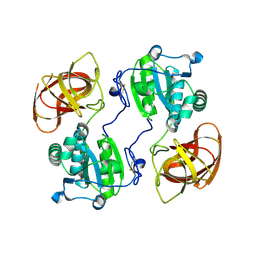
THE STRUCTURE OF 2ZN PIG INSULIN CRYSTALS AT 1.5 ANGSTROMS RESOLUTION
Descriptor: INSULIN (CHAIN A), INSULIN (CHAIN B), ZINC ION
Authors:Dodson, G.G, Dodson, E.J, Hodgkin, D.C, Isaacs, N.W, Vijayan, M.
Deposit date:1989-07-10
Release date:1990-04-15
Last modified:2017-11-29
Method:X-RAY DIFFRACTION (1.5 Å)
Cite:The structure of 2Zn pig insulin crystals at 1.5 A resolution.
Philos.Trans.R.Soc.London,Ser.B, 319, 1988
9PAP
DownloadVisualize
BU of 9pap by Molmil
STRUCTURE OF PAPAIN REFINED AT 1.65 ANGSTROMS RESOLUTION
Descriptor: METHANOL, PAPAIN
Authors:Kamphuis, I.G, Drenth, J.
Deposit date:1986-03-31
Release date:1986-10-24
Last modified:2017-11-29
Method:X-RAY DIFFRACTION (1.65 Å)
Cite:Structure of papain refined at 1.65 A resolution
J.Mol.Biol., 179, 1984
8G8P
DownloadVisualize
BU of 8g8p by Molmil
F420-2/GTP(GDP) complex of F420-gamma glutamyl ligase (CofE) from Archaeoglobus fulgidus
Descriptor: COENZYME F420, Coenzyme F420:L-glutamate ligase, GUANOSINE-5'-DIPHOSPHATE, ...
Authors:Bashiri, G, Squire, C.J.
Deposit date:2023-02-18
Release date:2024-02-28
Method:X-RAY DIFFRACTION (1.83 Å)
Cite:Poly-gamma-glutamylation of biomolecules.
Nat Commun, 15, 2024
2R61
DownloadVisualize
BU of 2r61 by Molmil
Crystal structure of the Staphylococcal superantigen-like protein SSL5 in complex with sialyl-Lewis X at pH 7.4
Descriptor: CHLORIDE ION, Exotoxin 3, GLYCEROL, ...
Authors:Baker, H.M.
Deposit date:2007-09-05
Release date:2007-11-27
Last modified:2023-10-25
Method:X-RAY DIFFRACTION (2.75 Å)
Cite:Crystal structures of the staphylococcal toxin SSL5 in complex with sialyl Lewis X reveal a conserved binding site that shares common features with viral and bacterial sialic acid binding proteins
J.Mol.Biol., 374, 2007
1OQH
DownloadVisualize
BU of 1oqh by Molmil
Crystal Structure of the R124A mutant of the N-lobe human transferrin
Descriptor: CARBONATE ION, FE (III) ION, POTASSIUM ION, ...
Authors:Baker, H.M, He, Q.-Y, Brigg, S.K, Mason, A.B, N Baker, E.
Deposit date:2003-03-10
Release date:2003-03-18
Last modified:2023-08-16
Method:X-RAY DIFFRACTION (2.4 Å)
Cite:Structural and functional consequences of binding site mutations in transferrin: crystal structures of the Asp63Glu and Arg124Ala mutants of the N-lobe of human transferrin
Biochemistry, 42, 2003
1DOT
DownloadVisualize
BU of 1dot by Molmil
CRYSTALLOGRAPHIC STRUCTURE OF DUCK OVOTRANSFERRIN AT 2.3 ANGSTROMS RESOLUTION
Descriptor: 2-acetamido-2-deoxy-beta-D-glucopyranose, CARBONATE ION, DUCK OVOTRANSFERRIN, ...
Authors:Rawas, A, Muirhead, H.
Deposit date:1995-08-03
Release date:1996-03-08
Last modified:2020-07-29
Method:X-RAY DIFFRACTION (2.35 Å)
Cite:Structure of diferric duck ovotransferrin at 2.35 A resolution.
Acta Crystallogr.,Sect.D, 52, 1996
7ULE
DownloadVisualize
BU of 7ule by Molmil
F420-1/GDP complex of F420-gamma glutamyl ligase (CofE) from Archaeoglobus fulgidus
Descriptor: (2~{S})-2-[[(2~{S})-2-[oxidanyl-[(2~{R},3~{S},4~{S})-2,3,4-tris(oxidanyl)-5-[2,4,8-tris(oxidanylidene)-1,9-dihydropyrimido[4,5-b]quinolin-10-yl]pentoxy]phosphoryl]oxypropanoyl]amino]pentanedioic acid, Coenzyme F420:L-glutamate ligase, GUANOSINE-5'-DIPHOSPHATE, ...
Authors:Bashiri, G, Squire, C.J.
Deposit date:2022-04-04
Release date:2023-04-12
Last modified:2023-10-25
Method:X-RAY DIFFRACTION (1.7 Å)
Cite:A Universal Mechanism for Poly-glutamylation
To Be Published
7ULD
DownloadVisualize
BU of 7uld by Molmil
GTP complex of F420-gamma glutamyl ligase (CofE) from Archaeoglobus fulgidus
Descriptor: CARBONATE ION, Coenzyme F420:L-glutamate ligase, GUANOSINE-5'-TRIPHOSPHATE, ...
Authors:Bashiri, G, Squire, C.J.
Deposit date:2022-04-04
Release date:2023-04-12
Last modified:2023-10-25
Method:X-RAY DIFFRACTION (1.3 Å)
Cite:A Universal Mechanism for Poly-glutamylation
To Be Published
7ULF
DownloadVisualize
BU of 7ulf by Molmil
l-glutamate/GTP complex of F420-gamma glutamyl ligase (CofE) from Archaeoglobus fulgidus
Descriptor: Coenzyme F420:L-glutamate ligase, GAMMA-L-GLUTAMIC ACID, GUANOSINE-5'-TRIPHOSPHATE, ...
Authors:Bashiri, G, Squire, C.J.
Deposit date:2022-04-04
Release date:2023-04-12
Last modified:2023-10-25
Method:X-RAY DIFFRACTION (1.61 Å)
Cite:A Universal Mechanism for Poly-glutamylation
To Be Published
1B0O
DownloadVisualize
BU of 1b0o by Molmil
BOVINE BETA-LACTOGLOBULIN COMPLEXED WITH PALMITATE, LATTICE Z
Descriptor: BETA-LACTOGLOBULIN, PALMITIC ACID
Authors:Wu, S.-Y, Sawyer, L.
Deposit date:1998-11-11
Release date:1999-02-02
Last modified:2023-08-02
Method:X-RAY DIFFRACTION (2.5 Å)
Cite:beta-lactoglobulin binds palmitate within its central cavity.
J.Biol.Chem., 274, 1999

219869

PDB entries from 2024-05-15

PDB statisticsPDBj update infoContact PDBjnumon