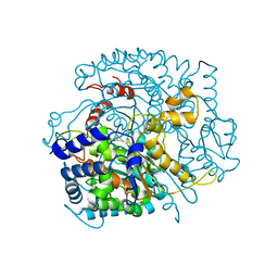
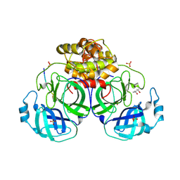
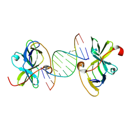
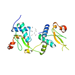
2PK2
 
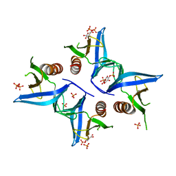
 | | Cyclin box structure of the P-TEFb subunit Cyclin T1 derived from a fusion complex with EIAV Tat | | 分子名称: | Cyclin-T1, Protein Tat | | 著者 | Anand, K, Schulte, A, Fujinaga, K, Scheffzek, K, Geyer, M. | | 登録日 | 2007-04-17 | | 公開日 | 2007-07-03 | | 最終更新日 | 2024-02-21 | | 実験手法 | X-RAY DIFFRACTION (2.67 Å) | | 主引用文献 | Cyclin Box Structure of the P-TEFb Subunit Cyclin T1 Derived from a Fusion Complex with EIAV Tat.
J.Mol.Biol., 370, 2007
|
|
2W2H
 | |
2X1V
 | |
2VB0
 | | Crystal structure of coxsackievirus B3 proteinase 3C | | 分子名称: | CHLORIDE ION, POLYPROTEIN 3BCD | | 著者 | Anand, K, Mesters, J.R, Goerlach, R, Zell, R, Hilgenfeld, R. | | 登録日 | 2007-09-05 | | 公開日 | 2008-10-28 | | 最終更新日 | 2019-07-24 | | 実験手法 | X-RAY DIFFRACTION (2.4 Å) | | 主引用文献 | Crystal Structure of Coxsackie Virus B3 Proteinase 3C
To be Published
|
|
2WU8
 | |
1LVO
 | | Structure of coronavirus main proteinase reveals combination of a chymotrypsin fold with an extra alpha-helical domain | | 分子名称: | (4R)-2-METHYLPENTANE-2,4-DIOL, 1,4-DIETHYLENE DIOXIDE, Replicase, ... | | 著者 | Anand, K, Palm, G.J, Mesters, J.R, Siddell, S.G, Ziebuhr, J, Hilgenfeld, R. | | 登録日 | 2002-05-29 | | 公開日 | 2002-07-17 | | 最終更新日 | 2024-03-13 | | 実験手法 | X-RAY DIFFRACTION (1.96 Å) | | 主引用文献 | Structure of coronavirus main proteinase reveals combination of a chymotrypsin fold with an extra alpha-helical domain.
EMBO J., 21, 2002
|
|
3DGV
 | | Crystal structure of thrombin activatable fibrinolysis inhibitor (TAFI) | | 分子名称: | 2-acetamido-2-deoxy-alpha-D-glucopyranose-(1-4)-2-acetamido-2-deoxy-beta-D-glucopyranose, 2-acetamido-2-deoxy-beta-D-glucopyranose, 2-acetamido-2-deoxy-beta-D-glucopyranose-(1-4)-2-acetamido-2-deoxy-beta-D-glucopyranose, ... | | 著者 | Anand, K, Pallares, I, Valnickova, Z, Christensen, T, Schreuder, H, Enghild, J. | | 登録日 | 2008-06-16 | | 公開日 | 2008-07-22 | | 最終更新日 | 2023-11-01 | | 実験手法 | X-RAY DIFFRACTION (2.5 Å) | | 主引用文献 | The crystal structure of thrombin-activable fibrinolysis inhibitor (TAFI) provides the structural basis for its intrinsic activity and the short half-life of TAFIa.
J.Biol.Chem., 283, 2008
|
|
4A6K
 | | Crystal structure of Slm1-PH domain in complex with D-myo-Inositol-4- phosphate | | 分子名称: | D-MYO-INOSITOL-4-PHOSPHATE, PHOSPHATE ION, PHOSPHATIDYLINOSITOL 4,5-BISPHOSPHATE-BINDING PROTEIN SLM1 | | 著者 | Anand, K, Maeda, K, Gavin, A.C. | | 登録日 | 2011-11-04 | | 公開日 | 2012-06-13 | | 実験手法 | X-RAY DIFFRACTION (1.8 Å) | | 主引用文献 | Structural Analyses of Slm1-Ph Domain Demonstrate Ligand Binding in the Non-Canonical Site
Plos One, 7, 2012
|
|
4A6H
 | | Crystal structure of Slm1-PH domain in complex with Inositol-4- phosphate | | 分子名称: | D-MYO-INOSITOL-4-PHOSPHATE, PHOSPHATE ION, PHOSPHATIDYLINOSITOL 4,5-BISPHOSPHATE-BINDING PROTEIN SLM1 | | 著者 | Anand, K, Maeda, K, Gavin, A.C. | | 登録日 | 2011-11-03 | | 公開日 | 2012-06-13 | | 最終更新日 | 2015-03-25 | | 実験手法 | X-RAY DIFFRACTION (1.449 Å) | | 主引用文献 | Structural Analyses of Slm1-Ph Domain Demonstrate Ligand Binding in the Non-Canonical Site
Plos One, 7, 2012
|
|
4A6F
 | | Crystal structure of Slm1-PH domain in complex with Phosphoserine | | 分子名称: | PHOSPHATE ION, PHOSPHATIDYLINOSITOL 4,5-BISPHOSPHATE-BINDING PROTEIN SLM1, PHOSPHOSERINE | | 著者 | Anand, K, Maeda, K, Gavin, A.C. | | 登録日 | 2011-11-02 | | 公開日 | 2012-06-13 | | 最終更新日 | 2023-12-20 | | 実験手法 | X-RAY DIFFRACTION (1.68 Å) | | 主引用文献 | Structural Analyses of Slm1-Ph Domain Demonstrate Ligand Binding in the Non-Canonical Site
Plos One, 7, 2012
|
|
4A5K
 | |
1P9S
 | | Coronavirus Main Proteinase (3CLpro) Structure: Basis for Design of anti-SARS Drugs | | 分子名称: | 1,4-DIETHYLENE DIOXIDE, Replicase polyprotein 1ab | | 著者 | Anand, K, Ziebuhr, J, Wadhwani, P, Mesters, J.R, Hilgenfeld, R. | | 登録日 | 2003-05-12 | | 公開日 | 2003-05-20 | | 最終更新日 | 2023-11-15 | | 実験手法 | X-RAY DIFFRACTION (2.54 Å) | | 主引用文献 | Coronavirus Main Proteinase (3CLpro) Structure: Basis for Design of anti-SARS Drugs
Science, 300, 2003
|
|
1P9U
 | | Coronavirus Main Proteinase (3CLpro) Structure: Basis for Design of anti-SARS Drugs | | 分子名称: | (4R)-2-METHYLPENTANE-2,4-DIOL, PHQ-VNSTLQ-CHLOROMETHYLKETONE INHIBITOR, SULFATE ION, ... | | 著者 | Anand, K, Ziebuhr, J, Wadhwani, P, Mesters, J.R, Hilgenfeld, R. | | 登録日 | 2003-05-12 | | 公開日 | 2003-05-20 | | 最終更新日 | 2017-10-11 | | 実験手法 | X-RAY DIFFRACTION (2.37 Å) | | 主引用文献 | Coronavirus Main Proteinase (3CLpro) Structure: Basis for Design of anti-SARS Drugs
Science, 300, 2003
|
|
8PYR
 | | Crystal structure of the dual T-loop phosphorylated Cdk7/CycH/Mat1 complex | | 分子名称: | 1,2-ETHANEDIOL, CDK-activating kinase assembly factor MAT1, Cyclin-H, ... | | 著者 | Anand, K, Duster, R, Geyer, M. | | 登録日 | 2023-07-26 | | 公開日 | 2024-03-06 | | 実験手法 | X-RAY DIFFRACTION (2.15 Å) | | 主引用文献 | Structural basis of Cdk7 activation by dual T-loop phosphorylation.
Biorxiv, 2024
|
|
8P81
 | | Crystal structure of human Cdk12/Cyclin K in complex with inhibitor SR-4835 | | 分子名称: | Cyclin-K, Cyclin-dependent kinase 12, ~{N}-[[5,6-bis(chloranyl)-1~{H}-benzimidazol-2-yl]methyl]-9-(1-methylpyrazol-4-yl)-2-morpholin-4-yl-purin-6-amine | | 著者 | Anand, K, Schmitz, M, Geyer, M. | | 登録日 | 2023-05-31 | | 公開日 | 2023-11-22 | | 最終更新日 | 2023-12-27 | | 実験手法 | X-RAY DIFFRACTION (2.68 Å) | | 主引用文献 | The reversible inhibitor SR-4835 binds Cdk12/cyclin K in a noncanonical G-loop conformation.
J.Biol.Chem., 300, 2023
|
|
7NXJ
 | | Crystal structure of human Cdk13/Cyclin K in complex with the inhibitor THZ531 | | 分子名称: | Cyclin-K, Cyclin-dependent kinase 13, N-[4-[(3R)-3-[[5-chloranyl-4-(1H-indol-3-yl)pyrimidin-2-yl]amino]piperidin-1-yl]carbonylphenyl]-4-(dimethylamino)butanamide | | 著者 | Anand, K, Greifenberg, A.K, Kaltheuner, I.H, Geyer, M. | | 登録日 | 2021-03-18 | | 公開日 | 2021-05-12 | | 最終更新日 | 2024-01-31 | | 実験手法 | X-RAY DIFFRACTION (2.36 Å) | | 主引用文献 | Structure-activity relationship study of THZ531 derivatives enables the discovery of BSJ-01-175 as a dual CDK12/13 covalent inhibitor with efficacy in Ewing sarcoma.
Eur.J.Med.Chem., 221, 2021
|
|
7NXK
 | | Crystal structure of human Cdk12/Cyclin K in complex with the inhibitor BSJ-01-175 | | 分子名称: | (E)-N-[4-[(1R,3R)-3-[[5-chloranyl-4-(1H-indol-3-yl)pyrimidin-2-yl]amino]cyclohexyl]oxyphenyl]-4-(dimethylamino)but-2-enamide, Cyclin-K, Cyclin-dependent kinase 12 | | 著者 | Anand, K, Dust, S, Kaltheuner, I.H, Geyer, M. | | 登録日 | 2021-03-18 | | 公開日 | 2021-05-12 | | 最終更新日 | 2024-01-31 | | 実験手法 | X-RAY DIFFRACTION (3 Å) | | 主引用文献 | Structure-activity relationship study of THZ531 derivatives enables the discovery of BSJ-01-175 as a dual CDK12/13 covalent inhibitor with efficacy in Ewing sarcoma.
Eur.J.Med.Chem., 221, 2021
|
|
7BAH
 | |
7BAI
 | |
6Z2G
 | | Crystal structure of human NLRP9 PYD | | 分子名称: | NACHT, LRR and PYD domains-containing protein 9 | | 著者 | Marleaux, M, Anand, K, Geyer, M. | | 登録日 | 2020-05-15 | | 公開日 | 2020-06-10 | | 最終更新日 | 2024-01-24 | | 実験手法 | X-RAY DIFFRACTION (1.95 Å) | | 主引用文献 | Crystal structure of the human NLRP9 pyrin domain suggests a distinct mode of inflammasome assembly.
Febs Lett., 594, 2020
|
|
5EFQ
 | | Crystal structure of human Cdk13/Cyclin K in complex with ADP-aluminum fluoride | | 分子名称: | ADENOSINE-5'-DIPHOSPHATE, ALUMINUM FLUORIDE, Cyclin-K, ... | | 著者 | Hoenig, D, Greifenberg, A.K, Anand, K, Geyer, M. | | 登録日 | 2015-10-24 | | 公開日 | 2015-12-30 | | 最終更新日 | 2024-01-10 | | 実験手法 | X-RAY DIFFRACTION (2 Å) | | 主引用文献 | Structural and Functional Analysis of the Cdk13/Cyclin K Complex.
Cell Rep, 14, 2016
|
|
5NUI
 | | Crystal structure of SIVmac239 Nef in an ExxxLM endocytic sorting motif bound state | | 分子名称: | Protein Nef, SER-GLN-ILE-LYS-ARG-LEU-LEU-SER | | 著者 | Manrique, S, Horenkamp, F.A, Anand, K, Geyer, M. | | 登録日 | 2017-04-30 | | 公開日 | 2017-08-16 | | 最終更新日 | 2024-01-17 | | 実験手法 | X-RAY DIFFRACTION (2.5 Å) | | 主引用文献 | Endocytic sorting motif interactions involved in Nef-mediated downmodulation of CD4 and CD3.
Nat Commun, 8, 2017
|
|
5NUH
 | |
4NST
 | | Crystal structure of human Cdk12/Cyclin K in complex with ADP-aluminum fluoride | | 分子名称: | 1,2-ETHANEDIOL, ADENOSINE-5'-DIPHOSPHATE, ALUMINUM FLUORIDE, ... | | 著者 | Boesken, C.A, Farnung, L, Anand, K, Geyer, M. | | 登録日 | 2013-11-29 | | 公開日 | 2014-03-26 | | 最終更新日 | 2023-09-20 | | 実験手法 | X-RAY DIFFRACTION (2.2 Å) | | 主引用文献 | The structure and substrate specificity of human Cdk12/Cyclin K.
Nat Commun, 5, 2014
|
|
7O7I
 | |


