Loading
PDBj
MenuPDBj@FacebookPDBj@TwitterPDBj@YouTubewwPDB FoundationwwPDB
RCSB PDBPDBeBMRBAdv. SearchSearch help
Search by PDB author
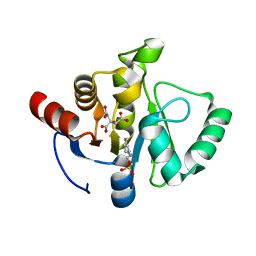
7TWO
DownloadVisualize
BU of 7two by Molmil
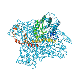
Crystal structure of SARS-CoV-2 NSP3 macrodomain at pH 6 (P43 crystal form)
Descriptor: 2-[N-CYCLOHEXYLAMINO]ETHANE SULFONIC ACID, CITRIC ACID, Non-structural protein 3
Authors:Correy, G.J, Fraser, J.S.
Deposit date:2022-02-07
Release date:2022-02-23
Last modified:2023-10-18
Method:X-RAY DIFFRACTION (0.9 Å)
Cite:The mechanisms of catalysis and ligand binding for the SARS-CoV-2 NSP3 macrodomain from neutron and x-ray diffraction at room temperature.
Sci Adv, 8, 2022
4PDJ
DownloadVisualize
BU of 4pdj by Molmil
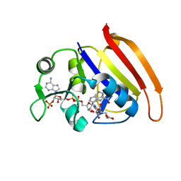
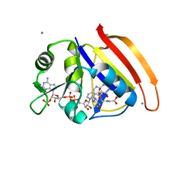
Neutron crystal Structure of E.coli Dihydrofolate Reductase complexed with folate and NADP+
Descriptor: DIHYDROFOLIC ACID, Dihydrofolate reductase, MANGANESE (II) ION, ...
Authors:Wan, Q, Kovalevsky, A.Y, Wilson, M, Langan, P, Dealwis, C, Bennett, B.
Deposit date:2014-04-18
Release date:2015-04-15
Last modified:2024-04-03
Method:NEUTRON DIFFRACTION (1.599 Å), X-RAY DIFFRACTION
Cite:Toward resolving the catalytic mechanism of dihydrofolate reductase using neutron and ultrahigh-resolution X-ray crystallography.
Proc.Natl.Acad.Sci.USA, 111, 2014
4QEE
DownloadVisualize
BU of 4qee by Molmil
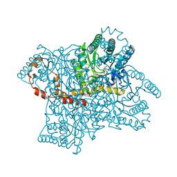
Room temperature X-ray structure of D-xylose isomerase in complex with two Ni2+ ions and L-ribose
Descriptor: NICKEL (II) ION, Xylose isomerase, alpha-L-ribofuranose
Authors:Kovalevsky, A.Y, Langan, P.
Deposit date:2014-05-15
Release date:2014-09-03
Last modified:2024-02-28
Method:X-RAY DIFFRACTION (1.6 Å)
Cite:L-Arabinose Binding, Isomerization, and Epimerization by D-Xylose Isomerase: X-Ray/Neutron Crystallographic and Molecular Simulation Study.
Structure, 22, 2014
4QDW
DownloadVisualize
BU of 4qdw by Molmil
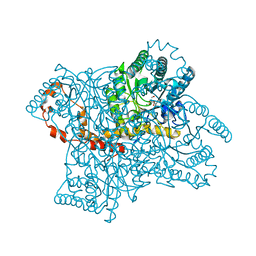
Joint X-ray and neutron structure of Streptomyces rubiginosus D-xylose isomerase in complex with two Ni2+ ions and linear L-arabinose
Descriptor: L-arabinose, NICKEL (II) ION, Xylose isomerase
Authors:Kovalevsky, A.Y, Langan, P.
Deposit date:2014-05-14
Release date:2014-09-03
Last modified:2024-02-28
Method:NEUTRON DIFFRACTION (1.8 Å), X-RAY DIFFRACTION
Cite:L-Arabinose Binding, Isomerization, and Epimerization by D-Xylose Isomerase: X-Ray/Neutron Crystallographic and Molecular Simulation Study.
Structure, 22, 2014
4QE5
DownloadVisualize
BU of 4qe5 by Molmil
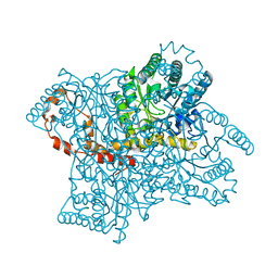
Room temperature X-ray structure of D-xylose isomerase in complex with two Mg2+ ions and L-ribulose
Descriptor: MAGNESIUM ION, Xylose isomerase, alpha-L-ribulofuranose
Authors:Kovalevsky, A.Y, Langan, P.
Deposit date:2014-05-15
Release date:2014-09-03
Last modified:2024-02-28
Method:X-RAY DIFFRACTION (1.56 Å)
Cite:L-Arabinose Binding, Isomerization, and Epimerization by D-Xylose Isomerase: X-Ray/Neutron Crystallographic and Molecular Simulation Study.
Structure, 22, 2014
4QEH
DownloadVisualize
BU of 4qeh by Molmil
Room temperature X-ray structure of D-xylose isomerase in complex with two Mg2+ ions and L-ribose
Descriptor: MAGNESIUM ION, Xylose isomerase, beta-L-ribofuranose
Authors:Kovalevsky, A.Y, Langan, P.
Deposit date:2014-05-16
Release date:2014-09-03
Last modified:2024-02-28
Method:X-RAY DIFFRACTION (1.55 Å)
Cite:L-Arabinose Binding, Isomerization, and Epimerization by D-Xylose Isomerase: X-Ray/Neutron Crystallographic and Molecular Simulation Study.
Structure, 22, 2014
4QE4
DownloadVisualize
BU of 4qe4 by Molmil
Room temperature X-ray structure of D-xylose isomerase in complex with two Ni2+ ions and L-ribulose
Descriptor: NICKEL (II) ION, Xylose isomerase, beta-L-ribulofuranose
Authors:Kovalevsky, A.Y, Langan, P.
Deposit date:2014-05-15
Release date:2014-09-03
Last modified:2024-02-28
Method:X-RAY DIFFRACTION (1.7 Å)
Cite:L-Arabinose Binding, Isomerization, and Epimerization by D-Xylose Isomerase: X-Ray/Neutron Crystallographic and Molecular Simulation Study.
Structure, 22, 2014
4QDP
DownloadVisualize
BU of 4qdp by Molmil
Joint X-ray and neutron structure of Streptomyces rubiginosus D-xylose isomerase in complex with two Cd2+ ions and cyclic beta-L-arabinose
Descriptor: CADMIUM ION, Xylose isomerase, beta-L-arabinopyranose
Authors:Kovalevsky, A.Y, Langan, P.
Deposit date:2014-05-14
Release date:2014-09-03
Last modified:2024-02-28
Method:NEUTRON DIFFRACTION (2 Å), X-RAY DIFFRACTION
Cite:L-Arabinose Binding, Isomerization, and Epimerization by D-Xylose Isomerase: X-Ray/Neutron Crystallographic and Molecular Simulation Study.
Structure, 22, 2014
4QE1
DownloadVisualize
BU of 4qe1 by Molmil
Room temperature X-ray structure of D-xylose isomerase in complex with two Cd2+ ions and L-ribulose
Descriptor: CADMIUM ION, Xylose isomerase, alpha-L-ribulofuranose
Authors:Kovalevsky, A.Y, Langan, P.
Deposit date:2014-05-15
Release date:2014-09-03
Last modified:2024-02-28
Method:X-RAY DIFFRACTION (1.55 Å)
Cite:L-Arabinose Binding, Isomerization, and Epimerization by D-Xylose Isomerase: X-Ray/Neutron Crystallographic and Molecular Simulation Study.
Structure, 22, 2014
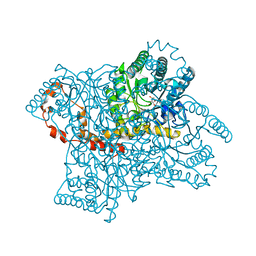
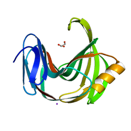
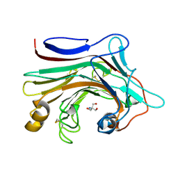
6K9R
DownloadVisualize
BU of 6k9r by Molmil
Crystal Structure Analysis of Endo-beta-1,4-xylanase II Complexed with Xylotriose
Descriptor: Endo-1,4-beta-xylanase 2, IODIDE ION, beta-D-xylopyranose-(1-4)-beta-D-xylopyranose-(1-4)-beta-D-xylopyranose
Authors:Li, C, Wan, Q.
Deposit date:2019-06-17
Release date:2020-07-08
Last modified:2023-11-22
Method:X-RAY DIFFRACTION (1.3 Å)
Cite:Studying the Role of a Single Mutation of a Family 11 Glycoside Hydrolase Using High-Resolution X-ray Crystallography.
Protein J., 39, 2020
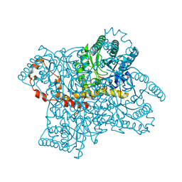
6K9O
DownloadVisualize
BU of 6k9o by Molmil
Crystal Structure Analysis of Protein
Descriptor: Endo-1,4-beta-xylanase 2, GLYCEROL, IODIDE ION
Authors:Li, C, Wan, Q.
Deposit date:2019-06-17
Release date:2020-06-17
Last modified:2023-11-22
Method:X-RAY DIFFRACTION (1.06 Å)
Cite:Studying the Role of a Single Mutation of a Family 11 Glycoside Hydrolase Using High-Resolution X-ray Crystallography.
Protein J., 39, 2020
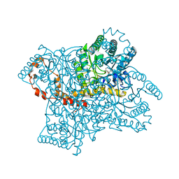
4HKW
DownloadVisualize
BU of 4hkw by Molmil
Crystal Structures of Mutant Endo-beta-1,4-xylanase II Complexed with Substrate and Products
Descriptor: 2-AMINO-2-HYDROXYMETHYL-PROPANE-1,3-DIOL, CALCIUM ION, Endo-1,4-beta-xylanase 2, ...
Authors:Kovalevsky, A.Y, Wan, Q, Langan, P, Coates, L.
Deposit date:2012-10-15
Release date:2014-01-08
Last modified:2020-07-29
Method:X-RAY DIFFRACTION (1.65 Å)
Cite:X-ray crystallographic studies of family 11 xylanase Michaelis and product complexes: implications for the catalytic mechanism.
Acta Crystallogr.,Sect.D, 70, 2014
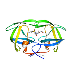
5T8H
DownloadVisualize
BU of 5t8h by Molmil
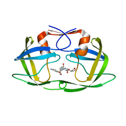
Joint X-ray/neutron structure of HIV-1 protease triple mutant (V32I,I47V,V82I) with amprenavir at pH 6.0
Descriptor: Protease, {3-[(4-AMINO-BENZENESULFONYL)-ISOBUTYL-AMINO]-1-BENZYL-2-HYDROXY-PROPYL}-CARBAMIC ACID TETRAHYDRO-FURAN-3-YL ESTER
Authors:Kovalevsky, A.Y, Gerlits, O.O.
Deposit date:2016-09-07
Release date:2017-03-01
Last modified:2024-04-03
Method:NEUTRON DIFFRACTION (1.85 Å), X-RAY DIFFRACTION
Cite:Room Temperature Neutron Crystallography of Drug Resistant HIV-1 Protease Uncovers Limitations of X-ray Structural Analysis at 100 K.
J. Med. Chem., 60, 2017
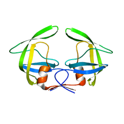
7N6T
DownloadVisualize
BU of 7n6t by Molmil
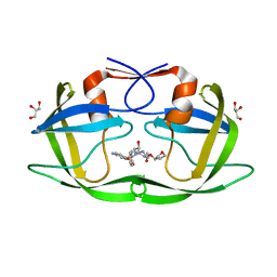
Crystal structure of inhibitor-free HIV-1 PRS17 revertant mutant PRS17 V48G
Descriptor: Protease
Authors:Burnaman, S.H, Wang, Y.-F, Weber, I.T.
Deposit date:2021-06-09
Release date:2021-09-08
Last modified:2023-10-18
Method:X-RAY DIFFRACTION (1.32 Å)
Cite:Revertant mutation V48G alters conformational dynamics of highly drug resistant HIV protease PRS17.
J.Mol.Graph.Model., 108, 2021
7N6X
DownloadVisualize
BU of 7n6x by Molmil
Crystal structure of HIV-1 Protease multiple mutants PRS17 bound to inhibitor Amprenavir
Descriptor: Protease, {3-[(4-AMINO-BENZENESULFONYL)-ISOBUTYL-AMINO]-1-BENZYL-2-HYDROXY-PROPYL}-CARBAMIC ACID TETRAHYDRO-FURAN-3-YL ESTER
Authors:Burnaman, S.H, Wang, Y.-F, Weber, I.T.
Deposit date:2021-06-09
Release date:2021-09-08
Last modified:2023-10-18
Method:X-RAY DIFFRACTION (1.47 Å)
Cite:Revertant mutation V48G alters conformational dynamics of highly drug resistant HIV protease PRS17.
J.Mol.Graph.Model., 108, 2021
7N6V
DownloadVisualize
BU of 7n6v by Molmil
Crystal structure of HIV-1 Protease multiple mutants PRS17 with Revertant mutation V48G bound to inhibitor Amprenavir
Descriptor: GLYCEROL, Protease, {3-[(4-AMINO-BENZENESULFONYL)-ISOBUTYL-AMINO]-1-BENZYL-2-HYDROXY-PROPYL}-CARBAMIC ACID TETRAHYDRO-FURAN-3-YL ESTER
Authors:Burnaman, S.H, Wang, Y.-F, Weber, I.T.
Deposit date:2021-06-09
Release date:2021-09-08
Last modified:2023-10-18
Method:X-RAY DIFFRACTION (1.39 Å)
Cite:Revertant mutation V48G alters conformational dynamics of highly drug resistant HIV protease PRS17.
J.Mol.Graph.Model., 108, 2021
7D3Z
DownloadVisualize
BU of 7d3z by Molmil
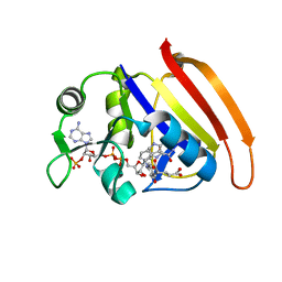
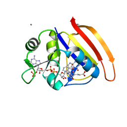
X-ray crystal Structure of E.coli Dihydrofolate Reductase complexed with folate and NADP+ at pH4.5
Descriptor: Dihydrofolate reductase, FOLIC ACID, NADP NICOTINAMIDE-ADENINE-DINUCLEOTIDE PHOSPHATE
Authors:Wan, Q, Dealwis, C.
Deposit date:2020-09-21
Release date:2021-06-09
Last modified:2023-11-29
Method:X-RAY DIFFRACTION (1.65 Å)
Cite:Capturing the Catalytic Proton of Dihydrofolate Reductase: Implications for General Acid-Base Catalysis
Acs Catalysis, 11, 2021
7D49
DownloadVisualize
BU of 7d49 by Molmil
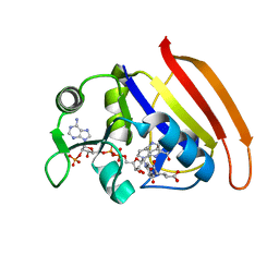
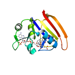
X-ray crystal Structure of E.coli Dihydrofolate Reductase complexed with folate and NADP+ at pH4.5
Descriptor: Dihydrofolate reductase, FOLIC ACID, NADP NICOTINAMIDE-ADENINE-DINUCLEOTIDE PHOSPHATE
Authors:Wan, Q, Dealwis, C.
Deposit date:2020-09-23
Release date:2021-06-09
Last modified:2023-11-29
Method:X-RAY DIFFRACTION (1.651 Å)
Cite:Capturing the Catalytic Proton of Dihydrofolate Reductase: Implications for General Acid-Base Catalysis
Acs Catalysis, 11, 2021
7D4X
DownloadVisualize
BU of 7d4x by Molmil
X-ray crystal Structure of E.coli Dihydrofolate Reductase complexed with folate and NADP+ at pH7.0
Descriptor: Dihydrofolate reductase, FOLIC ACID, NADP NICOTINAMIDE-ADENINE-DINUCLEOTIDE PHOSPHATE
Authors:Wan, Q, Dealwis, C.
Deposit date:2020-09-24
Release date:2021-06-09
Last modified:2023-11-29
Method:X-RAY DIFFRACTION (1.599 Å)
Cite:Capturing the Catalytic Proton of Dihydrofolate Reductase: Implications for General Acid-Base Catalysis
Acs Catalysis, 11, 2021
7D6G
DownloadVisualize
BU of 7d6g by Molmil
Neutron crystal Structure of E.coli Dihydrofolate Reductase complexed with folate and NADP+ at pH4.5
Descriptor: Dihydrofolate reductase, FOLIC ACID, MANGANESE (II) ION, ...
Authors:Wan, Q, Dealwis, C.
Deposit date:2020-09-30
Release date:2021-06-09
Last modified:2024-04-03
Method:NEUTRON DIFFRACTION (1.65 Å), X-RAY DIFFRACTION
Cite:Capturing the Catalytic Proton of Dihydrofolate Reductase: Implications for General Acid-Base Catalysis
Acs Catalysis, 11, 2021
7D4L
DownloadVisualize
BU of 7d4l by Molmil
X-ray crystal Structure of E.coli Dihydrofolate Reductase complexed with folate and NADP+ at pH7.0
Descriptor: Dihydrofolate reductase, FOLIC ACID, MANGANESE (II) ION, ...
Authors:Wan, Q, Dealwis, C.
Deposit date:2020-09-24
Release date:2021-06-09
Last modified:2023-11-29
Method:X-RAY DIFFRACTION (1.6 Å)
Cite:Capturing the Catalytic Proton of Dihydrofolate Reductase: Implications for General Acid-Base Catalysis
Acs Catalysis, 11, 2021
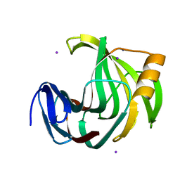
7EO6
DownloadVisualize
BU of 7eo6 by Molmil
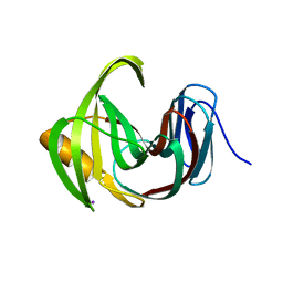
X-ray structure analysis of xylanase
Descriptor: Endo-1,4-beta-xylanase, IODIDE ION
Authors:Wan, Q, Yi, Y, Xu, S.
Deposit date:2021-04-21
Release date:2021-10-13
Last modified:2023-11-29
Method:X-RAY DIFFRACTION (1.9 Å)
Cite:Characterization and structural analysis of a thermophilic GH11 xylanase from compost metatranscriptome.
Appl.Microbiol.Biotechnol., 105, 2021
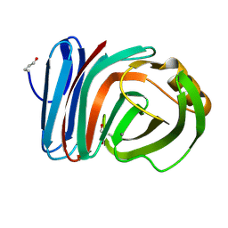
7EO3
DownloadVisualize
BU of 7eo3 by Molmil
X-ray structure analysis of beita-1,3-glucanase
Descriptor: 1,3-beta-glucanase, 2-AMINO-2-HYDROXYMETHYL-PROPANE-1,3-DIOL, MAGNESIUM ION
Authors:Wan, Q, Feng, J, Xu, S.
Deposit date:2021-04-21
Release date:2022-03-02
Last modified:2023-11-29
Method:X-RAY DIFFRACTION (1.141 Å)
Cite:Identification and structural analysis of a thermophilic beta-1,3-glucanase from compost
Bioresour Bioprocess, 8, 2021
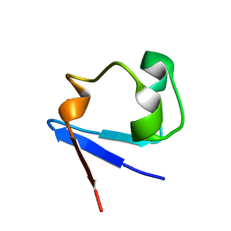
4K9F
DownloadVisualize
BU of 4k9f by Molmil
Neutron structure of Perdeuterated Rubredoxin refined against 1.75 resolution data collected on the new IMAGINE instrument at HFIR, ORNL
Descriptor: FE (III) ION, Rubredoxin
Authors:Munshi, P, Meilleur, F, Myles, D.
Deposit date:2013-04-19
Release date:2013-12-04
Last modified:2023-09-20
Method:NEUTRON DIFFRACTION (1.75 Å)
Cite:The IMAGINE instrument: first neutron protein structure and new capabilities for neutron macromolecular crystallography.
Acta Crystallogr.,Sect.D, 69, 2013
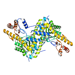
5TOQ
DownloadVisualize
BU of 5toq by Molmil
High resolution crystal structure of AAT
Descriptor: Aspartate aminotransferase, cytoplasmic
Authors:Mueser, T.C, Dajnowicz, S, Kovalevsky, A.
Deposit date:2016-10-18
Release date:2017-03-01
Last modified:2023-11-15
Method:X-RAY DIFFRACTION (1.2 Å)
Cite:Direct evidence that an extended hydrogen-bonding network influences activation of pyridoxal 5'-phosphate in aspartate aminotransferase.
J. Biol. Chem., 292, 2017

219515

PDB entries from 2024-05-08

PDB statisticsPDBj update infoContact PDBjnumon