Loading
PDBj
MenuPDBj@FacebookPDBj@TwitterPDBj@YouTubewwPDB FoundationwwPDB
RCSB PDBPDBeBMRBAdv. SearchSearch help
Search by PDB author
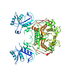
3HZG
DownloadVisualize
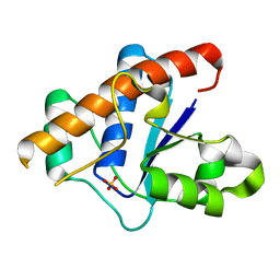
BU of 3hzg by Molmil
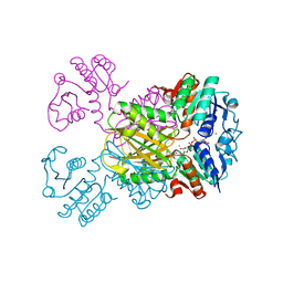
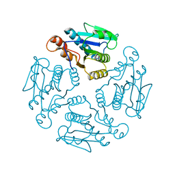
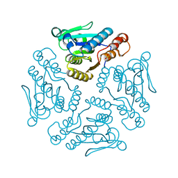
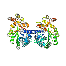
Crystal structure of mycobacterium tuberculosis thymidylate synthase X bound with FAD
Descriptor: FLAVIN-ADENINE DINUCLEOTIDE, GLYCEROL, PHOSPHATE ION, ...
Authors:Staker, B.L, Rathod, P, Hunter, J, Seattle Structural Genomics Center for Infectious Disease (SSGCID)
Deposit date:2009-06-23
Release date:2009-07-07
Last modified:2023-09-06
Method:X-RAY DIFFRACTION (2.45 Å)
Cite:Increasing the structural coverage of tuberculosis drug targets.
Tuberculosis (Edinb), 95, 2015
3I3R
DownloadVisualize
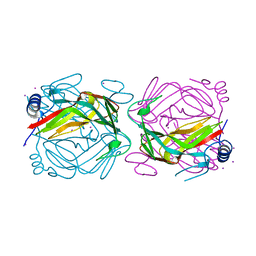
BU of 3i3r by Molmil
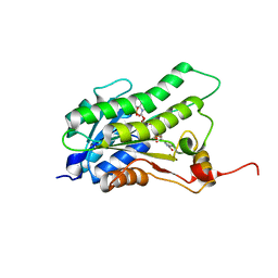
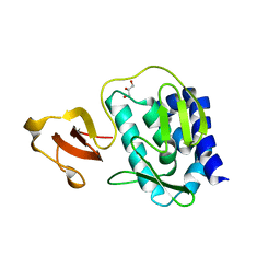
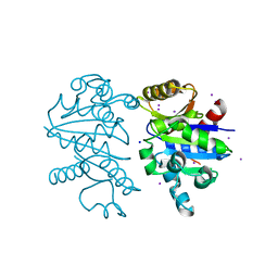
X-ray structure dihydrofolate reductase/thymidylate synthase from babesia bovis at 2.35A resolution
Descriptor: CHLORIDE ION, Dihydrofolate reductase/thymidylate synthase
Authors:Seattle Structural Genomics Center for Infectious Disease (SSGCID), Seattle Structural Genomics Center for Infectious Disease (SSGCID)
Deposit date:2009-06-30
Release date:2009-08-18
Last modified:2023-09-06
Method:X-RAY DIFFRACTION (2.35 Å)
Cite:Inhibitor-bound complexes of dihydrofolate reductase-thymidylate synthase from Babesia bovis.
Acta Crystallogr.,Sect.F, 67, 2011
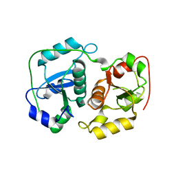
3HWI
DownloadVisualize
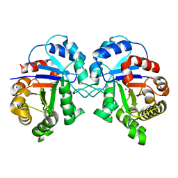
BU of 3hwi by Molmil
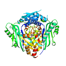
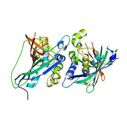
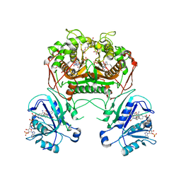
Crystal structure of probable thiosulfate sulfurtransferase Cysa2 (Rhodanese-like protein) from Mycobacterium tuberculosis
Descriptor: Thiosulfate sulfurtransferase
Authors:Seattle Structural Genomics Center for Infectious Disease (SSGCID)
Deposit date:2009-06-17
Release date:2009-06-30
Last modified:2023-09-06
Method:X-RAY DIFFRACTION (2.29 Å)
Cite:Increasing the structural coverage of tuberculosis drug targets.
Tuberculosis (Edinb), 95, 2015
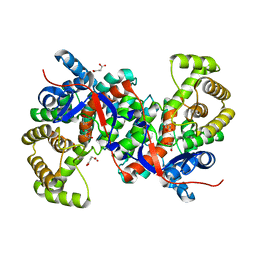
3HWK
DownloadVisualize
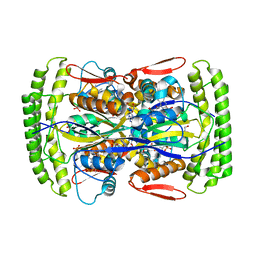
BU of 3hwk by Molmil
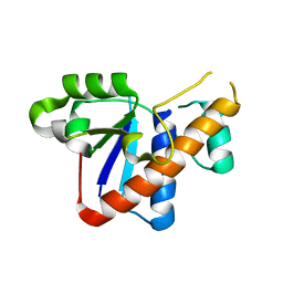
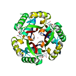
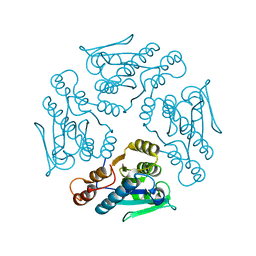
Crystal structure of methylcitrate synthase from Mycobacterium tuberculosis
Descriptor: Methylcitrate synthase, SUCCINIC ACID
Authors:Seattle Structural Genomics Center for Infectious Disease (SSGCID)
Deposit date:2009-06-17
Release date:2009-06-30
Last modified:2023-09-06
Method:X-RAY DIFFRACTION (2.3 Å)
Cite:Increasing the structural coverage of tuberculosis drug targets.
Tuberculosis (Edinb), 95, 2015
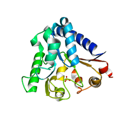
3ICO
DownloadVisualize
BU of 3ico by Molmil
Crystal structure of 6-phosphogluconolactonase from Mycobacterium tuberculosis
Descriptor: 6-phosphogluconolactonase, SULFATE ION
Authors:Seattle Structural Genomics Center for Infectious Disease (SSGCID)
Deposit date:2009-07-17
Release date:2009-07-28
Last modified:2023-09-06
Method:X-RAY DIFFRACTION (2.15 Å)
Cite:Increasing the structural coverage of tuberculosis drug targets.
Tuberculosis (Edinb), 95, 2015
3IJP
DownloadVisualize
BU of 3ijp by Molmil
Crystal structure of dihydrodipicolinate reductase from bartonella henselae at 2.0A resolution
Descriptor: CHLORIDE ION, Dihydrodipicolinate reductase, NADP NICOTINAMIDE-ADENINE-DINUCLEOTIDE PHOSPHATE, ...
Authors:Seattle Structural Genomics Center for Infectious Disease (SSGCID)
Deposit date:2009-08-04
Release date:2009-08-11
Last modified:2023-09-06
Method:X-RAY DIFFRACTION (2.3 Å)
Cite:The crystal structure of dihydrodipicolinate reductase from the human-pathogenic bacterium Bartonella henselae strain Houston-1 at 2.3 angstrom resolution.
Acta Crystallogr F Struct Biol Commun, 72, 2016
5BT9
DownloadVisualize
BU of 5bt9 by Molmil
Crystal Structure of FolM Alternative dihydrofolate reductase 1 from Brucella canis complexed with NADP
Descriptor: 3-oxoacyl-(Acyl-carrier-protein) reductase, NADP NICOTINAMIDE-ADENINE-DINUCLEOTIDE PHOSPHATE
Authors:Seattle Structural Genomics Center for Infectious Disease, Seattle Structural Genomics Center for Infectious Disease (SSGCID)
Deposit date:2015-06-02
Release date:2015-07-29
Last modified:2023-09-27
Method:X-RAY DIFFRACTION (1.5 Å)
Cite:Crystal structures of FolM alternative dihydrofolate reductase 1 from Brucella suis and Brucella canis.
Acta Crystallogr.,Sect.F, 78, 2022
3IML
DownloadVisualize
BU of 3iml by Molmil
Crystal Structure Of S-Adenosylmethionine Synthetase From Burkholderia Pseudomallei
Descriptor: S-adenosylmethionine synthetase
Authors:Staker, B.L, Seattle Structural Genomics Center for Infectious Disease (SSGCID)
Deposit date:2009-08-10
Release date:2009-09-22
Last modified:2023-09-06
Method:X-RAY DIFFRACTION (2.35 Å)
Cite:Combining functional and structural genomics to sample the essential Burkholderia structome.
Plos One, 8, 2013
3ILY
DownloadVisualize
BU of 3ily by Molmil
Apo crystal structure of protein tyrosine phosphatase from Entamoeba histolytica featuring a disordered active site
Descriptor: Protein tyrosine phosphatase, putative
Authors:Seattle Structural Genomics Center for Infectious Disease (SSGCID)
Deposit date:2009-08-07
Release date:2009-08-25
Last modified:2023-09-06
Method:X-RAY DIFFRACTION (2.2 Å)
Cite:Crystal structure and putative substrate identification for the Entamoeba histolytica low molecular weight tyrosine phosphatase.
Mol.Biochem.Parasitol., 193, 2014
3IDO
DownloadVisualize
BU of 3ido by Molmil
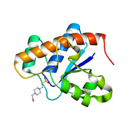
Crystal structure of protein tyrosine phosphatase from Entamoeba histolytica with a phosphotyrosine crude mimic HEPES molecule in the active site
Descriptor: 4-(2-HYDROXYETHYL)-1-PIPERAZINE ETHANESULFONIC ACID, Protein tyrosine phosphatase
Authors:Seattle Structural Genomics Center for Infectious Disease (SSGCID)
Deposit date:2009-07-21
Release date:2009-07-28
Last modified:2023-09-06
Method:X-RAY DIFFRACTION (2.2 Å)
Cite:Crystal structure and putative substrate identification for the Entamoeba histolytica low molecular weight tyrosine phosphatase.
Mol.Biochem.Parasitol., 193, 2014
3JS5
DownloadVisualize
BU of 3js5 by Molmil
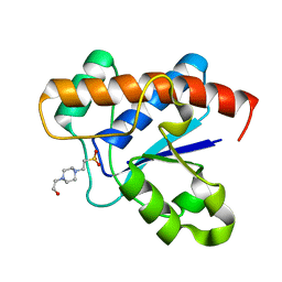
Crystal structure of protein tyrosine phosphatase from Entamoeba histolytica with Hepes in the active site. High resolution, alternative crystal form with 1 molecule in asymmetric unit
Descriptor: 4-(2-HYDROXYETHYL)-1-PIPERAZINE ETHANESULFONIC ACID, Protein tyrosine phosphatase, SODIUM ION
Authors:Seattle Structural Genomics Center for Infectious Disease (SSGCID)
Deposit date:2009-09-09
Release date:2009-09-22
Last modified:2023-09-06
Method:X-RAY DIFFRACTION (1.94 Å)
Cite:Crystal structure and putative substrate identification for the Entamoeba histolytica low molecular weight tyrosine phosphatase.
Mol.Biochem.Parasitol., 193, 2014
3KC6
DownloadVisualize
BU of 3kc6 by Molmil
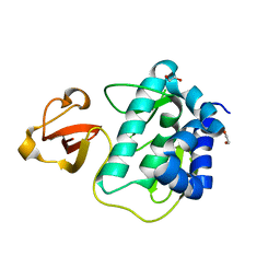
Crystal structure of the large c-terminal domain of polymerase basic protein 2 from influenza virus a/viet nam/1203/2004 (h5n1)
Descriptor: 1,2-ETHANEDIOL, Polymerase PB2
Authors:Seattle Structural Genomics Center for Infectious Disease (SSGCID)
Deposit date:2009-10-20
Release date:2010-03-09
Last modified:2023-09-06
Method:X-RAY DIFFRACTION (2.05 Å)
Cite:Biological and structural characterization of a host-adapting amino acid in influenza virus.
Plos Pathog., 6, 2010
1X9G
DownloadVisualize
BU of 1x9g by Molmil
PUTATIVE MAR1 RIBONUCLEASE FROM LEISHMANIA DONOVANI
Descriptor: PUTATIVE MAR1
Authors:Caruthers, J, Merritt, E.A, Structural Genomics of Pathogenic Protozoa Consortium (SGPP)
Deposit date:2004-08-20
Release date:2004-09-07
Last modified:2024-02-14
Method:X-RAY DIFFRACTION (2.41 Å)
Cite:Crystal structures and proposed structural/functional classification of three protozoan proteins from the isochorismatase superfamily.
Protein Sci., 14, 2005
1YZV
DownloadVisualize
BU of 1yzv by Molmil
HYPOTHETICAL PROTEIN FROM TRYPANOSOMA CRUZI
Descriptor: HYPOTHETICAL PROTEIN, SULFATE ION
Authors:Caruthers, J, Merritt, E.A, Structural Genomics of Pathogenic Protozoa Consortium (SGPP)
Deposit date:2005-02-28
Release date:2005-03-08
Last modified:2024-02-14
Method:X-RAY DIFFRACTION (2.001 Å)
Cite:Crystal structures and proposed structural/functional classification of three protozoan proteins from the isochorismatase superfamily.
Protein Sci., 14, 2005
3KHW
DownloadVisualize
BU of 3khw by Molmil
Crystal structure of the large c-terminal domain of polymerase basic protein 2 from influenza virus a/mexico/indre4487/2009(h1n1)
Descriptor: GLYCEROL, Polymerase PB2
Authors:Seattle Structural Genomics Center for Infectious Disease (SSGCID)
Deposit date:2009-10-31
Release date:2010-03-09
Last modified:2023-09-06
Method:X-RAY DIFFRACTION (2.1 Å)
Cite:Biological and structural characterization of a host-adapting amino acid in influenza virus.
Plos Pathog., 6, 2010
3KHP
DownloadVisualize
BU of 3khp by Molmil
Crystal structure of a possible dehydrogenase from Mycobacterium tuberculosis at 2.3A resolution
Descriptor: CHLORIDE ION, L(+)-TARTARIC ACID, MaoC family protein
Authors:Seattle Structural Genomics Center for Infectious Disease (SSGCID), Seattle Structural Genomics Center for Infectious Disease (SSGCID)
Deposit date:2009-10-30
Release date:2009-12-01
Last modified:2023-09-06
Method:X-RAY DIFFRACTION (2.3 Å)
Cite:Increasing the structural coverage of tuberculosis drug targets.
Tuberculosis (Edinb), 95, 2015
3KE1
DownloadVisualize
BU of 3ke1 by Molmil
Crystal structure of 2C-methyl-D-erythritol 2,4-cyclodiphosphate synthase from Burkholderia pseudomallei in complex with a fragment-nucleoside fusion D000161829
Descriptor: 2-C-methyl-D-erythritol 2,4-cyclodiphosphate synthase, 5'-deoxy-5'-[(pyridin-4-ylcarbonyl)amino]cytidine, POTASSIUM ION, ...
Authors:Seattle Structural Genomics Center for Infectious Disease (SSGCID)
Deposit date:2009-10-23
Release date:2009-11-10
Last modified:2023-09-06
Method:X-RAY DIFFRACTION (2.05 Å)
Cite:Cytidine derivatives as IspF inhibitors of Burkolderia pseudomallei.
Bioorg.Med.Chem.Lett., 8, 2013
3KRB
DownloadVisualize
BU of 3krb by Molmil
Structure of Aldose Reductase from Giardia Lamblia at 1.75A Resolution
Descriptor: 1,2-ETHANEDIOL, Aldose reductase, NADP NICOTINAMIDE-ADENINE-DINUCLEOTIDE PHOSPHATE, ...
Authors:Seattle Structural Genomics Center for Infectious Disease (SSGCID)
Deposit date:2009-11-18
Release date:2009-12-01
Last modified:2023-09-06
Method:X-RAY DIFFRACTION (1.75 Å)
Cite:Structure of aldose reductase from Giardia lamblia.
Acta Crystallogr.,Sect.F, 67, 2011
3K9G
DownloadVisualize
BU of 3k9g by Molmil
CRYSTAL STRUCTURE OF A PLASMID PARTITION PROTEIN FROM BORRELIA BURGDORFERI AT 2.25A RESOLUTION, iodide soak
Descriptor: IODIDE ION, PF-32 protein, SODIUM ION
Authors:Seattle Structural Genomics Center for Infectious Disease (SSGCID)
Deposit date:2009-10-15
Release date:2009-11-03
Last modified:2024-02-21
Method:X-RAY DIFFRACTION (2.25 Å)
Cite:SAD phasing using iodide ions in a high-throughput structural genomics environment.
J Struct Funct Genomics, 12, 2011
3K2H
DownloadVisualize
BU of 3k2h by Molmil
Co-crystal structure of dihydrofolate reductase/thymidylate synthase from Babesia bovis with dUMP, Pemetrexed and NADP
Descriptor: 1,2-ETHANEDIOL, 2'-DEOXYURIDINE 5'-MONOPHOSPHATE, 2-{4-[2-(2-AMINO-4-OXO-4,7-DIHYDRO-3H-PYRROLO[2,3-D]PYRIMIDIN-5-YL)-ETHYL]-BENZOYLAMINO}-PENTANEDIOIC ACID, ...
Authors:Seattle Structural Genomics Center for Infectious Disease (SSGCID)
Deposit date:2009-09-30
Release date:2009-10-13
Last modified:2023-09-06
Method:X-RAY DIFFRACTION (2.2 Å)
Cite:Inhibitor-bound complexes of dihydrofolate reductase-thymidylate synthase from Babesia bovis.
Acta Crystallogr.,Sect.F, 67, 2011
1XN4
DownloadVisualize
BU of 1xn4 by Molmil
PUTATIVE MAR1 RIBONUCLEASE FROM LEISHMANIA MAJOR
Descriptor: ribonuclease MAR1
Authors:Caruthers, J, Merritt, E.A, Structural Genomics of Pathogenic Protozoa Consortium (SGPP)
Deposit date:2004-10-04
Release date:2004-10-12
Last modified:2023-08-23
Method:X-RAY DIFFRACTION (3.8 Å)
Cite:Crystal structures and proposed structural/functional classification of three protozoan proteins from the isochorismatase superfamily.
Protein Sci., 14, 2005
3JVI
DownloadVisualize
BU of 3jvi by Molmil
Product state mimic crystal structure of protein tyrosine phosphatase from Entamoeba histolytica
Descriptor: Protein tyrosine phosphatase, SULFATE ION
Authors:Seattle Structural Genomics Center for Infectious Disease (SSGCID)
Deposit date:2009-09-16
Release date:2009-09-22
Last modified:2023-09-06
Method:X-RAY DIFFRACTION (1.8 Å)
Cite:Crystal structure and putative substrate identification for the Entamoeba histolytica low molecular weight tyrosine phosphatase.
Mol.Biochem.Parasitol., 193, 2014
3KM3
DownloadVisualize
BU of 3km3 by Molmil
Crystal structure of eoxycytidine triphosphate deaminase from anaplasma phagocytophilum at 2.1A resolution
Descriptor: Deoxycytidine triphosphate deaminase, IODIDE ION
Authors:Seattle Structural Genomics Center for Infectious Disease (SSGCID)
Deposit date:2009-11-09
Release date:2009-11-24
Last modified:2024-02-21
Method:X-RAY DIFFRACTION (2.1 Å)
Cite:SAD phasing using iodide ions in a high-throughput structural genomics environment.
J.Struct.Funct.Genom., 12, 2011
3KRS
DownloadVisualize
BU of 3krs by Molmil
Structure of Triosephosphate Isomerase from Cryptosporidium Parvum at 1.55A Resolution
Descriptor: SODIUM ION, Triosephosphate isomerase, UNKNOWN ATOM OR ION
Authors:Seattle Structural Genomics Center for Infectious Disease (SSGCID)
Deposit date:2009-11-19
Release date:2009-12-01
Last modified:2023-09-06
Method:X-RAY DIFFRACTION (1.55 Å)
Cite:Structure of triosephosphate isomerase from Cryptosporidium parvum.
Acta Crystallogr.,Sect.F, 67, 2011
3KW3
DownloadVisualize
BU of 3kw3 by Molmil
Crystal structure of alanine racemase from Bartonella henselae with covalently bound pyridoxal phosphate
Descriptor: Alanine racemase
Authors:Seattle Structural Genomics Center for Infectious Disease (SSGCID)
Deposit date:2009-11-30
Release date:2009-12-22
Last modified:2023-11-22
Method:X-RAY DIFFRACTION (2.04 Å)
Cite:SAD phasing using iodide ions in a high-throughput structural genomics environment.
J.Struct.Funct.Genom., 12, 2011

222415

PDB entries from 2024-07-10

PDB statisticsPDBj update infoContact PDBjnumon