Loading
PDBj
MenuPDBj@FacebookPDBj@TwitterPDBj@YouTubewwPDB FoundationwwPDB
RCSB PDBPDBeBMRBAdv. SearchSearch help
Search by PDB author
2RE3
DownloadVisualize
BU of 2re3 by Molmil
CRYSTAL STRUCTURE OF a DUF1285 family protein (SPO_0140) FROM SILICIBACTER POMEROYI DSS-3 AT 2.50 A RESOLUTION
Descriptor: GLYCEROL, Uncharacterized protein
Authors:Joint Center for Structural Genomics (JCSG)
Deposit date:2007-09-25
Release date:2007-10-09
Last modified:2023-01-25
Method:X-RAY DIFFRACTION (2.5 Å)
Cite:Structures of the first representatives of Pfam family PF06938 (DUF1285) reveal a new fold with repeated structural motifs and possible involvement in signal transduction.
Acta Crystallogr.,Sect.F, 66, 2010
2ETS
DownloadVisualize
BU of 2ets by Molmil
CRYSTAL STRUCTURE OF A BACTERIAL DOMAIN OF UNKNOWN FUNCTION FROM DUF1798 FAMILY (MW1337) FROM STAPHYLOCOCCUS AUREUS SUBSP. AUREUS AT 2.25 A RESOLUTION
Descriptor: CHLORIDE ION, PHOSPHATE ION, hypothetical protein
Authors:Joint Center for Structural Genomics (JCSG)
Deposit date:2005-10-27
Release date:2005-11-08
Last modified:2023-01-25
Method:X-RAY DIFFRACTION (2.25 Å)
Cite:Crystal structures of MW1337R and lin2004: representatives of a novel protein family that adopt a four-helical bundle fold.
Proteins, 71, 2008
2FG0
DownloadVisualize
BU of 2fg0 by Molmil
Crystal structure of a putative gamma-d-glutamyl-l-diamino acid endopeptidase (npun_r0659) from nostoc punctiforme pcc 73102 at 1.79 A resolution
Descriptor: COG0791: Cell wall-associated hydrolases (invasion-associated proteins), GLYCEROL
Authors:Joint Center for Structural Genomics (JCSG)
Deposit date:2005-12-20
Release date:2006-01-10
Last modified:2023-01-25
Method:X-RAY DIFFRACTION (1.79 Å)
Cite:Structural Basis of Murein Peptide Specificity of a gamma-D-Glutamyl-L-Diamino Acid Endopeptidase.
Structure, 17, 2009
2EVR
DownloadVisualize
BU of 2evr by Molmil
CRYSTAL STRUCTURE OF A PUTATIVE GAMMA-D-GLUTAMYL-L-DIAMINO ACID ENDOPEPTIDASE (NPUN_R0659) FROM NOSTOC PUNCTIFORME PCC 73102 AT 1.60 A RESOLUTION
Descriptor: 1,2-ETHANEDIOL, CHLORIDE ION, COG0791: Cell wall-associated hydrolases (invasion-associated proteins), ...
Authors:Joint Center for Structural Genomics (JCSG)
Deposit date:2005-10-31
Release date:2005-11-22
Last modified:2023-01-25
Method:X-RAY DIFFRACTION (1.6 Å)
Cite:Structural Basis of Murein Peptide Specificity of a gamma-D-Glutamyl-L-Diamino Acid Endopeptidase.
Structure, 17, 2009
2FEA
DownloadVisualize
BU of 2fea by Molmil
Crystal structure of MtnX phosphatase from Bacillus Subtilis at 2.00 A resolution
Descriptor: 1,2-ETHANEDIOL, 2-hydroxy-3-keto-5-methylthiopentenyl-1-phosphate phosphatase, MAGNESIUM ION, ...
Authors:Joint Center for Structural Genomics (JCSG)
Deposit date:2005-12-15
Release date:2005-12-27
Last modified:2023-01-25
Method:X-RAY DIFFRACTION (2 Å)
Cite:Crystal structure of MtnX phosphatase from Bacillus subtilis at 2.0 A resolution provides a structural basis for bipartite phosphomonoester hydrolysis of 2-hydroxy-3-keto-5-methylthiopentenyl-1-phosphate.
Proteins, 69, 2007
2F46
DownloadVisualize
BU of 2f46 by Molmil
Crystal structure of a putative phosphatase (nma1982) from neisseria meningitidis z2491 at 1.41 A resolution
Descriptor: CHLORIDE ION, UNKNOWN LIGAND, hypothetical protein
Authors:Joint Center for Structural Genomics (JCSG)
Deposit date:2005-11-22
Release date:2006-02-07
Last modified:2023-01-25
Method:X-RAY DIFFRACTION (1.41 Å)
Cite:Crystal structure of NMA1982 from Neisseria meningitidis at 1.5 A resolution provides a structural scaffold for nonclassical, eukaryotic-like phosphatases.
Proteins, 69, 2007
2FNA
DownloadVisualize
BU of 2fna by Molmil
Crystal structure of an archaeal aaa+ atpase (sso1545) from sulfolobus solfataricus p2 at 2.00 A resolution
Descriptor: 1,2-ETHANEDIOL, ADENOSINE-5'-DIPHOSPHATE, Conserved hypothetical protein, ...
Authors:Joint Center for Structural Genomics (JCSG)
Deposit date:2006-01-10
Release date:2006-02-07
Last modified:2023-01-25
Method:X-RAY DIFFRACTION (2 Å)
Cite:Crystal structure of a novel archaeal AAA+ ATPase SSO1545 from Sulfolobus solfataricus.
Proteins, 74, 2009
2G36
DownloadVisualize
BU of 2g36 by Molmil
Crystal structure of Tryptophanyl-tRNA synthetase (EC 6.1.1.2) (Tryptophan-tRNA ligase)(TrpRS) (tm0492) from THERMOTOGA MARITIMA at 2.50 A resolution
Descriptor: IRON/SULFUR CLUSTER, TRYPTOPHAN, Tryptophanyl-tRNA synthetase
Authors:Joint Center for Structural Genomics (JCSG)
Deposit date:2006-02-17
Release date:2006-03-28
Last modified:2024-04-03
Method:X-RAY DIFFRACTION (2.5 Å)
Cite:Structure of a tryptophanyl-tRNA synthetase containing an iron-sulfur cluster.
Acta Crystallogr.,Sect.F, 66, 2010
2FNO
DownloadVisualize
BU of 2fno by Molmil
Crystal structure of a glutathione s-transferase (atu5508) from agrobacterium tumefaciens str. c58 at 2.00 A resolution
Descriptor: AGR_pAT_752p, THIOCYANATE ION
Authors:Joint Center for Structural Genomics (JCSG)
Deposit date:2006-01-11
Release date:2006-02-14
Last modified:2023-01-25
Method:X-RAY DIFFRACTION (2 Å)
Cite:Comparative structural analysis of a novel glutathioneS-transferase (ATU5508) from Agrobacterium tumefaciens at 2.0 A resolution.
Proteins, 65, 2006
3CM1
DownloadVisualize
BU of 3cm1 by Molmil
Crystal structure of SsgA-like sporulation-specific cell division protein (YP_290167.1) from Thermobifida fusca YX-ER1 at 2.60 A resolution
Descriptor: SsgA-like sporulation-specific cell division protein
Authors:Joint Center for Structural Genomics (JCSG), Chruszcz, M, Minor, W, Wang, S.
Deposit date:2008-03-20
Release date:2008-04-01
Last modified:2023-02-01
Method:X-RAY DIFFRACTION (2.6 Å)
Cite:Structural and functional characterizations of SsgB, a conserved activator of developmental cell division in morphologically complex actinomycetes.
J.Biol.Chem., 284, 2009
3H50
DownloadVisualize
BU of 3h50 by Molmil
CRYSTAL STRUCTURE OF A TETRACENOMYCIN POLYKETIDE SYNTHESIS PROTEIN (TCMJ) FROM XANTHOMONAS CAMPESTRIS PV. CAMPESTRIS AT 1.60 A RESOLUTION
Descriptor: ACETATE ION, Tetracenomycin polyketide synthesis protein, ZINC ION
Authors:Joint Center for Structural Genomics (JCSG)
Deposit date:2009-04-21
Release date:2009-05-05
Last modified:2023-02-01
Method:X-RAY DIFFRACTION (1.6 Å)
Cite:Conformational changes associated with the binding of zinc acetate at the putative active site of XcTcmJ, a cupin from Xanthomonas campestris pv. campestris.
Acta Crystallogr.,Sect.F, 66, 2010
3IRB
DownloadVisualize
BU of 3irb by Molmil
Crystal structure of protein with unknown function from DUF35 family (13815350) from SULFOLOBUS SOLFATARICUS at 1.80 A resolution
Descriptor: ACETIC ACID, SULFATE ION, ZINC ION, ...
Authors:Joint Center for Structural Genomics (JCSG)
Deposit date:2009-08-21
Release date:2009-09-01
Last modified:2023-02-01
Method:X-RAY DIFFRACTION (1.8 Å)
Cite:The structure of SSO2064, the first representative of Pfam family PF01796, reveals a novel two-domain zinc-ribbon OB-fold architecture with a potential acyl-CoA-binding role.
Acta Crystallogr.,Sect.F, 66, 2010
3KHI
DownloadVisualize
BU of 3khi by Molmil
Crystal structure of a Putative Metal-dependent Hydrolase (YP_001336084.1) from Klebsiella pneumoniae subsp. pneumoniae MGH 78578 at 1.95 A resolution
Descriptor: 1,2-ETHANEDIOL, CHLORIDE ION, Putative Metal-dependent Hydrolase, ...
Authors:Joint Center for Structural Genomics (JCSG)
Deposit date:2009-10-30
Release date:2009-11-10
Last modified:2023-02-01
Method:X-RAY DIFFRACTION (1.95 Å)
Cite:The Structure of Mlc Titration Factor A (MtfA/YeeI) Reveals a Prototypical Zinc Metallopeptidase Related to Anthrax Lethal Factor.
J.Bacteriol., 194, 2012
3KW0
DownloadVisualize
BU of 3kw0 by Molmil
Crystal structure of Cysteine peptidase (NP_982244.1) from BACILLUS CEREUS ATCC 10987 at 2.50 A resolution
Descriptor: CHLORIDE ION, Cysteine peptidase, LYSINE
Authors:Joint Center for Structural Genomics (JCSG)
Deposit date:2009-11-30
Release date:2009-12-15
Last modified:2023-02-01
Method:X-RAY DIFFRACTION (2.5 Å)
Cite:Structural Analysis of Papain-Like NlpC/P60 Superfamily Enzymes with a Circularly Permuted Topology Reveals Potential Lipid Binding Sites.
Plos One, 6, 2011
3HN7
DownloadVisualize
BU of 3hn7 by Molmil
Crystal structure of a murein peptide ligase mpl (psyc_0032) from psychrobacter arcticus 273-4 at 1.65 A resolution
Descriptor: UDP-N-acetylmuramate-L-alanine ligase
Authors:Joint Center for Structural Genomics (JCSG)
Deposit date:2009-05-29
Release date:2009-06-16
Last modified:2023-02-01
Method:X-RAY DIFFRACTION (1.65 Å)
Cite:Structure and function of the first full-length murein Peptide ligase (mpl) cell wall recycling protein.
Plos One, 6, 2011
3L5O
DownloadVisualize
BU of 3l5o by Molmil
Crystal structure of protein with unknown function from DUF364 family (ZP_00559375.1) from Desulfitobacterium hafniense DCB-2 at 2.01 A resolution
Descriptor: 1,2-ETHANEDIOL, CHLORIDE ION, IMIDAZOLE, ...
Authors:Joint Center for Structural Genomics, Joint Center for Structural Genomics (JCSG)
Deposit date:2009-12-22
Release date:2010-02-02
Last modified:2023-02-01
Method:X-RAY DIFFRACTION (2.01 Å)
Cite:Structure of the first representative of Pfam family PF04016 (DUF364) reveals enolase and Rossmann-like folds that combine to form a unique active site with a possible role in heavy-metal chelation.
Acta Crystallogr.,Sect.F, 66, 2010
3LIU
DownloadVisualize
BU of 3liu by Molmil
Crystal structure of Putative cell adhesion protein (YP_001304840.1) from Parabacteroides distasonis ATCC 8503 at 2.05 A resolution
Descriptor: 1,2-ETHANEDIOL, CHLORIDE ION, GLYCEROL, ...
Authors:Joint Center for Structural Genomics (JCSG)
Deposit date:2010-01-25
Release date:2010-02-09
Last modified:2023-02-01
Method:X-RAY DIFFRACTION (2.05 Å)
Cite:A Distinct Type of Pilus from the Human Microbiome.
Cell, 165, 2016
2IAY
DownloadVisualize
BU of 2iay by Molmil
Crystal structure of a duf1831 family protein (lp2179) from lactobacillus plantarum at 1.20 A resolution
Descriptor: CHLORIDE ION, GLYCEROL, Hypothetical protein
Authors:Joint Center for Structural Genomics (JCSG)
Deposit date:2006-09-08
Release date:2006-10-10
Last modified:2023-01-25
Method:X-RAY DIFFRACTION (1.2 Å)
Cite:Structure of LP2179, the first representative of Pfam family PF08866, suggests a new fold with a role in amino-acid metabolism.
Acta Crystallogr.,Sect.F, 66, 2010
2GLZ
DownloadVisualize
BU of 2glz by Molmil
Crystal structure of a formylmethanofuran dehydrogenase subunit e-like protein (dhaf_2992) from desulfitobacterium hafniense dcb-2 at 1.45 A resolution
Descriptor: 1,2-ETHANEDIOL, NICKEL (II) ION, ZINC ION, ...
Authors:Joint Center for Structural Genomics (JCSG)
Deposit date:2006-04-05
Release date:2006-04-18
Last modified:2023-01-25
Method:X-RAY DIFFRACTION (1.45 Å)
Cite:Structures of three members of Pfam PF02663 (FmdE) implicated in microbial methanogenesis reveal a conserved alpha+beta core domain and an auxiliary C-terminal treble-clef zinc finger.
Acta Crystallogr.,Sect.F, 66, 2010
2GVI
DownloadVisualize
BU of 2gvi by Molmil
Crystal structure of a putative formylmethanofuran dehydrogenase subunit e (ta1109) from thermoplasma acidophilum at 1.87 A resolution
Descriptor: 1,2-ETHANEDIOL, ACETIC ACID, UNKNOWN LIGAND, ...
Authors:Joint Center for Structural Genomics (JCSG)
Deposit date:2006-05-02
Release date:2006-06-13
Last modified:2023-01-25
Method:X-RAY DIFFRACTION (1.87 Å)
Cite:Structures of three members of Pfam PF02663 (FmdE) implicated in microbial methanogenesis reveal a conserved alpha+beta core domain and an auxiliary C-terminal treble-clef zinc finger.
Acta Crystallogr.,Sect.F, 66, 2010
1G5P
DownloadVisualize
BU of 1g5p by Molmil
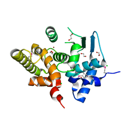
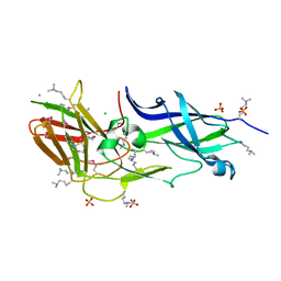
NITROGENASE IRON PROTEIN FROM AZOTOBACTER VINELANDII
Descriptor: IRON/SULFUR CLUSTER, NITROGENASE IRON PROTEIN
Authors:Strop, P, Takahara, P.M, Chiu, H.J, Angove, H.C, Burgess, B.K, Rees, D.C.
Deposit date:2000-11-01
Release date:2001-01-31
Last modified:2023-08-09
Method:X-RAY DIFFRACTION (2.2 Å)
Cite:Crystal structure of the all-ferrous [4Fe-4S]0 form of the nitrogenase iron protein from Azotobacter vinelandii.
Biochemistry, 40, 2001
4EPS
DownloadVisualize
BU of 4eps by Molmil
Crystal structure of a fimbrial protein (BACOVA_04982) from Bacteroides ovatus ATCC 8483 at 1.85 A resolution
Descriptor: CHLORIDE ION, GLYCEROL, SULFATE ION, ...
Authors:Joint Center for Structural Genomics (JCSG)
Deposit date:2012-04-17
Release date:2012-06-13
Last modified:2023-02-01
Method:X-RAY DIFFRACTION (1.85 Å)
Cite:A Distinct Type of Pilus from the Human Microbiome.
Cell, 165, 2016
7SUT
DownloadVisualize
BU of 7sut by Molmil
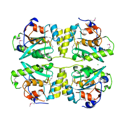
Light harvesting phycobiliprotein HaPE645 from the cryptophyte Hemiselmis andersenii CCMP644
Descriptor: (15,16)-DIHYDROBILIVERDIN (SINGLY LINKED), 2-[BIS-(2-HYDROXY-ETHYL)-AMINO]-2-HYDROXYMETHYL-PROPANE-1,3-DIOL, CHLORIDE ION, ...
Authors:Rathbone, H.W, Michie, K.A, Laos, A.L, Curmi, P.M.G.
Deposit date:2021-11-18
Release date:2023-10-25
Last modified:2023-11-29
Method:X-RAY DIFFRACTION (1.49 Å)
Cite:Molecular dissection of the soluble photosynthetic antenna from the cryptophyte alga Hemiselmis andersenii.
Commun Biol, 6, 2023
4DGU
DownloadVisualize
BU of 4dgu by Molmil
Crystal structure of a putative cell adhesion protein (BT0320) from Bacteroides thetaiotaomicron VPI-5482 at 2.37 A resolution
Descriptor: CHLORIDE ION, ZINC ION, putative cell adhesion protein
Authors:Joint Center for Structural Genomics (JCSG)
Deposit date:2012-01-26
Release date:2012-02-15
Last modified:2023-02-01
Method:X-RAY DIFFRACTION (2.37 Å)
Cite:A Distinct Type of Pilus from the Human Microbiome.
Cell, 165, 2016
3UFI
DownloadVisualize
BU of 3ufi by Molmil
Crystal structure of a putative cell adhesion protein (BACOVA_04980) from Bacteroides ovatus ATCC 8483 at 2.18 A resolution
Descriptor: CHLORIDE ION, SULFATE ION, hypothetical protein BACOVA_04980
Authors:Joint Center for Structural Genomics (JCSG)
Deposit date:2011-11-01
Release date:2011-12-14
Last modified:2023-02-01
Method:X-RAY DIFFRACTION (2.18 Å)
Cite:A Distinct Type of Pilus from the Human Microbiome.
Cell, 165, 2016

223790

PDB entries from 2024-08-14

PDB statisticsPDBj update infoContact PDBjnumon